

- About Us
- Information
-

The Author ensures that the research has been conducted responsibly and ethically with adherence to all relevant regulations. read more..
- For Authors
- For Reviewer
- Manuscript Guidelines
- Membership
- Publication Ethics
-
- Journals
- Reprints
- e-Books
- Videos
- Policies
- Contact Us
 COVID-19
- Submissions



Full Text
Techniques in Neurosurgery & Neurology
Utility of Tomographic Planning in Transpedicular Thoracolumbar Percutaneous Fixation
Molina Choez DD1*, Arteaga JA2, Enríquez Viteri JC3, Sacoto CB3 and Alava KS3
1 Department of Neurosurgery, Novaclínica Santa Cecilia, Hospital Sta Margarita, Universidad San Francisco de Quito, Ecuador
2 Department of Neurosurgery, Hospital Clínica Kennedy, Universidad Espíritu Santo, Ecuador
3 Universidad San Francisco de Quito, Ecuador
*Corresponding author:Duval D Molina Choeza, Department of Neurosurgery, Novaclínica Santa Cecilia, Quito-Ecuador, Hospital Sta Margarita, Portoviejo- Ecuador, Universidad San Francisco de Quito, Quito, Ecuador
Submission: June 18, 2026;Published: July 21, 2026

ISSN 2637-7748
Volume6 Issue2
Abstract
Background: Transpedicular fixation is useful to resolve various degenerative and traumatic diseases of
the thoracolumbar spine. Minimally Invasive Percutaneous Transpedicular Fixation (MIPTF) is preferred
by many spine surgeons for its advantages related to less tissue damage, however, a constant concern of
surgeons is the precision in percutaneous screw placement. The purpose of the study is to illustrate the
utility of performing preoperative tomographic planning by analyzing the morphometric characteristics
of each individual vertebra to increase the accuracy of thoracolumbar percutaneous transpedicular screw
placement.
Methods: Preoperative Computed Tomography (CT) of 96 patients with MIPTF between April 2016 and
December 2024 were included. 34 patients with traumatic disease and 62 patients with degenerative
disease. In all cases, a postoperative tomography was obtained to verify the location of the screws. OsiriX
DICOM Viewer program was used to perform the analysis of each fixed vertebra. No Intraoperative
Neurophysiological Monitoring (INM) was used. We used conventional C-arm fluoroscopy.
Result: 438 screws were placed by two experienced surgeons at four medical centers. 433 (98.85%)
screws with intrapedicular position and 5 (1.14%) screws with extrapedicular position showing eruption
of the cortical vertebral body and external cortical pedicle that involved less than 50% of its diameter in
the five cases. The positioning accuracy was graded using the Gertzbein and Robbins classification.
Conclusion: Performing preoperative morphometric analysis with CT prior to thoracolumbar
percutaneous transpedicular fixation is an acceptable conduct to increase fixation accuracy in absence
of INM.
Keywords:Planning CT pedicle screw; Percutaneous transpedicular fixation
Introduction
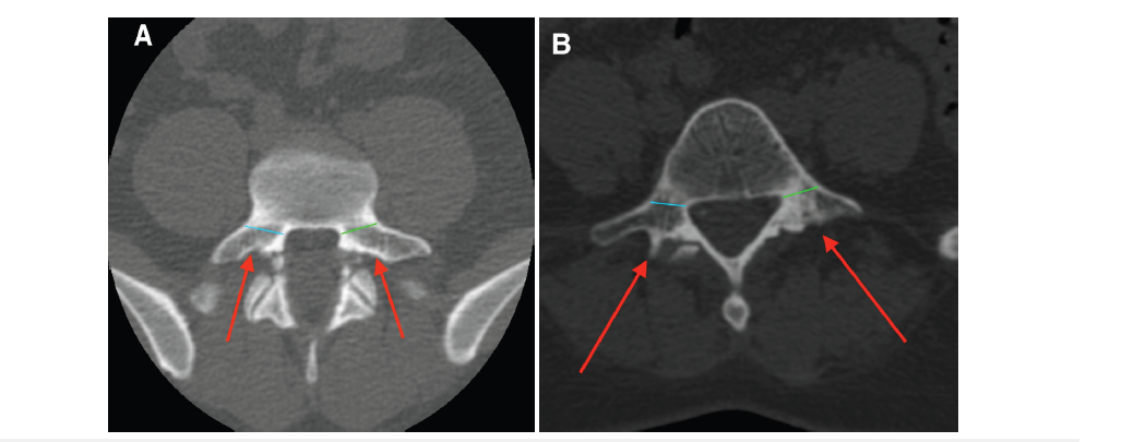
Spine degenerative and traumatic pathologies have a considerably high impact in cost and economic resources. Wide spectrum of these pathologies demands advance techniques that includes complex reconstructions of the anterior and posterior spine. One of the most widely tools used for its adequate stabilization is transpedicular fixation, which proven biomechanical advantages. During the last decade with the advent of minimally invasive techniques includes thoracolumbar fractures and various degenerative processes of the lumbar spine that are managed through the placement of the transpedicular percutaneous screws, and offers benefits related to less muscle manipulation, blood stray minor, shorter recovery period and better cosmetics results. For the precise placement of pedicle screws by percutaneous technique, is requires a training in minimally invasive techniques including deep anatomical knowledge with adequate radiological correlation and careful preoperative planning. In this study, we propose to plan the precision placement of the transpedicular percutaneous screws in a case series from 4 hospitals using a basic planning tool as fine section tomography of the spine segment, focusing on the level that will be instrumented considering all the morphological and dimensions of each vertebra with their own characteristic. Several studies that demonstrate the utility of preoperative morphometric analysis to improve the precision of spinal instrumentation at cervical, thoracic, and lumbosacral spine levels and the variability of the morphometric characteristics of each vertebra (Figure 1) & [1-10].
Figure 1:Axial sections of two L5 vertebrae. A. 43-year-old male. B. 39-year-old female. Differences are observed in the shape (ovoid in A and conical in B) and angulation required for pedicle screw placement.
Material and Methods
Figure 2:Gertzbein & Robbins CT classification [11]. Grade 1 (0mm), Grade 2 (<2mm), Grade 3 (2-4mm), and Grade 4 (>4mm).
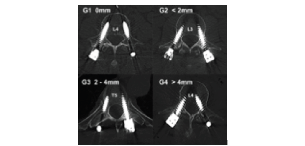
Observational experimental study of 96 consecutive cases operated by two Spine Surgeons between April 2016 to December 2024 in 4 Hospitals in Ecuador have been analyzed. All cases were operated by transpedicular percutaneous fixation with or without arthrodesis of the anterior spine according to the individual requirement of each patient. 62 cases of lumbar spine degenerative pathology and 34 cases of thoracic or lumbar fractures were treated. All the cases operated by transpedicular percutaneous fixation and non-monitored Jamshidi needle by cannulation technique (without neurophysiological monitoring support). In all cases we used conventional C-arm fluoroscopy with anteroposterior and lateral projections. Each case had a fine section computed tomography of the vertebral segment focusing on the vertebra that will be fixed. With the tomography we proceed to measurement of the dorso-ventral and lateral-medial diameter of each pedicle, the angle of the lateral medial trajectory (inclination in the axial plane) was performed, as well as a morphometric analysis of each vertebral body to optimize the precision of the diameter and length of each screw applied, the initial location in the anteroposterior view of each Jamshidi needle, and its approximate angle of incidence during cannulation in the sagittal and axial planes. The Osirix (TR) program was used to perform the measurements and planning (Figure 2). Postoperative tomographies were performed within the first 72 hours to verify the location of each screw in the axial, sagittal, coronal planes, and 3D reconstruction. The positioning accuracy was graded using the Gertzbein and Robbins classification [11]. A clinical correlation of the postoperative tomographic findings was performed, verifying the absence of radiculopathy. In all cases, biplanar fluoroscopy was used as an intraoperative instrumentation tool. Intraoperative neurophysiological monitoring and other precision tools such as conventional navigation were not used.
Result
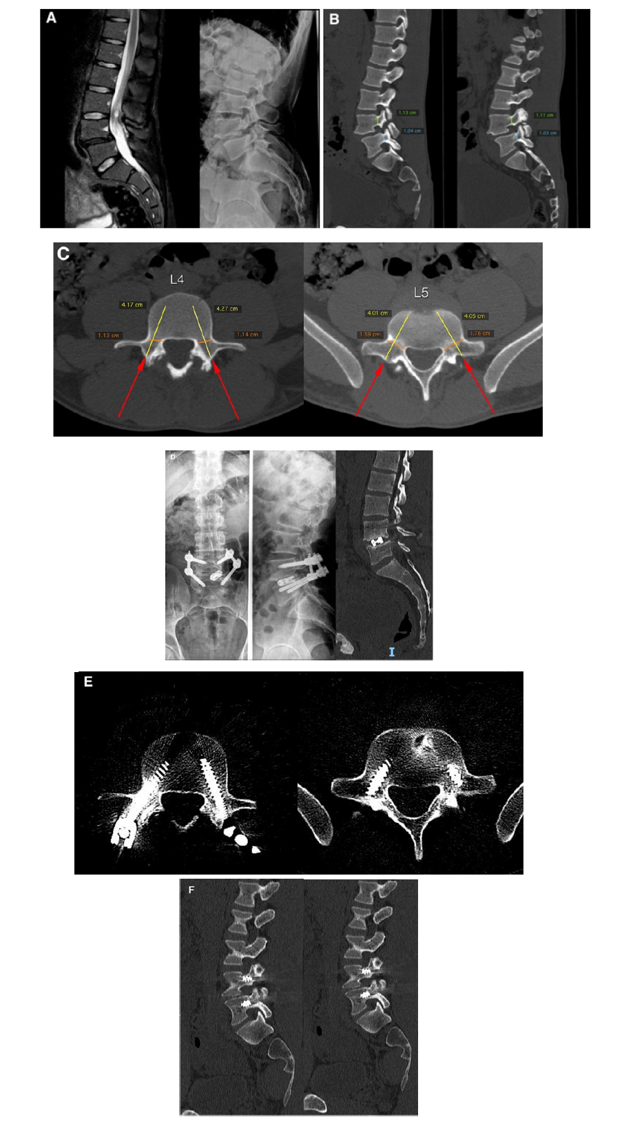
The postoperative tomography of the 96 operated cases was analyzed (Table 1). The positioning accuracy was graded using the Gertzbein and Robbins classification [11]; in the 62 cases with degenerative pathology, a precise placement of the screws was observed in the axial plane (without injury to the external or internal cortical pedicles or vertebral bodies) in 61 (98.38%) cases and in 1 (1.61%) case, an eruption of the cortical vertebral body and external cortical pedicle was observed by a single screw (unilateral) that involved less than 50% of its diameter (Figure 3). In the 34 cases with traumatic pathology, a correct placement was found in 32 (94.11%) cases in 2 (5.88%) cases there was evidence of an eruption of the cortical vertebral body and external cortical pedicle by two screws in both cases with involvement of less than 50% diameter. In 96 (100%) cases of the study, a correct postoperative location was observed in the tomographic sagittal plane (without eruption of the upper and lower pedicle cortical). In any case (0%) was the appearance of radiculopathy (or worsening of the previous) or neurological deficit associates with the placement of the pedicle screws.
Table 1:Clinical outcomes, surgical parameters, and pedicle screw placement accuracy across 96 surgical cases (438 screws).
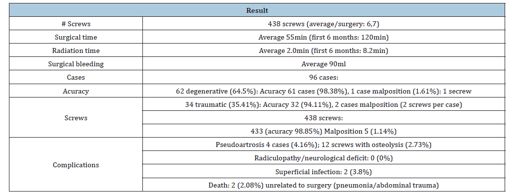
Figure 3:Tomographic planning in spondylolisthesis L4-L5 case (A) Planning in right and left sagittal (B) and axial (C) planes, with graphic planning of the potential directions, length and diameter of the screws. Radiographic and tomographic results (D, E, F) according to the previous planning.
Discussion
The relevance of preoperative planning in anterior and posterior instrumentation of the spine has been demonstrated by Abuzayed et al. [12] found differences compared to a number of previous morphometric studies performed mainly on a white population. Also, documented the individual morphometric differences of the same parameters in the same subgroups. These results emphasize the importance of preoperative computed tomography and conventional radiography of each patient in planning a surgical procedure and selecting the appropriate size of the instruments, thus avoiding possible postoperative complication related to implants [12]. Precision in the placement of transpedicular screws continues to be a concern in spine surgeons; anatomical knowledge, surgical technique and radiological relationships are crucial to achieve an effective trajectory in this type of instrumentation. Anatomical characteristics can vary between different pathologies (degenerative, traumatic, etc) and patients and even from one population to another. These morphological considerations are analyzed in several studies. Kaur et al. [1] concluded that a knowledge of the anatomical and radiological characteristics of the spine is essential to safely guide surgical and anesthetic procedures in the Indian population. Computerized software can help appropriately select implant sizes during spinal surgery [1]. Yoshida et al. [2] reported the accuracy of percutaneous thoracolumbar screw placement using fluoroscopy in oblique projections based on preoperative tomographic analysis; they concluded that minimally invasive lumbosacral Percutaneous Pedicle Screws (PPS) placement using the conventional fluoroscopic technique carries an increased risk of inaccurate screw placement and resultant neurological deficits, compared with that of the open procedure. Inaccuracy tended to be distributed between medial and lateral perforations of the L5 pedicle, as a result of pedicle morphology and the PPS pathway. Oblique fluoroscopic views, based on CT measurement, may allow accurate PPS insertion with a shorter fluoroscopy time [2].
Our study uses preoperative CT analysis, making measurements of each vertebra, identifying its morphology and variables that may be present not only at the pedicle level but also in the morphology of the vertebral body and pedicle direction, which may affect the trajectory of screw placement. This allows for individualized placement of percutaneous thoracolumbar screws in each case, increasing accuracy. This approach is supported by other studies that analyze the morphological characteristics of pedicles extensively in animal and human models to improve pedicle screw placement, whether conventional or percutaneous [5,6,8,9,13]. Liljenqvist et al. [14] concluded that morphometry in scoliotic vertebrae is substantially different from that of vertebrae in normal spines, with an asymmetrical intravertebral deformity shown in scoliotic vertebrae. Pedicle screw instrumentation on the concavity in the apical region of thoracic curves appears critical because of the small endosteal pedicle width; confirming the need for detailed planning [14]. This procedure has also been used for cervical pedicle screw and cervicothoracic junction insertion, improving accuracy and reducing the number of complications [3,4,7]. On the other hand, preoperative morphometric analysis has improved the accuracy of iliosacral screw placement in several reports [15,16]. It is essential that surgeons involved in these minimally invasive techniques acquire experience in percutaneous screw placement and that their learning curves be improved through the application of precise techniques [11,17].
An interesting study by İştemen et al. [18] supports the utility of preoperative prone position computed tomography in free hand subaxial cervical pedicular screwing reduces malposition and complications; this could be replicated at the lumbar level [18]. A cadaveric study by Kim SB et al. [19] reported the potential utility of patient-specific drill template with a preplanned trajectory has been considered a promising solution that may reduce the rate of spinal screw malposition without additional intraoperative radiation exposure. They used cortical bone trajectory screw technique that yields effective mechanical and clinical results, improving the holding screw strength with less invasive exposure. This cadaveric report could be interesting if the screws could be placed percutaneously; therefore, similar studies are needed. The use of current technology such as navigation and robotic arms improves the accuracy of percutaneous pedicle screw placement. Groisser BM et al. [20] reported the first description of a fully automated algorithm for determination of the technical accuracy of pedicle screw placement that does not require human intervention in the rigid-body alignment or measurement process; the technical accuracy results from this study of the Mazor X Stealth robotic system were equivalent to or better than reported accuracy from any prior in vivo or cadaver study [20]. We consider this study optimize the learning curve for surgeons that start their experience with percutaneous transpedicular techniques because it provides a preliminary tomographic view of the anatomy of each individual vertebra. This approach could be enhanced by using monitored Jamshidi needles when is possible. It is likely at the beginning of the learning curve of surgeons will tend to place screws with less convergence due to concerns about neurological damage, especially in pedicles that present greater technical difficult, such as the midthoracic pedicles.
Conclusion
a) The meticulous elaboration of an adequate tomographic
planning for the execution of a transpedicular percutaneous
fixation is a useful tool in the management of traumatic and
degenerative thoracolumbar pathologies.
b) Preoperative tomographic planning allows obtaining
adequate levels of precision in percutaneous thoracolumbar
transpedicular fixation, showing special importance in cases
where certain current precision technological elements such as
O-ARM or conventional navigation and even neurophysiological
intraoperative monitoring are not available.
c) We consider that our behavior potentially benefits
adding the use of monitored Jamshidi needles (intraoperative
neurophysiological monitoring).
d) It is likely that the rotational component of certain
fractures adds a greater degree of difficulty in the intraoperative
cannulation of the pedicles with laborious dynamics in the
incidence angles of the C-arm.
e) The collaboration of a technician trained in the use of the
C-arm during these procedures is crucial.
f) There could be an inconclusive tendency to direct the
screws in a lateral trajectory at the beginning of the learning
curve, it could be due to the inherent fear of avoiding a medial
trajectory toward the canal.
g) Another aspect that could improve the behavior is to use
an extrapedicular technique in certain thoracic pedicles the
merit it, such as bicorticals.
Acknowledgement
To my wife Kariam for accompanying me in every challenge I start.
References
- Kaur K, Singh R, Prasath V, Magu S, Tanwar M (2016) Computed tomographic-based morphometric study of thoracic spine and its relevance to anaesthetic and spinal surgical procedures. J Clin Orthop Trauma 7(2): 101-108.
- Yoshida G, Sato K, Kanemura T, Iwase T, Togawa D, et al. (2016) Accuracy of percutaneous lumbosacral pedicle screw placement using the oblique fluoroscopic view based on computed tomography evaluations. Asian Spine J 10(4): 630-638.
- Onibokun A, Khoo LT, Bistazzoni S, Chen NF, Sassi M (2009) Anatomical considerations for cervical pedicle screw insertion: The use of multiplanar computerized tomography measurements in 122 consecutive clinical cases. Spine J 9(9): 729-734.
- Chazono M, Soshi S, Inoue T, Kida Y, Ushiku C (2006) Anatomical considerations for cervical pedicle screw insertion: The use of multiplanar computerized tomography reconstruction measurements. J Neurosurg Spine 4(6): 472-477.
- Liau KM, Yusof MI, Abdullah MS, Abdullah S, Yusof AH (2006) Computed tomographic morphometry of thoracic pedicles: Safety margin of transpedicular screw fixation in Malaysian malay population. Spine (Phila Pa 1976) 31(16): E545-E550.
- Chen Y, Zeng J, Guan J, Guo Y, Wang X, et al. (2010) Reformatted computed tomographic evaluation of the thoracic pedicle in a Chinese population for the surgical application of transpedicular screw placement. Surg Radiol Anat 32(5): 463-468.
- Faghih-Jouibari M, Moazzeni K, Amini-Navai A, Hanaei S, Abdollahzadeh S, et al. (2016) Anatomical considerations for insertion of pedicular screw in cervicothoracic junction. Iran J Neurol 15(4): 228-231.
- Zheng C, Huang Q, Hu Y, Wang X, Chen W (2009) Computed tomographic morphometry of thoracic pedicles: Safety pedicle parameter measurement of the Chinese immature thoracic spine. Int Orthop 33(6): 1663-1668.
- Pai BS, Gangadhara, Nirmala S, Muralimohan S, Varsha SM (2010) Morphometric analysis of the thoracic pedicle: An anatomico-radiological study. Neurol India 58(2): 253-258.
- Devoto PR, Minghinelli FE, Mezzadri JJ, Pipolo DO, Lacsi MF, et al. (2024) Pedicle morphometry of the C7 and T1 vertebrae in an argentine population. Surg Neurol Int 15: 295.
- Gertzbein SD, Robbins SE (1990) Accuracy of pedicular screw placement in vivo. Spine (Phila Pa 1976) 15(1): 11-14.
- Abuzayed B, Tutunculer B, Kucukyuruk B, Tuzgen S (2010) Anatomic basis of anterior and posterior instrumentation of the spine: morphometric study. Surg Radiol Anat 32(1): 75-85.
- Mageed M, Berner D, Jülke H, Hohaus C, Brehm W, et al. (2013) Morphometrical dimensions of the sheep thoracolumbar vertebrae as seen on digitised CT images. Lab Anim Res 29(3): 138-147.
- Liljenqvist UR, Link TM, Halm HF (2000) Morphometric analysis of thoracic and lumbar vertebrae in idiopathic scoliosis. Spine (Phila Pa 1976) 25(10): 1247-1253.
- Han X, Fu Q, Yuan X, Wang W (2024) A new approach for percutaneous ilio-sacral screw fixation: CT-based pre-operative planning with conventional fluoroscopy to reduce malposition rate and operating time. Chin J Traumatol 28(5): 342-351.
- Krappinger D, Lindtner RA, Benedikt S (2019) Preoperative planning and safe intraoperative placement of iliosacral screws under fluoroscopic control. Oper Orthop Traumatol 31(6): 465-473.
- Landriel F, Hem S, Rasmussen J, Vecchi E, Yampolsky C (2018) [Learning curve of minimally invasive pedicle screw placement2QA. Surg Neurol Int 9(Suppl 2): S43-S49.
- İştemen İ, Arslan A, Olguner SK, Afşer KA, Açık V, et al. (2021) Significance of preoperative prone position computed tomography in free hand subaxial cervical pedicular screwing. J Korean Neurosurg Soc 64(2): 247-254.
- Kim SB, Rhee JM, Lee GS, Lee HY, Kim T, et al. (2018) Computer-assisted patient-specific prototype template for thoracolumbar cortical bone trajectory screw placement: A cadaveric study. Tech Orthop 33(4): 246-250.
- Groisser BN, Thakur A, Hillstrom HJ, Adhiyaman A, Zucker C, et al (2024) Fully automated determination of robotic pedicle screw accuracy and precision utilizing computer vision algorithms. J Robot Surg 18(1): 278.
© 2026 Molina Choez DD. This is an open access article distributed under the terms of the Creative Commons Attribution License , which permits unrestricted use, distribution, and build upon your work non-commercially.
Editor In Chief







.jpg)































Signup for Newsletter

Quick Links
 Editorial Board Registrations
 Submit your Article
 Best Paper of the Volume
 Reprints
 Refer a Friend
 Advertise With Us
Our Recent Edition

.jpg)



Top Editors




.jpg)

















.bmp)
.jpg)
.png)
.jpg)













.jpg)






.png)

.png)




.png)




Financial Support

Sponsors

Latest e-Books

Latest Video

 a Creative Commons Attribution 4.0 International License. Based on a work at www.crimsonpublishers.com.
Best viewed in
a Creative Commons Attribution 4.0 International License. Based on a work at www.crimsonpublishers.com.
Best viewed in 
 | Above IE 9.0 version