- Submissions

Full Text
Techniques in Neurosurgery & Neurology
Our Experience Cervical Interbody Fusion Using Improve Cylindrical Cage by Anterior Approach with Complication Cervical Disc Herniation
Sattarov AR1, Saidov SS2*, Shodmonov BR2
1The National Centre of Rehabilitation and Prosthetics of Person with Disabled’s, Uzbekistan
2Department of Vertebrology, Uzbekistan
*Corresponding author: Saidov SS, Senior Consultant of the Department of Vertebrology, National Centre of Rehabilitation and Prosthetics of Person with Disabled’s, Uzbekistan
Submission: August 01, 2022;Published: August 23, 2022

ISSN 2637-7748
Volume5 Issue2
Abstract
The aim of this study was to analyze treatment results of patients who underwent cervical fusion using cylindrical cage. 16 titanium cages were implanted to 12 patients with degenerative disease of cervical spine. Assessment of results was performed by neurologic examination, neovascularization data, visual analogue pain scale. Mean duration of surgery was 45-90min., blood loss-30-60ml, length of hospital stay-4-6 days. Substantial decrease of frequency and intensity of neck and arm pain was observed after surgery in 89% of patients. When properly performed, anterior cervical interbody fusion applying cylindrical cage is a simple and effective method.
Keywords: Hernia disk cervical division of the spine; Improve cylindrical titan cage; Micro discectomy; ACDF
Introduction
Deformation of the spinal canal, compression of the spinal cord, overstretching or compression of the vertebral vessels in the deformed vertebral foramina, instability in the spinal motion segments - all this can cause compression of the spinal cord and/or its roots. Given the anatomical and physiological features of this area, the presence of a compressing pathological process poses a great danger for the development of myelopathy with varying degrees of severity of neurological symptoms, which, if not treated in time, significantly worsens the quality of life and leads to disability and disability [1-10]. This pathological process in the cervical spine is an indication for surgical treatment [1,3].
Osteochondrosis of the cervical spine occurs mainly in the age group of 35-60 years, ranking second after the lumbar [1,2]. Anterior cervical discectomy with fixation of an autologous bone taken from the iliac crest is a proven effective treatment for radiculo- and myelopathy due to cervical spondylosis. Nevertheless, the authors [6] report the development of complications in the donor site up to 22%. To avoid such complications, many alternative materials for corporodesis have been proposed: homografts, xenografts, demineralized bone matrix, and cages. Currently, for interbody fusion, the following are used: bone autograft, titanium, carbon, PEEK cages, titanium plate, cage together with a plate, combined plate cages and cylindrical titanium cages. There is no consensus regarding the choice of the method of anterior fixation at the cervical level. A recent systematic review found that the choice of stabilization (graft, cage, plate) has little effect on pain symptom relief [10]. This is logical, since the “pain relief” stage of the operation is, first of all, a well-performed decompression of the nerve structures.
At the same time, low-level evidence is established that autografts cause more complications than cages [10]. This gives a minimal advantage in favor of implants. There is no consensus on the advisability of using plates for additional fixation of the cagestabilized segment. According to some authors, the additional use of a plate for one- and two-level cervical fusion in degenerative diseases of the cervical spine can improve vertebral union, reduce the incidence of subsidence and complications, ultimately improving clinical outcomes. Using a computer model of fusion at the C5-C6 level, showed that the placement of the anterior cervical plates in addition to the cage leads to a redistribution of tissue stress and a decrease in its value on each of the implants separately. Nevertheless, many authors [5] manage to achieve good results only with the use of cages, without additional fixation with a plate. Another interesting technical solution-the combination of a plate and a cage in one implant also has its supporters [5-9]. The aim of the work is to improve the results of surgical treatment of patients with cervical disc herniation using an improved cylindrical cage from the anterior parapharyngeal approach.
Material and Methods
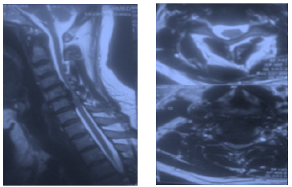
The results of treatment of 12 patients (7 women, 5 men) aged 18 to 62 years (mean age 32.4 years) with compression of the spinal cord and/or its roots by soft and/or hard (osteophytes) disc herniations were analyzed., who were implanted with 16 cylindrical titanium cages (patients with severe neurological disorders were excluded). All patients were operated on in 2021 at the “Vertebrology Department” of the National Centre of Rehabilitation and Prosthetics of Person with Disabled’s. Complaints, anamnesis, neurological status were assessed in the preoperative period. The severity of pain syndrome in the pre- and postoperative periods was assessed using a Visual Analog Pain Scale (VAPS). In addition, the duration of the operation, the volume of blood loss, the duration of postoperative bed rest, and the length of stay of patients in the hospital were determined. To verify the affected spinal motion segment, standard spondylography was performed, supplemented by functional tests in 9 patients; magnetic resonance imaging (MRI)-in 12 all patients; Multislice Computed Tomography (MSCT)-in 7; electroneuromyography (ENMG) of the upper limbsin 10 patients. All patients with discoradicular conflict underwent standard conservative treatment for 6-12 weeks before surgery at the place of residence and in our center in particular, which was not sufficiently effective. The most frequently involved vertebral segment in the pathological process was C5-C6 (Figure 1). Operations were performed at one level in 8 (66.6%) patients, and in 4 (33.4%) patients, at two adjacent levels.
Figure 1:MRI images of the cervical region (patient A), herniated discs at the level of VC5-6, VC6-7 with gross
compression of the spinal cord, and a focus of myelomalacia.

Surgical Technique
Patients of the analyzed group in all cases under endotracheal anesthesia underwent left-sided parapharyngeal access to the anterior surface of the spinal column according to Cloward. At the first stage, microsurgical decompression was performed by discectomy and resection of posterior exostoses using operating binoculars with a head light Karl Zeiss (Germany, with 3,5 times magnification and 500mm distance) and a standard set of microsurgical instruments. Rectangular excision with a scalpel of the anterior part of the annulus fibrosis was performed with the removal of the degenerated disc with the help of curettes and nippers up to the end plates and microsurgical decompression of the dural sac and roots by removing the disc herniation or resection of the posterior osteophytes from the central and lateral canals. At the second stage, the height of the cage was determined, an implant of the required diameter was installed with the holder and fixed by screwing into the bodies of adjacent vertebrae. After that, we used self-tapping screws to firmly stabilize the installed advanced cylindrical cages. All stages of stabilization were performed under X-ray control using a Brevis EVO image intensifier (France). All patients underwent external immobilization with a semi-rigid collar within 1 month from the day of surgery.
Results
When analyzing the results of surgical treatment, the following values were determined: the operation time was from 45 to 90 minutes (median 60 minutes), the volume of blood loss was from 30 to 60ml (median 45 ml), the activation of patients occurred on the day of surgery in the evening (since the improved a cylindrical cage is made with a thread and is screwed into the bodies of adjacent vertebrae - the value of the point of contact between the implant and the bone is large, after which self-tapping screws were screwed in for strong stabilization), the number of bed-days after the operation varied from 4 to 6 (median 5). After the operation, there was a significant decrease in the intensity of the pain syndrome both in the cervical spine and in the upper limbs. Assessment by VAPS Figure 2 revealed positive dynamics in the form of a decrease in the intensity of pain in the cervical spine from 9-10 points to 2-3 points, as well as the intensity of pain in the upper limb from 8 to 2 points (p< 0.05).
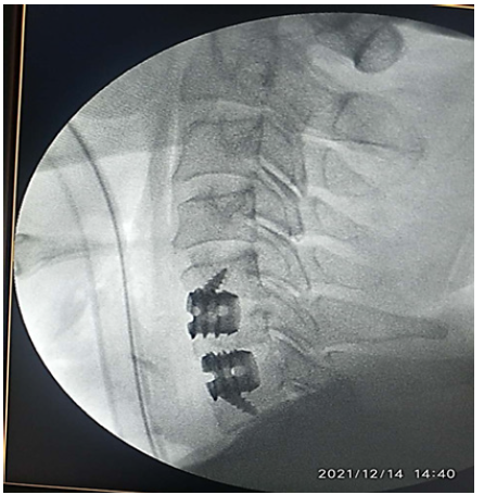
Figure 2:Postoperative x-rays of the cervical region of the same patient, cylindrical titanium cages are installed at
the level of VC5-6, VC6-7.

Analysis of the results of surgical treatment through standard periods of examination of patients (2, 6 and 12 months after surgery) showed that in all patients after surgery there was a complete regression of sensory and motor disorders. An analysis of labor rehabilitation showed that out of all operated patients, 8 (66,6%) patients returned to their previous work 2 months after the operation, 3 (25%) switched to light work after 2 months, and 1 (8,4%) became able-bodied 6 months after the operation. surgical treatment. In the postoperative period, all patients underwent a control x-ray of the cervical region in 2 projections, 8 (66,6%) patients underwent MRI or MSCT control within 2 to 6 months (8,2 months on average)-data on recurrence of discordicular or disco medullary conflict not received, the condition of the cages is satisfactory. Rigid fixation was achieved in all patients. A comparative analysis of the neurophysiological parameters of ENMG in the postoperative period showed that in the period from 3 to 6 months, the restoration of impulse conduction along previously compressed nerve structures occurred in 8 (66,6%) operated patients. Considering the absence of complaints, neurological deficit, radiological signs of instability of the operated segment and endoscopic signs of esophageal compression, it was decided not to perform a second surgery.
Clinical Example
Patient A. was admitted to the department with a diagnosis
of osteochondrosis of the cervical spine. Herniated discs C5-C6,
C6-C7. Radiculopathy C6, C7 root on the right with a slight distal
monoparesis in the hand. Pronounced reflex-tonic syndrome in the
acute stage. The patient complained of severe pain in the cervical
spine, aggravated by dynamic loads, radiating to the supraclavicular
region, to the right shoulder blade, to the right shoulder (along the
outer-lateral surface), in the forearm, to the feeling of “crawling”
in the arm, to numbness in the area of pain syndrome, weakness
of the right hand. From the anamnesis: that pains in the cervical
spine have been disturbing for 2 years, after hypothermia and an
incorrect sharp movement in the neck. Six months ago, the pain
syndrome intensified, there was irradiation of pain in the right
hand. She took painkillers at home. The last exacerbation began
2 weeks ago. During neurological examination, movements in
the cervical spine are limited when bending forward and to the right, painful. Pain on palpation of the spinous processes of C5-
C7, tension of the paravertebral muscles. Reflexes from the biceps
D=S alive, from the triceps D
Hypesthesia in dermatomes C6, C7. There are no pelvic disorders. Results of instrumental examination: X-ray of the cervical spine with functional tests: limitation of flexion and extension with signs of instability in the spinal motion segments C5-C6, C6-C7. MRI of the cervical spine: osteochondrosis of the cervical spine. Herniated discs C5-C6, C6-C7. ENMG of the upper extremities: conduction disturbance along the C6, C7 roots on the right (signs of axonopathy). The patient underwent microsurgical removal of herniated discs C5-C6, C6-C7 by anterior parapharyngeal approach, foraminotomy along the C6 and C7 roots on the right. Front corporodesis С5-С6, С6-С7 cylindrical titanium cage. After processing the surgical field with an antiseptic solution under endotracheal anesthesia in the supine position, an incision was made on the left along the medial edge of the digging muscle. Without technical difficulties, a typical left-sided parapharyngeal approach to the anterior edge of bodies C5, C6 and C7 was performed. X-ray image intensifier tube control with confirmation of the level of the lesion. Microdiscectomy was performed using a high-speed drill, supplemented by microsurgical removal of the posterior exostoses of the C5, C6, C7 bodies and foraminotomy for the roots of C6, C7 on the right. Anterior corporodesis C5-C6, C6-C7 was performed with cages. Cage diameter on moderate C5-C6 distraction was 5mm (correction of kyphotic deformity), C6–C7 cage size was 5.5mm. X-ray control: the position of the cages is correct. Hemostasis. Layered suturing of the surgical wound. Aseptic bandage. External fixation of the cervical spine with a Philadelphia collar.
Discussion
8 (66.6%) patients who underwent a control MRI or MSCT study in the long-term period. When assessing the pain syndrome according to VAS, we did not obtain fundamental differences. The results indicate the completeness and adequacy of the choice of surgical intervention. The advantage of using an improved cylindrical titanium cage, in our opinion, is the simplicity of design with a minimum number of surgical instruments used for implantation, the possibility of instant reliable rigid interbody stabilization of the operated spinal motion segment (there is no need to wear a collar for a long time after surgery), the possibility of stabilization on two and more affected levels. Given the large volume of the contact point of the titanium implant and the working surface of the bodies of adjacent vertebrae, we obtained a strong stabilization in two levels of disc herniation without the use of titanium plates of the anterior surface of the vertebral bodies. The use of titanium material did not reveal any negative aspects and did not lead to the development of complications. There were no technical difficulties during the installation of the cage on our part.
Conclusion
A. Surgical treatment with implantation of improved cylindrical titanium cages in patients with discoradicular and disco medullary conflict at the cervical level allowed us to obtain good clinical and functional results in the immediate and long-term periods.
B. The method of anterior cervical corporodesis with improved cylindrical titanium cages is simple, effective and has minimal complications if performed correctly.
References
- Gushcha AO, Shevelev IN, Shakhnovich AR, Safronov VA, Arestov SO (2006) Differentiated surgical treatment of spinal canal stenosis at the cervical level. Spine Surgery 4: 47-54.
- Irger IM (1971) Neurosurgery. M Medicine 360-374.
- Sorokovikov VA, Byvaltsev VA, Kalinina SA, Gorbunov AV, Egorov AV, et al. (2010) Analysis of the results of decompressive operations using stabilization in degenerative-dystrophic diseases and traumatic injuries of the cervical spine. Topical Issues of Surgical Practice at the Eastern Railway, Irkutsk, Russia, pp. 59-62.
- Khelimsky AM (2000) Chronic discogenic pain syndromes of cervical and lumbar osteochondrosis. Khabarovsk, Russia, pp. 139-140.
- Benezech J (2001) Cervical fusion with monocomponent PCB plate-cage. In: Szpalski M, Gunzburg R (Eds.), De-generative Cervical Spine, Lip-Pincott Williams & Wilkins, Philadelphia, USA, pp. 265-273.
- Castro FP, Holt RT, Majd M, White TS (2000) A cost analysis of two anterior cervical fusion procedures. J Spinal Dis 13(6): 511-514.
- Fernandes PC, Fernandes PR, Folgado JO, Melancia LJ (2011) Biomechanical analysis of the anterior cervical fusion. Comput Methods Biomech Biomed Engin 15(12): 1337-1346.
- Jacobs W, Willems PC, Kruyt M, Limbeek J, Anderson PG, et al. (2011) Systematic review of anterior interbody fusion tech-niques for single- and double-level cervical degenerative disc disease. Spine 36: 950-960.
- Kyung S, Cyrus E, Taghavi, Margaret S, Kwang B (2010) Plate augmentation in anterior cervical discectomy and fusion with cage for degenerative cervical spinal disorders. Eur Spine J 19(10): 1677-1683.
© 2022 Saidov SS. This is an open access article distributed under the terms of the Creative Commons Attribution License , which permits unrestricted use, distribution, and build upon your work non-commercially.
 a Creative Commons Attribution 4.0 International License. Based on a work at www.crimsonpublishers.com.
Best viewed in


.jpg)


 Editorial Board Registrations
 Submit your Article
 Refer a Friend
 Advertise With Us
.jpg)


.jpg)


.bmp)
.jpg)
.png)
.jpg)


.jpg)


.png)

.png)


.png)


