

- About Us
- Information
-

The Author ensures that the research has been conducted responsibly and ethically with adherence to all relevant regulations. read more..
- For Authors
- For Reviewer
- Manuscript Guidelines
- Membership
- Publication Ethics
-
- Journals
- Reprints
- e-Books
- Videos
- Policies
- Contact Us
 COVID-19
- Submissions



Full Text
Researches in Arthritis & Bone Study
Case Reports & Review of Non-Operative Managing of Injuries Around Ankle
Suresh Kishanrao*
Family Physician & Public Health Consultant, India
*Corresponding author:Suresh Kishanrao, Family Physician & Public Health Consultant, India
Submission: October 13, 2026;Published: October 26, 2026

Volume2 Issue4October 26, 2026
Abstract
Introduction: The ankle joint complex is a highly intricate, weight-bearing structure formed at the
meeting point of the lower ends of the tibia and fibula, along with the small tarsal bones. It supports body
weight and allows foot movement. Ankle injuries including sprain, tendon tears & fractures ranging from
isolated lateral malleolar breaks to complex trimalleolar disruptions involving syndesmotic & ligamentous
injuries are one of the frequent lower-limb injuries. A twisted ankle might seem like a minor “sprain,” but
could be a serious fracture in disguise. Inability to bear weight, as evidenced by struggling to take four
steps on the foot usually means a severe grade 3 sprain or a possible fracture requiring an X-ray. Swelling
& bruising signals rapid inflammation or a torn ligament or high ankle injury, which takes longer to heal
than a stretch. The key mechanisms of injury to ankle include i) twisting of the foot inward (inversion)
harming the outer ligaments, & ii) a rotational fall can cause a high ankle sprain. General epidemiological
research in India regarding foot and ankle injuries shows a higher overall prevalence and presentation
in males, largely driven by occupational exposure, road traffic accidents, and male-dominated sports
participation. However, distinct gender vulnerabilities of higher chronic ankle instability and lateral
laxity in females are observed. Diagnosis is based on clinical examination for local tenderness; inability to
walk a few steps to be confirmed by a plain Xray.
Materials and Methods: This article is based on two cases of fractures (One in an elderly male with
minimal mobility and another in a young lady with avulsion fracture), managed recently to highlight the
role of general practitioners in ankle injuries and modern approaches to fix the fractures and Sprains
Outcome: Both case management strategies were based on stability, fragment size and soft-tissue
condition. Unstable fractures needed open reduction and internal fixation (ORIF) using plates, screws but
managed with a plaster boot as the general condition was risky for the operative procedure. For Ligament
Tears & Sprains of soft-tissue, inversion, and high-ankle sprains involving the Anterior Talofibular
Ligament (ATFL) or Calcaneofibular Ligament (CFL), was managed by POLICE framework which mean
Protection by braces, crutches, optimal loading, cryotherapy, compression bandage and elevation of
the foot above heart level. Rehabilitation protocols include early mobilization and weight-bearing, to
accelerate return to function without compromising healing.
Keywords:Ankle joint; Ankle fracture; Avulsion fracture; ATFL (Anterior Talofibular Ligament); CFL (Calcaneofibular Ligament)
Introduction
The ankle joint complex is a highly intricate, weight-bearing structure formed at the meeting point of the lower leg bones and the foot. The lower ends of the tibia and fibula, along with the small tarsal bones around the ankle, form a stable Ankle Anatomy that supports body weight and allows foot movement. Ankle injuries including sprain, tendon tears & fractures ranging from isolated lateral malleolar breaks to complex trimalleolar disruptions, at the network of ligaments connecting the shinbone -tibia and the outer leg bone-fibula just above the ankle joint and ligamentous injuries are the most frequent lower-limb injuries [1]. While swelling & bruising signals rapid inflammation or a torn ligament or high ankle injury, Inability to bear weight, or struggling to take even four steps on the foot usually means a severe grade 3 sprain or a possible fracture requiring an X-ray to differentiate.
While globally Ottawa Ankle Rules guide classifying and managing the acute phase to rule out fractures Clinically by (i) pain in the malleolar zone plus (ii) inability to walk 4 steps unassisted immediately and in the clinic or ii) Bone tenderness at distal 6 cm of the posterior/tip of the lateral or medial malleolus or iii) Bone tenderness at the base of the 5th metatarsal or the navicular bone and confirm by an Xray, before diagnosing a sprain [2].
In India, there is no single, consolidated domestic “National Guideline” document that covers all three aspects of ankle injuries like sprains, ligament tears, and fractures. However, Top Indian orthopedic surgeons and institutions rely on the AO Trauma Fracture Management framework alongside standard international clinical practice guidelines adapted for Indian clinical settings [3]. For fractures the AO Manual of Fracture Management is recognized by the Indian Orthopedic Association (IOA). While classification uses the Danis-Weber system to determine stability, management focuses on Open Reduction and Internal Fixation (ORIF) for unstable fractures to restore joint congruity & prevent long-term post-traumatic osteoarthritis [3-5].
For Ligament Tears & Sprains of soft-tissue, inversion, and highankle sprains involving the Anterior Talofibular Ligament (ATFL) or Calcaneofibular Ligament (CFL), advocates moving away from rigid RICE approach (Rest, Ice application, compression bandage and elevation) to the POLICE framework which promotes i) Protection by using splints, functional braces, or crutches in the first 24-72 hours to prevent further mechanical stress, ii) introducing early, pain-free active range of motion and progressive weight-bearing to promote cellular healing and dynamic stabilization iii) Cryotherapy applied for 15-20 minutes every 2 hours during the first 48 hours to manage acute pain and swelling iv) Figure-of-eight elastic compression bandaging or supportive ankle binders to minimize interstitial edema and v) Keeping the foot elevated above the level of the heart to facilitate venous and lymphatic drainage [6].
This article is based on two cases of fractures managed recently to highlight the role of general practitioners in ankle injuries.
Case Reports:
Case 1. Elderly male with fracture lower and of fibula and navicular bone:
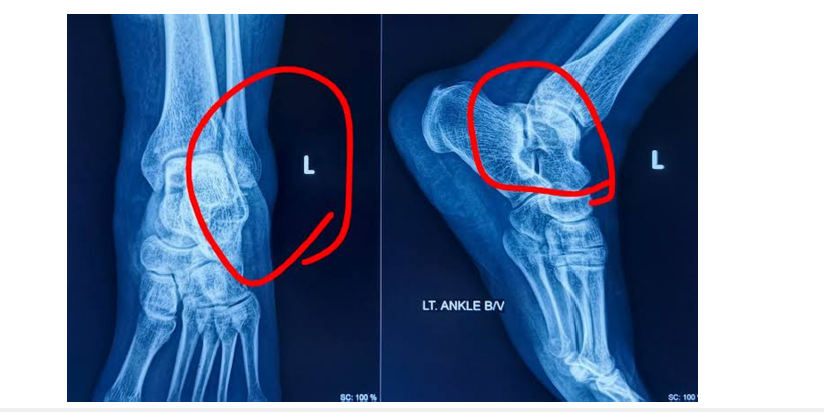
Mr. Chimanna aged about 76 years, known diabetic for 20 years, Hypertensive for 10 years and right sided stroke for 5 years, suffering from Bed-ridden syndrome for last 3 years slipped in bathroom, twisted his Right ankle on 10 September 2026. He was unable to bear weight on the right leg and managed to reach his bed with the support of his wife. By the end of the day, when his son returned from work his right ankle and lower leg were swollen. A quick series of Xray’s around 21:45 revealed Fracture Lower and of Fibula and Navicular bone. A surprising shadow behind the leg was ruled out as that of a plastic sheet given for support given before taking an Xray. Next morning basic blood investigations like TC, DC, RBC, PCV, MCV, MCH, MCHC counts, Blood group, Hb%, Blood Urea, Serum Electrolytes, Prothrombin time were done in preparation for surgical fixing using screws and wires. Except for Hb% =10.5 g/dl (13-16g/dl), RBC Count 3.61 (4.5-6.1 K) PCV=32.4% (37-47%) and Blood Urea 48 mg/dl (15-45 mg/dl) normal range in parenthesis were normal.
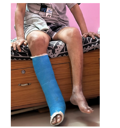
The family was informed about the chances of malunion if a simple plaster boot is put and nailing would cost about INR 300,000. The author was consulted, who, in consultation with the surgeon, doubting the fitness of the patient for surgical intervention and as patients was hardly to multiple health problems it was decided not to take any risk and discharge with a simple plaster boot to immobilize ankle joint and allow it to take care. After 4-6 weeks an Xray will be taken and see if there is any malunion. Current status after nearly 4 weeks, pain and swelling have disappeared, but the patient is unwilling to put any efforts to move the leg (Figure 1 & 2).
Figure 1:X-ray’s showing Fracture lower end of Fibula & Navicular bones
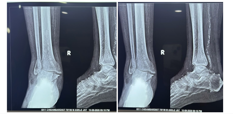
Figure 2:Same person with Plaster boot.
Case 2. Avulsion fracture in a young female:
In a friendly Activate which was fun, this young lady ended up twisting her left ankle towards the end. A local Dentist examined and opined it to be a bad sprain and advised keeping the foot elevated. That night she had some pain and swelling but rested well but had disturbed sleep despite avoiding weight on it. Being Left leg she felt it was easier to hop around on her dominant right foot. Ankle was swollen but pain was reduced by the morning thanks to ankle elevated all night and a combo tablet Tylenol (acetaminophen) & Advil (ibuprofen) over-the-counter medication to relieve pain. By the next morning, she felt better. However, to rule out any fracture she took an X Ray next day which showed Avulsion Fracture.
The course of management did not change After 4 days swelling was reduced and pain in putting on weight persists (Figure 3-5).
Figure 3:Sprained foot on day 1.
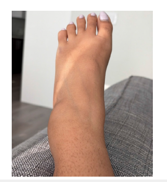
Figure 4:Same foot after 48 hrs. of Ice application.
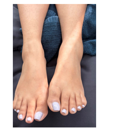
Figure 5:X Ray showing Avulsion Fracture.
Discussions
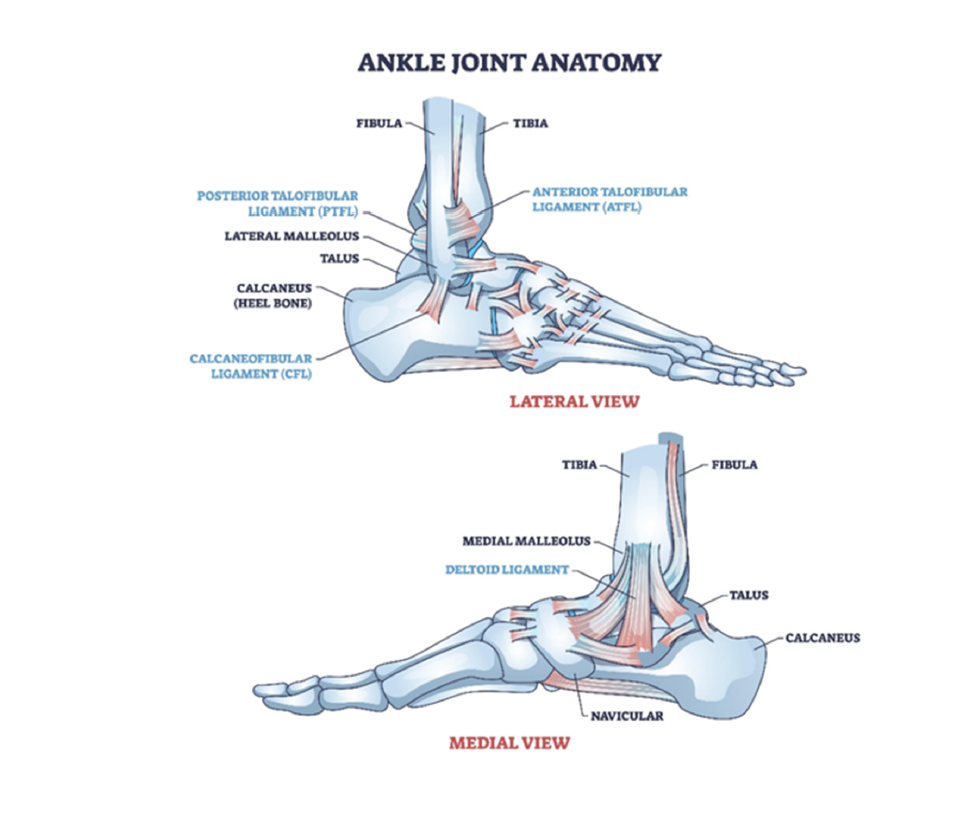
The lower ends of the tibia and fibula, along with the small tarsal bones around the ankle, form a stable Ankle Anatomy that supports body weight and allows foot movement [1].
Distal End of the Tibia has:
A. Medial Malleolus: The prominent bony bump on the inner
side of your ankle
B. Posterior Malleolus: The back part of the lower tibial rim
C. Inferior Articular Surface: The smooth bottom part that
rests directly on top of the main ankle bone to transmit weight
D. Fibular Notch: A rough indentation on the outer side of
the lower tibia where the lower fibula attaches [1].
Lower End (Distal End) of the Fibula has:
a) Lateral Malleolus: The large bony bump on the outer side
of our ankle.
b) Articular Facet of the Lateral Malleolus: The smooth inner
face that grips the side of the ankle bone for lateral stability
c) Malleolar Fossa: A small pit behind the articular facet
used for heavy ligament attachment [1].
Tarsal Bones
Three are five small bones
A. Talus: The keystone dome-shaped bone sitting directly
under the tibia and fibula, receiving all leg weight
B. Calcaneus: The large heel bone situated directly beneath
the talus
C. Navicular Bone: A boat-shaped bone located on the inner
side of the foot in front of the talus
D. Cuboid Bone: A cube-shaped bone sitting on the outer
side of the foot opposite the navicular
E. Cuneiform Bones: Three small wedge-shaped bones
(medial, intermediate, and lateral) lying in front of the navicular
[1] (Figure 6).
Figure 6:Ankle Joint Anatomy.
Core Ligaments:
Ligaments connect bone to bone. The ankle relies on three
primary groups to remain stable
a) Lateral Ligament Complex (Outside Ankle): The most
frequently injured ligaments during a “rolled ankle”.
b) Anterior Talofibular Ligament (ATFL): Connects the fibula
to the front of the talus. It is the weakest and most sprained
ankle ligament
c) Calcaneofibular Ligament (CFL): Connects the fibula
down to the heel bone
d) Posterior Talofibular Ligament (PTFL): Connects the back
of the fibula to the talus
e) Deltoid Ligament Complex (Inside Ankle): A massive,
thick triangle-shaped band of ligaments on the inside of the
ankle, incredibly strong and rarely tears completely unless the
ankle bones fracture.
f) The Syndesmosis (High Ankle Ligaments): A collection
of tough fibrous tissue holding the lower tibia & fibula tightly
together just above the joint line. Injuries here are known as
“high ankle sprains.”
Approach to Injuries at Ankle
Ankle injuries are the most frequent musculoskeletal complaints evaluated in orthopedic and sports medicine settings. The complex biomechanical design of the ankle joint leaves it susceptible to a spectrum of trauma ranging from simple soft tissue stretches to complex intra-articular breaks. This joint is maintained by the anterior inferior tibiofibular ligament (AITFL), posterior inferior tibiofibular ligament (PITFL), transverse ligament, and interosseous ligament/membrane, which tear leading to abnormal separation between the tibia and fibula, known as diastasis, destabilizes the fork-shaped bony socket formed by the lower ends of the Tibia & Fibula, which cradles the dome of the Talus.
The same physical mechanism of an unnatural rolling or turning of the ankle joint begins with the same misstep but differs in spectrum of severity entirely in the type and extent of structural damage caused to the ankle. Ankle injuries occur when the foot is planted and forcibly twisted outwards or toes pulled up toward the shin, often seen in contact and cutting sports like football, soccer, and skiing. As a general practitioner or orthopedic practitioner, must follow assessment & diagnostic strategy and decide to either refer urgently to a specialist first. When a patient comes with an acute ankle injury, it is important to differentiate between severe soft-tissue trauma and fractures by eliciting tenderness at the posterior edges of the malleoli or observing client’s ability to bear weight asking him to walk for four steps. When either or both signs are there, it must trigger the need for standard AP, lateral, and mortise X-rays.
Physical examination must assess localized ligament pathology- The Anterior Drawer test for the Anterior Talofibular Ligament (ATFL), the Talar Tilt test for Calcaneofibular Ligament (CFL), and the Squeeze & External Rotation tests for syndesmotic “high ankle” integrity be applied to evaluate these ligaments.
MRI is reserved for suspected occult fractures, syndesmotic disruptions, osteochondral lesions, or cases presenting with chronic instability after 4-6 weeks of physical therapy. CT scans are primarily utilized for pre-operative 3D mapping of complex, comminuted fractures.
Ligament Tears & Sprains (Inversion/Eversion Injuries)
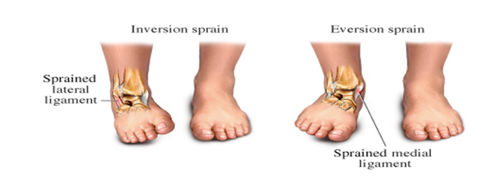
Ligamentous damage usually involves the lateral ligament complex (ATFL and CFL) caused by a sudden inversion mechanism. Less common are medial deltoid injuries or syndesmotic (high ankle) sprains (Figure 7).
Figure 7:
Grading Scale is used to determine such sprains [6].
A. Grade 1 (Mild): There is microscopic tearing or stretching
of the ligament fibers without loss of structural integrity and
patients usually present symptoms of mild pain, slight swelling,
minimal tenderness, and little or no bruising. Patients can
usually bear weight or move the joint with little to no difficulty.
Treatment to follow would be to Rest, apply ice and compression
bandage, and elevation (R.I.C.E.) of the foot above the heart
level, and over-the-counter anti-inflammatory medications if
required.
B. Grade 2 (Moderate): Such cases have moderate to severe
pain, noticeable swelling, localized bruising, tissue tenderness,
due to partial tearing of the ligament fibers & partially intact.
Joint stability is compromised, bearing weight or moving the
limb is difficult and painful
C. Grade 3 (Severe): These patients report with Intense,
severe pain initially or painless if nerves are damaged, heavy
swelling, widespread deep bruising, and severe joint instability.
Complete rupture or total tearing of the ligament into separate
pieces is the hall mark of such injuries. There is complete loss
of normal function; patient cannot bear weight or stabilize the
joint, needing Immobilization via a cast or boot. Some cases
may even need surgical reconstruction or repair, followed by
extensive rehabilitation.
Current Management Practices for Sprains and Ligament Tears [6-8]
Modern management protocols prioritize maintaining joint stability, minimizing prolonged immobilization, and initiating early functional rehabilitation [6,8]. Grade 1 sprains involve mild ligament stretching or minor fiber tears with minimal pain and 1-3-week recovery. Grade 2 sprains feature partial ligament tears with moderate instability and 4-8-week healing, sometimes longer. Grade 3 sprains mean complete ligament rupture with severe instability requiring 8-12 weeks, minimum, 3-6 months for full recovery. Proper grading determines treatment approach and prevents long-term joint problems
a) Conservative Control: The traditional Rest, Ice,
Compression, Elevation (RICE) remains the foundational
approach for the first 48-72 hours to control swelling and acute
pain as was done in our second case [7]
b) Early Mobilization: Long-term cast immobilization for
acute ligament tears is not practiced. For Grade I and II sprains,
immediate weight-bearing is encouraged using a semi-rigid
brace or elastic support. For severe Grade III injuries, a brief
period of immobilization (5- 10 days) in a below-knee cast or
walking boot is used to allow initial tissue alignment, followed
immediately by functional bracing [6,7].
c) Functional Rehabilitation: Progressive physical therapy
is critical. Programs focus initially on restoring range of
motion, strengthening the peroneal muscles, implementing
neuromuscular/proprioceptive training to prevent recurrent
sprains and chronic instability.
d) Surgical Interventions: Routine acute surgery is
considered for high-demand elite athletes or cases of Chronic
Ankle Instability (CAI), after 3-6 months of comprehensive
physical therapy fails. Procedures like the Brostrom-Gould
anatomic reconstruction (tightening and re-attaching the
native ligaments) or tendon-graft reconstructions are utilized.
High ankle syndesmotic tears may require dynamic suturebutton
fixation or traditional syndesmotic screw stabilization
to stop diastasis (widening) of the tibia and fibula [6-8].
Ankle Fractures
Conservative Treatment for Stable, non-displaced fractures (such as Danis-Weber Type A or selected Type B) are managed with a cast or boot for roughly 6 weeks, paired with early guided activity. Unstable or displaced fractures (such as Danis-Weber Type C or unstable Type B) require Open Reduction and Internal Fixation (ORIF) or intramedullary nailing.
Ankle fractures disrupt the bony architecture of the ankle mortise and are classified structurally via the Danis-Weber system (based on the level of the fibular break relative to the syndesmosis) or anatomically by the number of malleoli involved (isolated, bimalleolar, Trimalleolar [9].
Danis-Weber Classification:
Type A (Infrasyndesmotic): Fracture below the joint line; stable syndesmosis.
Type B (Transsyndesmotic): Fracture at the level of the syndesmosis; variable joint instability.
Type C (Suprasyndesmotic): Fracture above the joint; highly unstable; frequent syndesmotic rupture.
Uni-malleolar Fracture, in which usually the lateral malleolus on the outside or only one of the other malleoli is broken. They are usually stable and treated with a walking boot or cast if the bones haven’t shifted.
Bimalleolar Fractures where two malleoli are broken are unstable and mostly taken for surgical realignment using plates and screws.
Tri malleolar Fracture, where the lateral, medial, and posterior malleoli are broken and joint is unstable, surgical restoration of structural alignment is done.
Avulsion Fracture, in which a ligament or tendon pulls a tiny piece of bone away from the main bone, during sudden twist, fall or sports move. It is a minor, stable injury and treated much like a sprain.
Maisonneuve Fracture is a severe spiral fracture of the upper neck of the fibula combined with a tear of the syndesmosis and a medial ankle injury, usually missed because the pain is felt high up the leg, but the ankle joint itself is highly unstable and it requires surgery.
Pilon Fracture, a high-energy crush injury where the talus rams upward, shattering the tibial plafond usually caused by car accidents or falls from heights, that requires complex surgical reconstruction [9,10].
Conclusion
Ankle injuries including sprain, tendon tears and fractures are one of the most frequent lower-limb injuries worldwide, with patterns ranging from isolated lateral malleolar breaks to complex trimalleolar disruptions involving syndesmotic and ligamentous injuries. In India non-operative management of injures around ankles is like that in the developed world when compared many other developing countries.
Initial assessment combines clinical examination with radiographic and three-dimensional imaging to classify fractures according to systems such as AO/OTA and Danis-Weber. Overall strategies balance operative and non-operative approaches, that address stability, fragment size and soft-tissue condition. Unstable fractures typically undergo open reduction & internal fixation (ORIF) using plates, screws or intramedullary devices. Recent innovations include minimally invasive fibular nailing & anti-glide plating guided by detailed fracture mapping. Syndesmotic injuries are stabilized with traditional screws or dynamic suture-button constructs, each offering distinct biomechanical benefits.
Rehabilitation protocols include early mobilization & weightbearing, to accelerate return to function without compromising healing. A comprehensive understanding of fracture morphology, fixation biomechanics and patient-centered outcomes underpins the Indian effort to optimize ankle fracture and injuries care to restore mobility, though controversies persist regarding fixation of small posterior malleolar fragments and indications for deltoid ligament repair.
References
- Types of lower end of tibia, fibula and small bones, ligaments & common sprain & fracture types around anklet.
- Gang Liu, Zhijian Sun, Minghui Yang, Xinbao Wu, Peter VG (2025) An overview on the management of ankle fractures in elderly patients aged 65 and over. Eur J Orthop Surg Traumatol 13:35(1): 396.
- Dave PK (2001) Standard-treatment-guidelines for orthopedics.
- Sonaram Jaat explains (2026) Sprain or fracture?
- Hans P, Karl Georg K, Wolf Christian P, Florian H, Ben O, at el. (2011) Diagnosis & treatment of acute ankle injuries: development of an evidence-based algorithm. Orthop Rev (Pavia) 4(1): e5.
- Ankle ligament tear causes and treatment explained.
- Rachel B, Daniel L, Victoria LS (2025) Acute Ankle Sprain.
- Ryan P McGovern, RobRoy LM (2016) Managing ankle ligament sprains and tears: current opinion. Open Access J Sports Med 2:7: 33-42.
- Ankle-fracture-management & treatment-techniques.
- Benjamin Taylor et.al, Ankle-fractures.
© 2026 Suresh Kishanrao. This is an open access article distributed under the terms of the Creative Commons Attribution License , which permits unrestricted use, distribution, and build upon your work non-commercially.
Editor In Chief







.jpg)































Signup for Newsletter

Quick Links
 Editorial Board Registrations
 Submit your Article
 Best Paper of the Volume
 Reprints
 Refer a Friend
 Advertise With Us
Our Recent Edition




Top Editors




.jpg)
















.bmp)
.jpg)
.png)
.jpg)













.jpg)






.png)

.png)




.png)




Financial Support

Sponsors

Latest e-Books

Latest Video

 a Creative Commons Attribution 4.0 International License. Based on a work at www.crimsonpublishers.com.
Best viewed in

 | Above IE 9.0 version