

- About Us
- Information
-

The Author ensures that the research has been conducted responsibly and ethically with adherence to all relevant regulations. read more..
- For Authors
- For Reviewer
- Manuscript Guidelines
- Membership
- Publication Ethics
-
- Journals
- Reprints
- e-Books
- Videos
- Policies
- Contact Us
 COVID-19
- Submissions



Full Text
Researches in Arthritis & Bone Study
The Osteoporosis Value Paradox: Compare Cost of Care for Cardiovascular Disease Versus Osteoporosis, Dual-Energy X-Ray Absorptiometry (DXA), Fragility Fractures and Implications for Economics and Secondary Prevention
John Abner Goldman1*, Patrice Gutentag2 and Glenn R Parris3
1Clinical Professor of Medicine, Rheumatology, Immunology, Emeritus, Emory School of Medicine, Georgia, Parris and Associates Georgia, Northside Hospital, Atlanta, Emory St. Josephs Hospital, Atlanta, USA
2Parris and Associates, Georgia
3Northside Hospital, Gwinnett, Parris and Associates, Georgia
*Corresponding author:John Abner Goldman, Clinical Professor of Medicine, Rheumatology, Immunology, Emeritus, Emory School of Medicine, Georgia, Parris and Associates Georgia, Northside Hospital, Atlanta, Emory St. Josephs Hospital, Atlanta, USA
Submission: July 06, 2026;Published: August 24, 2026

Volume2 Issue3August 24, 2026
Abstract
Background/Purpose: Cardiovascular Disease (CVD) and osteoporosis are leading contributors
to morbidity, mortality, and healthcare expenditures. While CVD Has a much larger population and
dominates total spending, osteoporosis-related fractures, especially hip fractures impose a larger
substantial expense per-event and long-term costs. This study compares the economic burden, cost
drivers, and prevention value of these conditions, with emphasis on gaps in osteoporosis care delivery.
Methods: A narrative economic analysis was conducted using published U.S. cost data, cost-effectiveness
studies, and implementation metrics for CVD and osteoporosis. Outcomes included total annual costs,
per-event costs, cost-effectiveness of preventive interventions, and real-world treatment rates following
sentinel clinical events. The effects of the Women’s Health Initiative (WHI) and the Dual-Energy X-Ray
Absorptiometry (DXA) pricing were incorporated.
Using the 2015 to March 2020 National Health and Nutrition Examination Survey and 2015 to 2019
Medical Expenditure Panel Survey, this data indicates the estimated trends in prevalence for cardiovascular
risk factors based on American Heart Association (AHA) adverse levels of Life’s Essential 8 and clinical
cardiovascular disease and stroke. It is projected for both contributors through 2050, overall and by age
and race and ethnicity, accounting for changes in disease prevalence and demographics. The Cochrane
Systemic Review also participated in some of the analyses.
Results: Government cutbacks on bone density readings include a professional component paying
only $37 for non-facility bone density studies and the impact of the Women’s Health Initiative had a
detrimental effect on osteoporosis and fractures. Annual costs for CVD exceed $500 billion, compared
with approximately $57 billion for osteoporosis-related fractures. However, per-event costs for hip
fracture ($47,000-$71,000) equal or exceed those for myocardial infarction ($18,000-$29,000) and
stroke ($15,000-$34,000). Hip fractures account for approximately 72% of osteoporosis-related
costs despite representing a minority of fractures. Both diseases demonstrate high-value preventive
interventions: statins and smoking cessation in CVD; DXA underpayment, anabolic and non-anabolic
therapy and Fracture Liaison Services (FLS) in osteoporosis. Anabolic therapy, which is more effective
but more expensive should also be included. Fracture Liaison Service (FLS) is cost-saving, with estimated
savings of $418 per patient and $418 million per 1 million Medicare beneficiaries treated, preventing
approximately 30,000 fractures. Despite this, implementation differs markedly: >90% of CVD patients
receive secondary prevention after myocardial infarction, whereas <20% of patients receive osteoporosis
treatment following hip fracture, but only ≈9% treated within 6 months.
Conclusion: Correcting underpayment and availability of bone DXA studies and understanding how
incorrect interpretation of the WHI led to a higher risk of fractures. Although CVD carries a greater total
economic burden, osteoporosis demonstrates comparable of greater cost intensity per clinical event and
possible but not obtained highly favorable prevention economics. The persistent post-fracture treatment
gap represents a major systems failure. FLS offers a scalable, cost-saving intervention that improves
outcomes and reduces downstream costs. Aligning osteoporosis care with established CVD prevention
frameworks represents a critical opportunity for value-based healthcare improvement.
Keywords:Osteoporosis; Fragility fracture; DXA; Cardiovascular disease; Cost-effectiveness; Fracture liaison service; Value-based care
Introduction
Cardiovascular Disease (CVD) and osteoporosis are major contributors to morbidity, mortality, and healthcare expenditures. While CVD, since it is a much larger proportion of the population, dominates total spending, fragility fractures impose substantial per-event costs, long-term disability, and mortality risk, yet remain underprioritized in prevention efforts [1-6]. Comparing the cost of care for cardiovascular disease versus the cost of care for osteoporosis and fractures, we find that Cardiovascular Disease (CVD) imposes a substantially larger economic burden than osteoporosis and fractures, with annual costs exceeding $500 billion versus approximately $57 billion for osteoporosis, yet both conditions demonstrate similar patterns of underinvestment in prevention relative to treatment costs and marked cost-effectiveness of targeted interventions when appropriately implemented. The comparison reveals critical differences in disease prevalence, acute care intensity, and prevention infrastructure that inform resource allocation decisions.
Comparative cost burden [1-6]
The CVD burden is approximately 9-fold larger in absolute dollars. However, this ratio obscures important clinical distinctions: CVD affects a larger population with an estimated 121.5 million adults (48% of U.S. adults ≥20 years), while osteoporosis affects 10.2 million adults ≥50 years with 2 million annual fractures [4- 6]. On a per-patient or per-event basis, osteoporotic fractures demonstrate comparable or greater cost intensity (Tables 1 & 2).
Table 1:CVD Cardiovascular Vascular Disease includes a larger population than the osteoporosis population and more projected costs than osteoporotic fractures.

Table 2:The individual cardiovascular events of a myocardial infarction or a cerebral vascular stroke cost per event is lower than hip fracture.

Per-event cost comparison
The cost per hip fracture exceeds that of many acute cardiovascular events, and hip fractures account for 72% of osteoporosis costs despite representing only 14% of fractures [5- 7]. Second fractures within three years incur incremental costs of $78,138 versus $44,467 for single fractures, demonstrating the compounding economic burden of inadequate secondary prevention [8].
Prevention Cost-Effectiveness Comparison
Both conditions demonstrate high-value preventive interventions, but implementation differs markedly:
Cardiovascular disease prevention
The preventive intervention for cardiovascular disease is prompter and more universal Life’s Essential 8 [9-15] (Figure 1 & Table 3). The components of Life’s Essential 8 include diet (updated), physical activity, nicotine exposure (updated), sleep health (new), body mass index, blood lipids (updated), blood glucose (updated), and blood pressure. This is provided by the American Heart Association [15].
Figure 1:

Table 3:

Osteoporosis prevention
An FLS targeting patients post-hip fracture should result in cost savings and reduced fractures under most scenarios (Table 4) [16- 21].
Table 4:The bone density test and osteoporosis prevention should be, but not enough, it is less likely to be initiated [21]. An FLS targeting patients post-hip fracture should result in cost savings and reduced fractures under most scenarios [16].

WHI - Women’s Health Initiative
Review the economic impact of the declining use of estrogen therapy on both cardiovascular and fracture outcomes since the WHI trials
The decline in estrogen therapy following the 2002 Women’s Health Initiative (WHI) trials created a profound economic and clinical trade-off: reduced cardiovascular and breast cancer events in some populations, but a substantial increase in osteoporotic fractures and associated costs, with net economic effects varying dramatically by age and time since menopause [22-25].
The WHI scenario resulted in 4.3 million fewer Combined Hormone Therapy (cHT) users, 126,000 fewer breast cancer cases, 76,000 fewer cardiovascular disease cases, 263,000 more fractures, 145,000 more quality-adjusted life-years, and expenditure savings of $35.2 billion. The corresponding net economic return of the trial was $37.1 billion ($140 per dollar invested in the trial) at a willingness-to-pay level of $100,000 per quality-adjusted life-year [22]. The WHI E+P trial made high-value use of public funds with a substantial return on investment. These results can contribute to discussions about the role of public funding for large, prospective trials with high potential for public health effects [22].
Over 13 years, the decline in ET utilization was associated with $4.1 billion expenditure for excess chronic diseases (37,549 excess events) among women in their 50s, compared to savings of $1.5 billion and $4.4 billion for diseases averted by lower ET utilization among women in their 60s (13,495 fewer events) and 70s (40,792 fewer events), respectively [23,24].
WHI data reveal that both combined Conjugated Equine Estrogen-Medroxyprogesterone Acetate (CEE-MPA) and CEE-only therapy significantly reduce hip, vertebral, and total fracture risk, with further skeletal protection from calcium and vitamin D coadministration [6,25,26]. Cardiovascular outcomes are strongly influenced by timing: initiation before age 60 or within 10 years of menopause may confer benefit, while delayed initiation (≥65 years) increases risks of coronary events and stroke, supporting the “window of opportunity” hypothesis [27]. CEE-MPA therapy increases invasive breast cancer incidence (especially in prior users), while estrogen-only therapy is associated with a marginal nonsignificant reduction in breast cancer risk. Both regimens lowered colorectal cancer incidence during active treatment. Early Menopausal hormone therapy (MHT) initiation has no effect on cognitive function, whereas late initiation increases dementia risk [24]. The WHI termination of estrogen in 2002 precipitated a 57% decline in hormone therapy [27]. Hormone use continued to decline through 2009-2010 across all patients’ demographic groups, with the current prevalence now at 4.7% (95% CI 3.3-6.1) overall, 2.7% (95% CI 1.9-3.4) for estrogen only, and 1.7% (95% CI 0.7-2.7) for estrogen plus progestin [28-30].
This decline was not accompanied by a proportional increase in alternative osteoporosis therapies-bisphosphonate prescribing continued its linear rise without acceleration, creating a treatment gap [26].
Economic impact by age: A heterogeneous picture
The economic consequences of reduced HT use diverged sharply by age group [30]. Decreased events were noted in older women ages 60 to 79 but increased in younger women ages 50 to 59 [25] (Table 5).
Table 5:

For women in their 50s-those most likely to experience, symptoms and within the “window of opportunity” for cardiovascular benefit-reduced HT use generated substantial net harm and costs. For older women, the risk-benefit calculus favored reduced use [28].
Estrogen plus progestin and estrogen alone decreased risk for fractures but increased risk for stroke, thromboembolic events, gallbladder disease, and urinary incontinence. Estrogen plus progestin increased risk for breast cancer and probable dementia, whereas estrogen alone decreased risk for breast cancer [29].
Fracture-Specific Economic Burden
The WHI definitively established HT’s antifracture efficacy: 44- 56 fewer osteoporotic fractures per 10,000 woman-years [29,30].
More than 2 million incident fractures at a cost of $17 billion were predicted for 2005. Total costs including prevalent fractures are more than $19 billion. Men account for 29% of fractures and 25% of costs. Total incident fractures by skeletal site were vertebral (27%), wrist (19%), hip (14%), pelvic (7%), and other (33%). Total costs by fracture type were vertebral (6%), hip (72%), wrist (3%), pelvic (5%), and other (14%). By 2025, annual fractures and costs were projected to rise by almost 50%. The most rapid growth is estimated for people 65-74 years of age, with an increase>87%. An increase of nearly 175% is projected for Hispanic and other subpopulations [29].
This study shows some differences and predicts the burden of incident osteoporosis-related fractures and costs in the United States, by sex, age group, race/ethnicity, and fracture type, from 2005 to 2025. Total fractures were >2 million, costing nearly $17 billion in 2005. Men account for >25% of the burden. Rapid growth in the disease burden is projected among nonwhite populations [30]. Mean all-cause costs were greater in the fracture vs nonfracture cohort ($47 163.25 vs $16 034.61) overall and for men ($52 273.79 vs $17 352.68). The highest mean costs were for skilled nursing facility ($29 216), inpatient costs ($24 190.19), and hospice care ($20 996.83). The highest incremental costs versus the nonfracture cohort were for hips ($71 057.83 vs $16 807.74), spine ($37 543.87 vs $16 860.49), and radius/ulna ($24 505.27 vs $14 673.86). Total medical and pharmacy costs for patients who experienced a second fracture were higher compared with those who did not ($78 137.59 vs $44 467.47). Proportionally more patients in the fracture versus nonfracture cohort died (18% vs 9.3%), with higher death rates among men (20% vs 11%) [30].
Both studies predict increase in fractures.
Menopausal hormone therapy has a complex pattern of risks and benefits. Findings from the intervention and extended postintervention follow-up of the two WHI hormone therapy trials do not support use of this therapy for chronic disease prevention, although it is appropriate for symptom management in some women [29,30]. This outcome was because of the multiple other medical issues patients had and ignores the fact that women who stayed on hormone therapy had less fractures. This manuscript was published in 2013 by the same author as the following manuscript [30,31].
WHI findings indicate important differences in HT-related clinical outcomes by age and time since menopause. Systemic HT has an acceptable safety profile for menopause management when initiated among healthy women who are younger (or recently menopausal) and not at elevated risk for cardiovascular disease or breast cancer. Initiation of treatment in older women who are distant from menopause onset, however, should be avoided. Other HT formulations and routes of delivery warrant further study [32]. This manuscript was published in 2020 and she is the lead author. This is when the different age group outcomes became more germane.
Post-WHI, the 263,000 additional fractures projected through 2012 carried substantial costs [23]. Hip fractures alone-accounting for 72% of osteoporosis costs-imposed $47,000-$71,000 per events [8].
Medicaid data revealed 664% increase in osteoporosis medication spending from 1995 to 2004, shifting from HT to bisphosphonates without improving population-level fracture rates [27]. No complete data on anabolic therapy which uses more effective therapy but at a higher cost [26].
Cardiovascular economic effects
The cardiovascular impact from WHI remains contentious and
age-dependent:
a) Overall WHI population: No reduction in CHD; but
increased stroke and venous thromboembolism [31-33].
b) Women 50-59 years or <10 years postmenopausal:
Trend toward reduced CHD risk (HR 0.76) and favorable allcause
mortality Earlier in the study it was reported that it
increased cardiovascular events, but latter it was found that
was not the cause [28,32].
c) Economic modelling: $35.2 billion in expenditure
savings from reduced HT use, but $37.1 billion net increase
return on WHI trial investment when QALYs valued at $100,000
[24].
The “timing hypothesis” suggests cardiovascular benefit when HT is initiated early, but economic analyses rarely incorporate this stratification, leading to aggregated estimates that obscure agespecific value [31].
There are a variety of issues regarding osteoporosis including anti resorptive and anabolic agents [33-36].
Alternative therapies: Cost implications
The shift to bisphosphonates and selective estrogen receptor modulators altered the cost structure while the anabolic teriparatide is more expensive (Table 6) [30-38]:
Table 6:
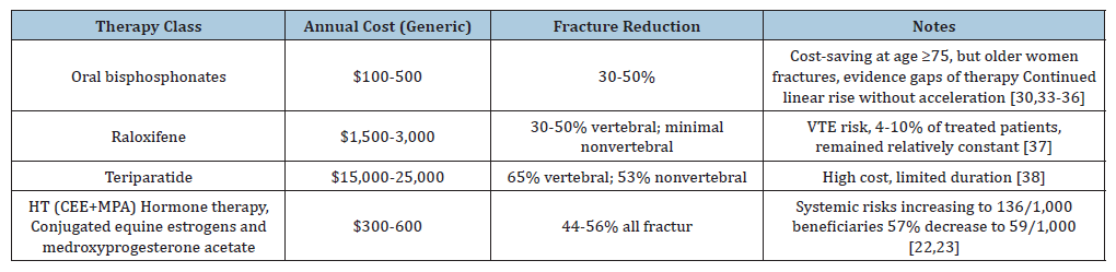
Although the data overall do not support the use of Menopausal Hormone Therapy (MHT) or selective estrogen receptor modulator for primary prevention of CVD, evidence is accumulating that careful use of MHT for perimenopausal symptoms may not carry CVD harm. Still MHT remained low and dropped [39].
Secondary fracture prevention intervention resulted in an average cost savings of $418 and an increase in QALYs of 0.0299 per patient over the lifetime; for 1 million patients who receive the intervention instead of usual care, expected cost savings for Medicare would be $418 million dollars. There was risk of cancer [40,41].
With loss of hormones bone density drops and women fracture. Women who discontinued postmenopausal HT had significantly increased risk of hip fracture and lower BMD compared with women who continued taking HT. The protective association of HT with hip fracture disappeared within 2 years of cessation of HT. [36].
Older women at high risk of fracture, 10 to 13 years of bisphosphonate use was associated with higher risk of any clinical fracture than 2 years of use. These results add to concerns about the benefit of very long-term bisphosphonate use [5,34].
Although the data overall do not support the use of Menopausal Hormone Therapy (MHT) or selective estrogen receptor modulator for primary prevention of CVD, evidence is accumulating that careful use of MHT for perimenopausal symptoms may not carry CVD harm. Still MHT remained low and dropped [39,42].
Data so far, favor transdermal estradiol over conventional-dose CEE with respect to CVD risk and oral estradiol over conventionaldose CEE with respect to stroke risk. Low-dose oral CEE may similarly have benefit over conventional-dose oral CEE for some CVD events. In addition, the transdermal route of delivery may avoid the excess risk of certain CVD events associated with MHT and lower doses of estrogen may have fewer adverse effects than the doses previously tested in WHI [32,39].
The absence of a low-cost, fracture-preventing alternative to HT for symptomatic women in their 50s represents a market failure with substantial economic consequences. However anabolic therapy is more effective but more expensive as noted [38].
Teriparatide recombinant human parathyroid hormone [1- 33] received FDA approval on November 26, 2002-just months after the July 2002 publication of the WHI estrogen-plus-progestin trial results that caused hormone therapy prescriptions to decline 57% by mid-2004. This timing positioned teriparatide as the first anabolic bone agent available to fill the emerging treatment gap [38]. Other formulations of HT such as topical and other formulations have been suggested [32,39].
Current economic paradox
Despite 20 years of follow-up data supporting age-stratified HT use for symptomatic women [27,28], economic analyses and guidelines remain anchored to the 2002 aggregate findings. The USPSTF maintains Grade D recommendations against HT for chronic disease prevention, but without any help since there is omission of noting adequate differentiation by age or indication [28,29].
Key economic insights from extended follow-up:
a. CEE-alone: 22% reduction in breast cancer with longterm
follow-up [40,41]
b. Younger women (50-59): 32% reduction in all-cause
mortality but early WHI data evaluation did not recognize the
benefit of CVD with estrogen-only therapy [32]
c. Removing CEE led to loss of symptom control which was
very important for these women [22-24,29]
d. HT therapy is given by the physician after evaluation of
the patient using the timing hypothesis [31,42]
e. One conclusion that fracture benefits persist after
HT discontinuation ignores the straight-line utilization of
bisphosphonates [35]. Older women paradoxically actually
had more fractures [35] This conclusion ignores the effective
use of hormones used in early -ages 50-59 or within 10 years
of menopause on bone mineral density of which the WHI
demonstrated. If hormone therapy is stopped there is rapid
loss of bone that additional bone directed medications need
to be used. Women who discontinued postmenopausal HT
had significantly increased risk of hip fracture and lower
BMD compared with women who continued taking HT
[43,44]. But even the low circulating estrogen levels present
in postmenopausal women have a significant impact on bone
turnover. The proportion of hip fracture patients treated with
osteoporosis drugs has increased, but remains low, with fewer
than one-third receiving pharmacotherapy. Of patients treated
before and after hip fracture, 18% changed therapy post
fracture. Significantly more patients changed therapy following
fracture if a different physician prescribed treatment (26%)
compared to those treated by the same physician pre- and postfracture
(13%; p < 0.0001) [34].
f. Teriparatide an anabolic was also used but had time limits
but did not have enthusiasm and length of therapy then as we
have now [38].
Policy and economic implications
A. Age-stratified coverage policies: Value based insurance
design (VBID) should favor HT for women <50 years or <10
years postmenopausal, with reduced copayments aligned with
cardiovascular and fracture prevention value [45].
B. Fracture Liaison Service (FLS) integration: Given
HT’s fracture prevention and the treatment gap post-WHI, FLS
programs offer cost-saving secondary prevention [16].
C. Revised economic models: Current cost-effectiveness
analyses should incorporate age-specific and time-sincemenopause
stratification rather than aggregate populations
[22-24].
D. Formulation considerations: Transdermal estradiol
and oral progesterone may offer favorable cardiovascular and
breast cancer profiles compared with CEE+MPA, with economic
implications requiring evaluation [32,39].
At elevated cardiovascular and breast cancer risk. The failure to implement age-stratified prescribing guidelines and economic models that capture heterogeneous effects perpetuates suboptimal resource allocation. Reframing HT as a time-sensitive intervention for appropriate candidates-rather than a uniformly harmful therapy-represents ignored finding a major opportunity for valuebased care improvement [31].
a) The issues with the WHI were more apparent to some than others [46,47]. The influence on CVD was initially misread and the menopausal symptoms were ignored. The ignored findings were that HT initiated in the younger menopause group 50 to 59 Years or within 10 years of menopause helps CVD, the bones and the symptoms of menopause.
Where are We Now?
Given an approximate gender distribution of 55% women and 45% men among adults aged ≥50 years, this corresponds to roughly 41 million women and 34 million men. Based on lifetime risk estimates, approximately one in two women and one in five men over age 50 will experience an osteoporosis-related fracture, translating to ≈20 million women and ≈7 million men, or ≈27 million individuals overall [48,49].
Health outcomes: Approximately 20-30% of older adults who sustain a hip fracture die within one year, most often due to postoperative or medical complications, with higher mortality observed in men [48,49].
Many more survivors face chronic pain, reduced mobility, a hunched posture, and long-term nursing: Many survivors experience chronic pain, reduced mobility, kyphotic deformity, loss of independence, and a high likelihood of long-term institutional care. These downstream consequences drive substantial healthcare utilization, with direct costs exceeding $20 billion annually in the United States and projected to increase markedly with population aging. Osteoporotic fractures lead to chronic pain, disability, loss of independence, and long-term institutionalization. Hip fractures result in major functional decline, with many patients requiring nursing home placement and failing to regain baseline function [50].
Sources
Economic context
The cost imbalance in osteoporosis care remains stark: Solomon et al. (2014) noted that if payers required demonstration of adequate post fracture osteoporosis care, such as bone mineral density testing and appropriate medication prescribing, “local health care systems would likely improve their internal communication among providers, and potentially adopt collaborative care models” [16]. Nayak et al. [5] demonstrated that secondary fracture prevention interventions for Medicare beneficiaries were “highly likely to be cost-saving, both improving future health outcomes and reducing healthcare spending compared with usual care” [5].
What about “they only pay you $ 37” per DXA examination?
Prevention of osteoporosis includes diagnosis. As a physician we can develop signs on our history and physical, including family history of osteoporosis and of fractures, previous fractures, current smoking, long term glucocorticoid steroid use, associated rheumatoid arthritis, excess alcohol consumption and physical findings of osteoporosis including dermatoporosis as found on the dorsum of the hands a sign of thin skin of collagen fragility associated with osteoporosis [51]. Other findings include clinical signs of a previous fracture, kyphosis or stooped back kyphosis/ thoracic deformity: Self-reported “humped back” or observed thoracic kyphosis increases suspicion for vertebral fractures; the wall-occiput distance >0 cm (LR+ 4.6) and rib-pelvis distance <2 fingerbreadths (LR+3.8) are associated with occult spinal fracture, vertebral tenderness suggesting a recent fracture, height loss suggested thresholds include >0.8 inches (2cm) over 1-3 years or >1.5 inches (4cm) from peak height at age 20 years, low body weight less than 112 lbs, Low body weight: Weight less than 51kg (112lb) carries the highest positive likelihood ratio (LR+7.3) for osteoporosis; low BMI (<20kg/m² or <127lb.) is a recognized clinical risk factor, low tooth count suggested less than 20 to reflecting alveolar mouth bone softness, Pain-related behavior: In patients with back pain, assessment of grimacing, sighing, difficulty turning prone, or need for assistance with positioning may help distinguish recent vertebral fracture from other causes of back pain [52].
Examination of the imaging findings by X-rays, DXA bone density (Dual-Energy X-ray Absorptiometry) (originally spelled DEXA but now DXA) and can also be found on a CT scan (computed tomography) and MRI (Magnetic Resonance Imaging). A new study called the biomechanical CT (BCT) is an approved test by Medicare measures both bone mass and bone strength. It has just been approved this year and by October 5th, 2026, will be fully covered is a bone mass measurement with the CPT codes 055T-058T. Initially its availability may be limited But DXA measures bone mineral density but this more effectively measures bone mineral density and bone strength [53].
DXA scan can quantify the level of bone mineral density which is a measure of bone quantity and identify osteopenia and osteoporosis. Sometimes bone quality can be further identified by the Trabecular Bone Score which is advanced imaging software that analyzes the texture and pixel variations of a standard lumbar spine DXA scan unlike a bone mineral density which measures bone quantity. The trabecular bone scan provides an indirect estimate of bone quality and microarchitecture on how solid or porous the inner bone structure is. It is a costly separate examination program for the usual DXA unit. This can also be measured by visualization by the MRI but includes additional evaluation that is usually not available by the standard MRI unit. David Dempster PhD has shown on bone biopsies that the trabecular bone with osteoporosis has a less solid microarchitecture structure than the normal bone as is demonstrated in these figures [52,54].
Loss of Horizontal Crossties why the Osteoporosis Bone Shows a risk of Fracture (Figure 2)
Figure 2:
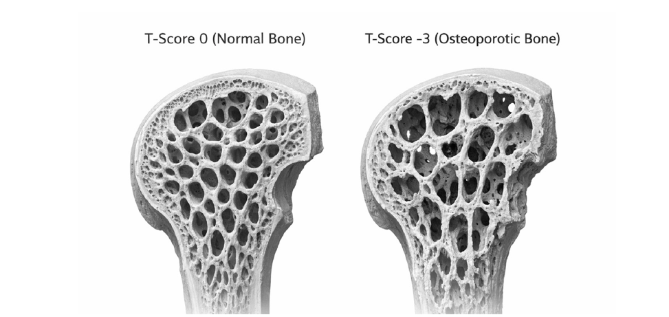
Normal Bone - thick connections
Osteoporosis Bone - loss of bone and connections
Model inspired by David Dempster [52].
Antiresorptive drugs maintain bone structure and primarily increase BMD by adding mineral to preexisting bone while anabolic drugs improve bone structure and primarily increase BMD by adding new, young bone to the skeleton
Dual - Energy X-ray Absorptiometry (DXA): The $37 government payment for the Professional component of a bone density test
The “$37 reimbursement” figure does not come from a single CMS rule document stated that way-it shows up in the medical literature describing the impact of Medicare payment cuts, particularly after the Deficit Reduction Act (DRA) of 2005 and subsequent Physician Fee Schedule changes [55-57].
The clearest place is explicitly stated:
A. A peer-reviewed review article in Journal of Clinical
Endocrinology & Metabolism notes that Medicare
reimbursement for office-based DXA fell to “approximately
$37 per test” after the cuts beginning in 2007[55]. The article
summarizes CMS policy effects, not issuing the rule itself [58].
B. This led to the loss of office-based non-facility DXA
locations which lead to loss of easy access DXAs for our patients
[50,56, 59-61].
C. The National coverage determination data is not clear
[58].
What actually happened (policy-wise):
a. The Deficit Reduction Act of 2005 capped imaging
reimbursement in physician offices starting in 2007 [57].
b. CMS then implemented this through the Medicare
Physician Fee Schedule, which progressively reduced DXA
payments [58].
c. Office-based DXA reimbursement dropped sharply
(roughly from ≈$130-140 in 2006 → ≈$80 → ≈$70 → and in
some analyses down to ≈$33-$37 effective payment levels
depending on component/accounting) [50,56,59-61].
d. Office-based non-facility DXA reimbursement dropped
substantially after 2007 and continued declining [50,56,59-61].
e. Overall bone densitometry reimbursement fell ≈70%
from 2007-2019.
f. Policy discussions and advocacy pieces later describe
typical reimbursement in the ≈$40 range, consistent with that
≈$37 figure.
g. In the United States, Medicare gradually reduced payments
for Dual-Energy X-ray Absorptiometry (DXA) performed at
physician offices (or other nonhospital settings, labeled non
facility) from an average (professional and technical fees) of
$139 in 2006 to about $82 in 2007 and 2008 and $72 in 2009.
The reduction in DXA reimbursement was associated with a
decrease in the number of DXAs performed in physician offices
and fewer physician offices could not afford to provided DXA
services [50,56,59-61].
Total DXA imaging -facility and non-facility (Table 7)
Table 7:
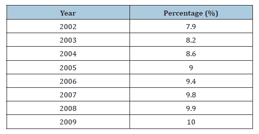
The measured percentage of DXA imaging increased steadily from 7.9% in 2002 to 10.0% in 2009, with the rate of increase slowing and approaching a plateau after 2007.
From 2002 to 2009, the proportion of non-facility care remained relatively stable at approximately 68-70% before declining slightly to 66%, while facility-based care (especially hospital) increased modestly from about 30-31% to 34% over the same period as facility reimbursement increased.
Proportions of facility and non-facility central DXA by calendar year [50,56,59-61] (Table 8).
Table 8:
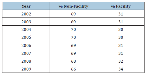
From 2002 to 2009, the proportion of non-facility care remained relatively stable at approximately 68-70% before declining slightly to 66%, while facility-based care increased modestly from about 30-31% to 34% over the same period.
From 2002 to 2004, there was a marked increase in practices initiating DXA services, reaching a peak in 2004, followed by a progressive decline; by 2007, the trend reversed, with discontinuations exceeding initiations and resulting in a sustained net loss of DXA-providing practices through 2009. This relates to our patients getting less DXAs
Net Change in DXA Practices (Table 9)
Table 9:
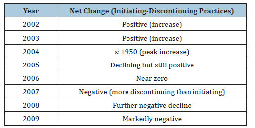
The difference between the numbers of physician office-based practices initiating and discontinuing DXA services by calendar year [50,56].
Between 1996 and 2002, the number of DXA scans performed on all Medicare patients increased by more than fourfold. In the same population, the percentage of scans performed by radiologists increased from less than one-third to almost one-half. Consequently, radiologists were steadily gaining market share of this procedure in the Medicare population [58,59]. The Medicare source did not give understandable specifics [58].
The DXA program is an important program to diagnose osteoporosis, but the government decided to decrease the payment to the point where the payment was beneath the cost it would take to do a DXA. When we look at this explanation below of the payment we see how DXA payment revolves [59-64] (Tables 10&11).
Table 10:Year-by-year Medicare reimbursement trends for CPT 77080 and 77081.
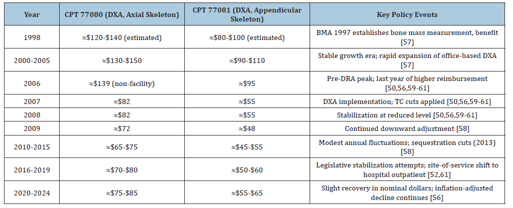
Notes on the table:
Values represent non-facility (physician office) national averages for total reimbursement (PC+TC)
Hospital outpatient payments remained substantially higher (≈$100-$120) throughout this period, driving the site shift
[56,59-61].
The 70.5% inflation-adjusted decrease for bone densitometry between 2007-2019 was the largest of any imaging modality:
Inflation-adjusted Medicare reimbursement for all imaging modalities decreased between 2007 and 2019. The greatest
mean decrease in reimbursement rates was observed for MRI (-$52.08), and the largest decrease in total percentage
change was seen for bone densitometry (-70.5%). Nuclear medicine demonstrated the smallest mean decreases in both
annual change (-$0.32) and total percentage change (-4.28%) [60].
The loss of bone density around the world with its other correlations was associated with increased death [50].
Table 11:Critical distinctions in DXA coding.

Critical Impact of Reimbursement Collapse
The payment reduction triggered profound access
consequences.
1) Office-based DXA discontinuations exceeded initiations
starting in 2007; by 2009, 1,876 practices discontinued versus
1,394 that initiated.
2) Site-of-service shift: Hospital-based DXA rose from 31%
to 34% of Medicare volume (2006-2009).
3) Screening rates plateaued: After 2006, annual growth in
DXA utilization dropped from 0.4% to 0.1%
4) Undertreatment cascade: Reduced DXA access correlated
with decreased osteoporosis diagnosis and bisphosphonate
prescriptions, followed by plateauing hip fracture rates after
2012.
“Medicare’s significant drop in payment rates for DXA from its high-water mark may have the appearance of a cost savings for that line item on CMS outlays, but it has generated MORE costs to the overall health system due to under diagnosing and under treating osteoporosis and the associated increase in fractures. The Increasing Access to Osteoporosis Testing for Medicare Beneficiaries Act of 2021 (S.1943) sought to restore DXA reimbursement to 70% of 2006 levels. The 2025 USPSTF systematic review confirmed screening reduces hip fractures (pooled RR 0.83) with 5-6 fewer fractures per 1,000 screened supporting the economic argument that upfront screening costs are offset by downstream fracture prevention [52,62].
Prevalence of osteoporosis
The most reliable U.S. estimates come from NHANES 2017- 2018 and the National Osteoporosis Foundation (NOF) [63-65].
Corrected epidemiologic figures (Table 12)
Table 12:
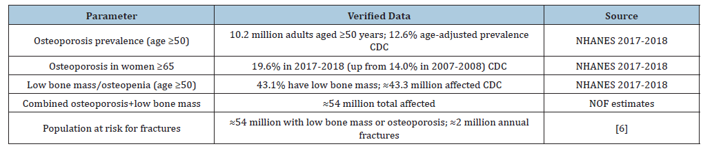
“Approximately 54 million Americans aged ≥50 years have low bone mass or osteoporosis-10.2 million with osteoporosis and 43.3 million with osteopenia. Osteoporosis-related fractures increase substantially after age 50, with ≈2 million incident fractures annually in the U.S.” [63-65].
“Medicare’s significant drop in payment rates for DXA from its high-water mark may have the appearance of a cost savings for that line item on CMS outlays, but it has generated MORE costs to the overall health system due to under diagnosing and treating osteoporosis and the associated increase in fractures need to be justified providing details on preventive screening guidelines (DXA scans) or lifestyle and medical treatments used to reduce fracture risks [66,67].
McAdam-Marx et al. [68] examined the effect of Medicare reimbursement reductions for imaging services on osteoporosis screening rates and found that while BMD screening rates did not substantially decline in the immediate 2 years after reimbursement cuts, the proportion of women whose first osteoporosis diagnosis occurred after a fracture increased-suggesting a shift toward diagnosis at the point of fracture rather than through preventive screening [68].
Miller et al. [55] explicitly identified “the decline in bone mineral density testing by dual-energy x-ray absorptiometry (DXA) in non-facility designated DXA sites (eg: private practices)” as one of three primary reasons for underdiagnosis and undertreatment of osteoporosis. The authors noted that Medicare reimbursement for DXA in non-facility settings had declined to approximately $37 per test by 2007, compared with $100 per test in facility settings, and that professional societies including the International Society for Clinical Densitometry supported legislation to establish “a flat and common floor for all DXA providers nationwide of $98/test” [55].
Tanner et al. [61] summarized that “patient access to DXA scans has been threatened by declining reimbursement and, therefore, access to diagnosis and fracture prevention,” and noted that “successful efforts to reverse this trend” had been made but that “the future remains uncertain” [61].
McPhee et al. [62] stated that “due to CMS reimbursement cuts for DXA scans, the number of providers who can perform DXA scans has dwindled and machines are no longer common in provider offices,” and explicitly endorsed the Increasing Access to Osteoporosis Testing for Medicare Beneficiaries Act of 2021 (S.1943), which aimed to restore reimbursement to 70% of 2006 levels [62].
Williams et al. (2021) documented that osteoporosis-related fractures in Medicare beneficiaries generated mean total all-cause healthcare costs more than 3-fold higher than matched nonfracture cohorts, with medical service costs 10-fold higher than pharmacy costs. They noted that “a decline in fracture rates between 2007 and 2013 followed by a subsequent rise in 2017... may be a result of changes in testing and treatment of osteoporosis during this time” [8].
Hsieh et al. [50] (cited in Lancet Rheumatology 2025) described associations between cuts in Medicare reimbursements for DXA and decreased provision of physician’s office-based DXA services and prescriptions for FDA-approved osteoporosis therapies 2 years later, with age-adjusted hip fracture rates plateauing from 2012 to 2015 following a decade of steady decreases [50].
Prevention
Osteoporosis is a skeletal disorder characterized by compromised bone strength, which predisposes a person to increased risk of fracture. In the United States, 26% of women aged > or =65 years and >50% of women aged > or =85 years have osteoporosis. Over 1.5 million fractures per year are attributable to osteoporosis; these fractures result in 500,000 hospitalizations, 800,000 emergency room visits, 2.6 million physician visits, 180,000 nursing home placements, and 12 billion dollars to 18 billion dollars in direct healthcare costs each year. Fracture also results in loss of function and has a negative impact on psychological status. In recognition of the importance of bone health, the US Surgeon General has, for the first time, issued a comprehensive report on bone health and treatment. The report recommends a pyramidal approach to osteoporosis treatment that includes calcium and vitamin D supplementation, physical activity, and fall prevention as the first line in fracture prevention. The second level consists of treating secondary causes of osteoporosis; the third and top level consists of pharmacotherapy. Pharmacotherapeutic interventions (e.g., bisphosphonates, selective estrogen receptor modulators, calcitonin, and anabolic like teriparatide) in women with postmenopausal osteoporosis provide substantial reduction in fracture risk over and above risk reduction with calcium and vitamin D supplementation alone. Despite the effectiveness of therapy, most patients who receive treatment do not remain on treatment for >1 year. An important approach to reducing the rate of fractures is first to target our treatments to patients at high risk for fractures and then to develop strategies to improve treatment continuation rates [69-71].
Participatory Learning for Action (PLA)
The Participatory Learning for Action (PLA) approach to CME planning is grounded in performance Improvement Continuing Medical Education (PI-CME) principles and Quality Improvement (QI) methodology, particularly the Plan-Do-Study-Act (PDSA) cycle [72]. The 2020 AAFP framework describes “Meta PI-CME” as continuous cycles of needs assessment, learning, implementation, and reassessment rather than “one and done” activities [72]. Greenspan et al. [73] demonstrated that a three-stage PI-CME initiative improved osteoporosis screening, fall-risk assessment, and treatment adherence through self-evaluation and goal setting [73]. Solomon et al. [74] showed that multifaceted interventions targeting primary care physicians and patients increased BMD testing and osteoporosis medication use, though absolute rates remained low [74]. The Asia Pacific Consortium on Osteoporosis (APCO) framework explicitly recommends “bottom-up” participatory approaches with healthcare providers conducting iterative benchmarking and quality improvement projects [75]. A 2026 ClinicalTrials.gov entry (NCT06682650) describes combining Normalization Process Theory (NPT) with PLA for implementation, using democratic techniques to identify barriers and prioritize action steps [75]. These sources support PLA-CME as an evidencebased strategy to translate osteoporosis knowledge into practice change.
Conclusion
Advocacy and attempted remediation
Professional societies {ISCD (International Society of Clinical Densitometry), ACR (American College of Rheumatology), ACCE, (American College of Clinical Endocrinologist) NOF (National Osteoporosis Foundation) CSRO (Coalition of State Rheumatology organizations)} have consistently advocated restoration to approximately $98 per test as a minimum viable reimbursement floor. (Still not enough to cover expenses) The “Increasing Access to Osteoporosis Testing for Medicare Beneficiaries Act” and similar legislation have been introduced repeatedly since 2010 to reverse DRA-mandated cuts, with partial success in stabilizing-but not restoring-payment levels [51,62,63].
We used to have many physician offices that were doing bone densities in Atlanta, but as you see this imploded. It still has not returned despite as noted many attempts. This is because the financial blockade has not been overridden [70].
The therapy for patients with osteoporosis now expresses the importance of an anabolic agent like teriparatide (Forteo), abaloparatide (Tymlos) and romosozumab (Evenity). (These are all injections) The scare of Osteonecrosis of the Jaw (ONJ) frightened patients from accepting bisphosphonate therapy like alendronate (Fosamax), risedronate Actonel,(These are all oral medication) ibandronate (Boniva) (available as oral or injection) zoledronate (Reclast) (Which is given by infusion), despite that ONJ is a very unusual episode in the course of therapy, much more in the high doses used for oncology and not the low doses used for osteoporosis. Patients did not want to take that chance of ONJ.
It appears that the episodes of government payment interference, Osteonecrosis of the Jaw (ONJ), misinformation spread by the Women’s Health Initiative, the decrease in testing since physicians we’re less likely to have a bone density unit in the office because of the cost of it, the cost of maintenance of it, the teaching of bone density technologists to facilitate performing the procedure. This has led to inappropriate concern. In some ways it was the government associated with the two-pronged attack whether they might have not known what that would do to our patients. We have now realized what the WHI and DXA reimbursement in the long run has cost our patients more.
The WHI E+P trial made high-value use of public funds with a substantial return on investment. These results can contribute to discussions about the role of public funding for large, prospective trials with high potential for public health effects [22].
We need to emphasize the importance and costs of osteoporosis and fractures and let people be aware of the need. The use of David Dempster models of the trabecular structure to assist in convicting people who don’t want to take osteoporosis therapy is important. Unless they have personal experience, they are often reluctant to do any of the testing and Osteoporosis therapy.
Cardiologists are on top of treating the risks for cardiovascular disease, both before and then Importantly after the myocardial infarction, especially with anti-lipid and other therapy including imaging and echoing is an example of the idea of what we can use for bone protection and prevention.
What do we do as osteoporosis providers? We need to spread the word to our primary care and orthopaedic colleagues of the care of people with risks of osteoporosis and risks of fracture and our desire to advise and assist them. There are low rates of osteoporosis testing and treatment initiation and then continue the therapy which is decreased. There is a high secondary fracture rate (particularly among patients at very high risk) highlight the need for better management of patients after a fracture [71]. These shadow the implementation gap. The Medicare and insurance obstruction is limiting our ability to do this and we need change.
We are not allowed to trying to prevent osteoporosis and we should stop fractures at earlier ages! This needs to be done but there is the implementation gap! The gap is both in the patient being prescribed the medicine at the appropriate time, ideally before the fracture when osteoporosis is discovered but at least at the time of the fracture and then should continue to stay on the medicine which is low. Obstruction at trying to make an earlier diagnosis of osteoporosis is inappropriate for our health systems.
For osteoporosis and fractures, we should proceed by the
example of cardiology.
1) We should be doing bone densities at an earlier age for
women and men and if we find that there is risk then we should
provide therapy. We should perform DXAs more often
2) We should be doing anti-osteoporosis therapy at an
earlier age, the so-called preventive stage
3) We should stop the ridiculous under payment of the
simple tests of a bone density to help identify women and men
who were at risk. To this we should use age 40 for women and
age 50 for men.
4) The federal mandate of underpaying has increased the risk
of osteoporosis fractures, and it needs to stop. We should take
this to our state legislators, our United States Congressional
legislators and the American Medical Association.
5) The rheumatologists in our community were performing
DXAs in their offices but they couldn’t afford to continue it
because the federal Medicare payment dropped as low as $37 a
test for a test which costs over $100- $150 or more for purchase
and maintenance of bone density units as well as education
for the providers who perform the testing of the precision
assessment.
6) We should develop this program immediately to help
our patients prevent the risk of fracture, and to help control
symptoms.
7) We need to confront the underpayment of our DXA exams
from our offices as non-facility sites compared to the facility
sites at the hospital. We don’t have control over how they’re
done in the hospital and very often we find corrections. Also,
in our office we have to do quality precision assessment to
control examinations which unfortunately are not performed
appropriately in most facility bone density exams
8) We are the champions of our patients, and we need to
correct how the regulators are treating our patients and our
physicians.
The moral of the story: Medicare’s significant drop in payment rates for DXA from its highwater mark may have the appearance of a cost savings for that line item on CMS outlays, but it has generated MORE costs to the overall health system due to underdiagnosing and undertreating osteoporosis and the associated increase in fractures. Penny wise and pound foolish.
References
- Didem Bernard, Zhengyi Fang (2019) Financial burdens and barriers to care among nonelderly adults with heart disease: 2010-2015. J Am Heart Assoc 8(24): e008831.
- Maedeh Zokaei N, Sundeep K, Marwa KM, Ashok Krishnaswami, Mehdi HS, et al. (2026) The economic burden of cardiovascular disease in the United States. Am J Cardiol 270: 172-179.
- Sandra BD, Olga AK, Tamilyn B, Gail Hunt, Rebecca AK, et al. (2018) Projected costs of informal caregiving for cardiovascular disease: 2015 to 2035: A policy statement from the American heart association. Circulation 137(19): e558-e577.
- Joseph SY, Nidhi GK, Michael GF, Donna GB, Matthew AF, et al. (2022) ACR Appropriateness criteria® osteoporosis and bone mineral density: 2022 update. J Am Coll Radiol 19(11S): S417-S432.
- Smita N, Andrea S, Susan LG (2021) Cost-effectiveness of secondary fracture prevention intervention for Medicare beneficiaries. J Am Geriatr Soc 69(12): 3435-3444.
- Russel B, Bess DH, Daniel HS, John BW, Alison K, et al. (2007) Incidence and economic burden of osteoporosis-related fractures in the United States, 2005-2025. J Bone Miner Res 22(3): 465-475.
- Gabriel ST, Natalia RN, Andrew EM, Zugui Z, Paul K, et al. (2024) Cost of cardiovascular disease event and cardiovascular disease treatment-related complication hospitalizations in the United States. Circ Cardiovasc Qual Outcomes 17(3): e009999.
- Setareh AW, Shanette GD, Richard W, Yamei W, Tarun A, et al. (2021) Economic burden of osteoporosis-related fractures in the US Medicare population. Ann Pharmacother 55(7): 821-829.
- Kohli-Lynch CN, Brandon KB, George T, Yiyi Z, Mark JP, et al. (2019) Cost-effectiveness of low-density lipoprotein Cholesterol Level-guided statin treatment in patients with borderline cardiovascular risk. JAMA Cardiol 4(10): 969-977.
- Richard K, Rose Marie R, Robert S, David E (2008) The impact of prevention on reducing the burden of cardiovascular disease. Diabetes Care 31(8): 1686-1696.
- https://www.cdc.gov/tobacco/hcp/patient-care-settings/cardiovascular.htm
- Michael W, Saman F, Gitanjali MS, Shahab K, Renata M, et al. (2017) Cost effectiveness of a government supported policy strategy to decrease sodium intake: Global analysis across 183 nations. BMJ 356: i6699
- Tzeyu L Michaud, Jean Abraham, Hawre J, Russell VL, Sue D, et al. (2015) Cost-effectiveness of a statewide campaign to promote aspirin use for primary prevention of cardiovascular disease. J Am Heart Assoc 4(12): e002321.
- American Heart Association, Dallas, Texas, USA.
- Life’s essentials 8TM, American Heart Association, Dallas, Texas, USA.
- Daniel HS, Amanda RP, John S, Elena L (2014) The potential economic benefits of improved post fracture care: A cost-effectiveness analysis of a fracture liaison service in the US health-care system. J Bone Miner Res 29(7): 1667-1674.
- Annalise NP, Santanu KD, Thomas JW, Louise CW, Colon-Emeric CS (2011) Cost-effectiveness of oral bisphosphonates for osteoporosis at different ages and levels of life expectancy. J Am Geriatr Soc 59(9): 1642-1649.
- Glenn EL, Scott M, Gregory JG (2011) Management of fragility hip fractures: Our institutional experience. Geriatr Orthop Surg Rehabil 10: 2151459319828618.
- Leon N, Audrey D, Mathieu G, Xavier D, Suzanne M, et al. (2013) An economic evaluation: Simulation of the cost-effectiveness and cost-utility of universal prevention strategies against osteoporosis-related fractures. J Bone Miner Res 28(2): 383-394.
- Giovanni A, Elena T, Maurizio R, Thomas FB, Roland C (2023) Patients with osteoporosis: Children of a lesser god. RMD Open 9(1): e002973.
- The International Society for Clinical Densitometry (ISCD), Connecticut, USA.
- Joshua AR, Ruth E, Teresa MW, Mary P, Jacques ER, et al. (2014) Economic return from the women's health initiative estrogen plus progestin clinical trial: A modeling study. Ann Intern Med 160(9): 594-602.
- Macarius M Donneyong, Teng-Jen C, Joshua AR, McKenna G, Daniel A, et al. (2020) The women's health initiative estrogen-alone trial had differential disease and medical expenditure consequences across age groups. Menopause 27(6): 632-639.
- Irene L, Eleni A, Nikoletta M, Panagiotis A (2026) Then and now: What we have learned from the WHI. J Clin Endocrinol Metab 111(4): e974-e994.
- Brian LS, Amy TD, Kathleen AC (2012) A sustained decline in postmenopausal hormone use: Results from the national health and nutrition examination survey, 1999-2010. Obstet Gynecol 120(3): 595-603.
- Jacob AU, Michael AF, M Alan B, Daniel HS, Niteesh KC (2006) Effect of the women's health initiative on osteoporosis therapy and expenditure in Medicaid. J Bone Miner Res 21(5): 765-771.
- Jacques ER, Ross LP, JoAnn EM, Lieling W, David B, et al. (2007) Postmenopausal hormone therapy and risk of cardiovascular disease by age and years since menopause. JAMA 297(13): 1465-1477.
- Heidi DN, Miranda W, Bernadette Z, Jennifer M (2012) Menopausal hormone therapy for the primary prevention of chronic conditions: A systematic review to update the U.S. preventive services task force recommendations. Ann Intern Med 157(2): 104-113.
- JoAnn EM, Rowan TC, Marcia LS, Aaron KA, Jacques ER, et al. (2013) Menopausal hormone therapy and health outcomes during the intervention and extended poststopping phases of the Women's Health Initiative randomized trials. JAMA 310(13): 1353-1368.
- JoAnn EM, Shari SB, Andrew MK, JoAnn VP (2020) The women's health initiative trials of menopausal hormone therapy: Lessons learned. Menopause 27(8): 918-928.
- S Mitchell H, Eric V, Eliot AB, Matthew JB, Marcelle IC, et al. (2011) Timing and duration of menopausal hormone treatment may affect cardiovascular outcomes. Am J Med 124(3): 199-205.
- Georgina EH, Chrisandra LS (2015) Hormone therapy in menopause: An update on cardiovascular disease considerations. Trends Cardiovasc Med 25(6): 540-549.
- Clifford JR, Sue AB (2005) A rational approach to evidence gaps in the management of osteoporosis. Am J Med 118(11): 1183-1189.
- Euni L, Mary KM, Anthony KW, Ilene HZ (2010) Long-term effect of the women's health initiative study on antiosteoporosis medication prescribing. J Womens Health (Larchmt) 19(5): 847-854.
- Rebecca LD, Andrea ZL, Shirley AAB, Denise MB, Charles K, et al. (2017) Long-term oral bisphosphonate therapy and fractures in older women: The women's health initiative. J Am Geriatr Soc 65(9): 1924-1931.
- Suzanne MC, Jeffrey NK, M Alan B, Raisa L, Margaret RS, et al. (2008) Trends in drug prescribing for osteoporosis after hip fracture, 1995-2004. J Rheumatol 35(2): 319-326.
- Wenger NK, Barrett-Connor E, Collins P, Grady D, Kornitzer M, et al. (2002) Baseline characteristics of participants in the Raloxifene Use for The Heart (RUTH) trial. Am J Cardiol 90(11): 1204-1210.
- Miller PD, Bilezikian JP, Deal C, Harriset ST, Pacifici R, et al. (2004) Clinical use of teriparatide in the real world: Initial insights. Endocr Practice 10(2): 139-148.
- Simon J (2014) What if the women’s health initiative had used transdermal estradiol and oral progesterone instead? Menopause 21(7): 769-783.
- Pan K, Lavasani S, Aragaki AK, Chlebowski RT (2022) Estrogen therapy and breast cancer in randomized clinical trials: A narrative review. Menopause 29(9): 1086-1092.
- Chlebowski RT, Aragaki AK (2023) The women's health initiative randomized trials of menopausal hormone therapy and breast cancer: Findings in context. Menopause 30(4): 454-461.
- Valdiviezo C, Lawson S, Ouyang P (2013) An update on menopausal hormone replacement therapy in women and cardiovascular disease. Curr Opin Endocrinol Diabetes Obes 20(2): 148-155.
- Karim R, Dell RM, Greene DF, Mack WJ, Gallagher JC, et al. 2011) Hip fracture in postmenopausal women after cessation of hormone therapy: Results from a prospective study in a large health management organization. Menopause 18(11): 1172-1177.
- Khosla S (2010) Update on estrogens and the skeleton. J Clin Endocrinol Metab 95(8): 3569-3577.
- Kelly AM, Cronin P, Carlos RC (2008) Introduction to value-based insurance design. J Am Coll Radiol 5(11): 1118-1124.
- Goldman JA (2004) The women's health initiative 2004-review and critique. Medscape General Medicine 6(3): 65.
- Bush TM, Bonomi AE, Nekhlyudov L, Ludman EJ, Reed SD (2007) How the Women's Health Initiative (WHI) influenced physicians' practice and attitudes. J Gen Intern Med 22(9): 1311-1316.
- Wang M, Seibel MJ (2023) Approach to the patient with bone fracture: Making the first fracture the last. J Clin Endocrinol Metab 108(12): 3345-3352.
- Harris K, Zagar CA, Lawrence KV (2023) Osteoporosis: Common questions and answers. American Family Physician 107(3): 238-246.
- Hsieh E, Bryazka D, Ong K, Rhinehart PA, Cousin E, et al. (2025) The global, regional, and national burden attributable to low bone mineral density, 1990-2020: An analysis of a modifiable risk factor from the global burden of disease study 2021. The Lancet Rheumatology 7(12): e873-e894.
- Goldman JA, Parris GR, Johnson M (2026) Dermatoporosis a clue to osteoporosis: With collagen fragility of aging. Res Arthritis Bone Study 2(3): 1-6.
- Camacho PM, Petak SM, Binkley N, Diab DL, Eldeiry LS, et al. (2020) American Association of Clinical Endocrinologists (AACE)/American college of endocrinology clinical practice guidelines for the diagnosis and treatment of postmenopausal osteoporosis-2020 update. Endocr Pract 26(Suppl 1): 1-46.
- Keaveny TM, Adams AL, Orwoll ES, Khosla S, McClung MR, et al. (2026) Improved prediction of hip fracture using multi-faceted biomechanical computed tomography, J Bone Miner Res 41(5): 507-520.
- Dempster DW, Shane E, Horbert W, Lindsay R (1986) A simple method for correlative light and scanning electron microscopy of human iliac crest bone biopsies: Qualitative observations in normal and osteoporotic subjects. J Bone Miner Res 1(1): 15-21.
- Miller PD (2016) Underdiagnosis and undertreatment of osteoporosis: The battle to be won. J Clin Endocrinol Metab 101(3): 852-859.
- Zhang J, Delzell E, Zhao H, Laster AJ, Saag KG, et al. (2012) Central DXA utilization shifts from office-based to hospital-based settings among Medicare beneficiaries in the wake of reimbursement changes. J Bone Miner Res 27(4): 858-864.
- Markus AR, Rosenbaum S (2005) Deficit reduction act of 2005: An overview of key Medicaid provisions and their implications for early childhood development services.
- Bone (Mineral) density studies. National Coverage Determination (NCD), USA.
- Intenzo CM, Parker L, Rao VM, Levin DC (2005) Changes in procedure volume and service provider distribution among radiologists and non-radiologists in dual-energy x-ray absorptiometry between 1996 and 2002. J Am Coll Radiol 2(8): 662-664.
- Kumar S, Khurana A, Haglin JM, Hidlay DT, Neville K, et al. (2020) Trends in diagnostic imaging medicare reimbursements: 2007 to 2019. J Am Coll Radiol 17(12): 1584-1590.
- Tanner SB (2011) Dual-energy X-ray absorptiometry in clinical practice: New guidelines and concerns. Curr Opin Rheumatol 23(4): 385-388.
- McPhee C, Aninye IO, Horan L (2022) Recommendations for improving women's bone health throughout the lifespan. J Womens Health (Larchmt) 31(12): 1671-1676.
- US Preventive Services Task Force, Silverstein M, Wong JB, Chelmow D, Coker TR, et al. (2025) Screening for osteoporosis to prevent fractures: US preventive services task force recommendation statement. JAMA 333(6): 498-508.
- The most reliable U.S. estimates come from NHANES 2017-2018 and the National Osteoporosis Foundation (NOF).
- Naso CM, Lin SY, Song G, Hong Xue (2025) Time trend analysis of osteoporosis prevalence among adults 50 years of age and older in the USA, 2005-2018. Osteoporosis Int 36(3): 547-554.
- Wright NC, Looker AC, Saag KG, Curtis JR, Delzell ES, et al. (2014) The recent prevalence of osteoporosis and low bone mass in the United States based on bone mineral density at the femoral neck or lumbar spine. J Bone Miner Res 29(11): 2520-25266.
- https://iscd.org/wp-content/uploads/2012/10/PreserveMedicareFundsforDXAImaging.pdf
- McAdam-Marx C, Unni S, Ye X, Nelson S, Nickman NA, et al. (2012) Effect of medicare reimbursement reduction for imaging services on osteoporosis screening rates. J Am Geriatr Soc 60(3): 511-516.
- Gass M, Dawson-Hughes B (2006) Preventing osteoporosis-related fractures: An overview. Am J Med 119(4 Suppl 1): S3-S11.
- Osteoporosis medications and medication-related osteonecrosis of the jaw, American Dental Association (ADA), USA.
- Singer AJ, Williams SA, Pearman L, Wang Y, Pyrih N, et al. (2023) An evaluation of treatment patterns for osteoporosis and outcomes after a fragility fracture in a real-world setting. J Orthop Trauma 37(4): e159-e164.
- Knight C (2020) The American academy of family physician's approach to developing and supporting the intrinsically motivated learner. J Am Board Fam Med 33(Suppl): S21-S23.
- Susan LG, John PB, Nelson BW, Carolyn AB, William AM, et al. (2013) A clinician performance initiative to improve quality of care for patients with osteoporosis. J Womens Health (Larchmt) 22(10): 853-861.
- Daniel HS, Jennifer MP, Margaret S, Colleen T, Laura B, et al. (2007) Improving care of patients at-risk for osteoporosis: A randomized controlled trial. J Gen Intern Med 2007 22(3): 362–367.
- Manju C, Peter RE, Paul JM, Tuan VN (2022) Harmonization of osteoporosis guidelines: Paving the way for disrupting the status quo in osteoporosis management in the Asia Pacific. J Bone Miner Res 37(4): 608-615.
© 2026 John Abner Goldman. This is an open access article distributed under the terms of the Creative Commons Attribution License , which permits unrestricted use, distribution, and build upon your work non-commercially.
Editor In Chief







.jpg)































Signup for Newsletter

Quick Links
 Editorial Board Registrations
 Submit your Article
 Best Paper of the Volume
 Reprints
 Refer a Friend
 Advertise With Us
Our Recent Edition
.jpg)



Top Editors




.jpg)

















.bmp)
.jpg)
.png)
.jpg)













.jpg)






.png)

.png)




.png)




Financial Support

Sponsors

Latest e-Books

Latest Video

 a Creative Commons Attribution 4.0 International License. Based on a work at www.crimsonpublishers.com.
Best viewed in
a Creative Commons Attribution 4.0 International License. Based on a work at www.crimsonpublishers.com.
Best viewed in 
 | Above IE 9.0 version