- Submissions

Full Text
Orthoplastic Surgery & Orthopedic Care International Journal
Screw fixation for Lateral Condylar Fracture of Humerus in Children
Byanjankar S1*, Amatya S2, Shrestha R1, Sharma JR3 and Chhetri S4
1Department of Orthopaedics and Traumatology, Lumbini Medical College & Teaching Hospital, Nepal
2Alka Hospital, Nepal
3Department of Orthopaedics and Traumatology, Anandaban Hospital, Nepal
4Department of Orthopaedics and Traumatology, Nepalgunj Medical College and Teaching Hospital, Kohalpur, Nepal
*Corresponding author: Byanjankar Subin, Department of Orthopaedics and Traumatology, Lumbini Medical College & Teaching Hospital, Palpa, Nepal
Submission: February 09, 2018;Published: February 26, 2018
ISSN: 2578-0069Volume1 Issue3
Abstract
Introduction: Lateral condyle fracture of humerus in children is the most common elbow fracture that involves the growth plate and the second most common elbow fracture children following supracondylar fracture of thehumerus. The purpose of the study was to evaluate the clinical outcome of operative treatment of displaced lateral condyle fractures using cannulated cancellous screws.
method:
We reviewed 37 lateral condyle fracture of humerus in children treated with screw fixation, from July 2014 to June 2016. Patient with age 5 to 12 years, closed fracture with displacement >2mm, fracture with enough metaphysical fragment for screw and fracture <2weeks were included. Patients with open fractures, anatomical elbow deformity, associated another injury in thesame joint were excluded. Preoperative X-rays were used to classify fracture according to Milch classification Postoperative X-rays were used to evaluate screw positions, fracture union, thepresence of growthplate arrest and avascular necrosis.Results: There were 26 boys and 6 girls in the study, with an average age of 7.7 (peak age mode 7, range 5 to 12 years). Average follow-up was 14months (range 8 to 24 months). The commonest mode of injury was fall from height (mainly tree) and it was seen in 24 patients (75%), and 8 patients (25%) had fractures caused by motor vehicle accidents. The final outcome was evaluated with Mayo Elbow Performance score. At final follow up 90.62% (n=29) had excellent outcome and 9% (n=3) had good outcome. There were no poor results.
Conclusion: Screw provides absolute stability which reduces the possibility of lateral prominence and promotes early fracture union. Absolute stability of fracture permits anearly range of motion with early return to pre-injury state.
Keywords: Lateral condyle; Fracture; Screw fixation
Introduction
Lateral condyle fracture of humerus in children is the most common elbow fracture that involves the growth plate and the second most common elbow fracture children following supracondylar fracture of the humerus [1]. It accounts for 10-20% of all elbow fractures in children [2].
Displaced fracture results in both physeal and articular incongruity. The goal of treatment is to re-establish the articular congruity to obtain near anatomical alignment. Various treatment modalities are available including surgical treatment with closed [3,4], arthroscopic assisted [5,6] or open methods versus nonoperative management with the cast. Operative treatment is considered for displaced fracture >2mm. Minimally displaced fractures are amenable to nonsurgical management. Flynn et al. [7] noted that minimally displaced fractures healed quickly with abundant callus formation. Non displaced fractures are usually treated with cast immobilization, but close follow-up is required as later fracture displacement is a well-documented complication [8].
Although K-wire is the most common metallic implant, a plaster splint or cast is required for a period of immobilization. Few authors suggest that screw fixation promotes fracture union without significant complications [9,10]. There have been very few published reports comparing cannulated screws to K-wires in the displaced lateral condyle fractures [11,12]. Regardless of treatment, temporary elbow stiffness invariably occurs after these injuries. However, screw fixation decreases immobilization period. The advantage of screw over K-wire is rotational stability, inter fragmentary compression at the fracture site, prevents secondary fracture displacement, decreases consolidation time and the risk of valgus deformity [13,14].
Screw fixation provides superior fixation compare to pin fixation, lower rates of lateral overgrowth, fixation loss and infection. Here, we retrospectively reviewed patients treated with a screw to evaluate the clinical outcome for the displaced lateral humeral condyle fractures in our set up.
Methodology
We performed an Institutional Review Board approved retrospective review of all the children with lateral condyle fracture treated with open reduction and screw fixation at our institution. We reviewed the medical records of 37 lateral condyle fracture of humerus in children treated with screw fixation, from July 2014 to June 2016. Patient with age 5 to 12 years, closed fracture with displacement >2mm, fracture with enough metaphysical fragment for screw and fracture <2weeks were included. Patients with open fractures, anatomical elbow deformity, associated another injury in the same joint were excluded.
Preoperative X-rays were used to classify fracture according to Milch [8] classification: type I - fracture line courses lateral to the trochlea through the capitulotrochlear groove and type II - fracture line extends into the apex of the trochlea. Postoperative X-rays were used to evaluate screw positions, fracture union, the presence of growth-plate arrest and a vascular necrosis.
Demographic data including the age of patients, gender, and mechanism of injury, clinical and radiological union, and complications of surgery were reviewed from patient charts. The mechanism of injury was classified as
i. Fall from height
ii. Motor vehicle accidents.
Surgical technique
General anesthesia was given to all the patients. The patient was placed supine on a radiolucent table. The affected limb was then prepped and draped free. Kocher’s approach was used in all the patients. The fracture was reduced and under C-arm, the guide wire was placed perpendicular to fracture line. With the confirmation of proper alignment, 4 mm cannulated screw was passed after drilling and tapping to compress the fracture site. Stability was checked with elbow movement.
Postoperative management and assessment
Postoperatively, the elbow was immobilized with long arm posterior slab for a week followed by anarm sling. Active elbow range of motion was started once the pain subsides. The suture was removed after 2 weeks. The screw was removed after 5-6 months of surgery. The patient was followed up at 6 weeks, 3 months 6 month and one year. Clinical outcomes were evaluated using Mayo elbow performance score.
Results
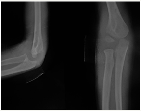
A total of 37 cases were enrolled in the study. Out of which 4 cases got lost till last follow up, 1 was excluded due to re-fracture at six weeks follow up period. Only 32 patients with lateral condyle fracture treated with open reduction and screw fixation met inclusion criteria. There were 26 boys and 6 girls in the study, with an average age of 7.7(peak age mode 7, range 5 to 12years). There was more left sided (18 patients) than right-sided (14 patients) fracture. Milch type II (28, 87.5%) was more common than Milch type I (4, 12.5%). In all the patients, screws were placed in the metaphysis. Average follow-up was 14 months (range 8 to 24 months). The commonest mode of injury was fall from height (mainly tree) and it was seen in 24 patients (75%), and 8 patients (25%) had fractures caused by motor vehicle accidents. The final outcome was evaluated with Mayo Elbow Performance score. At final follow up 90.62% (n=29) had excellent outcome and 9% (n=3) had good outcome. No patient had a poor score in any of the followups. All the patients had screw removal (Figures 1-4).
Figure 1: Preoperative.
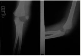
Figure 2: Postoperative.
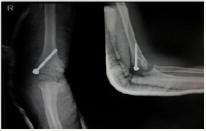
Figure 3: Five months follow up.
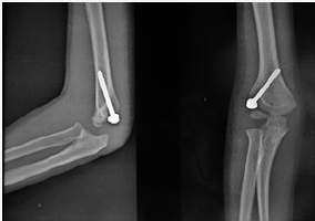
Figure 4: Post-implant removal.
Discussion
Open reduction and internal fixation are indicated for displaced lateral condyle fracture of humerus in children [13]. Internal fixation with K-wires or cannulated screws has been reported to stabilize the displaced lateral condyle fractures. K-wire fixation requires 4 to 8 weeks of postoperative elbow immobilization with a plaster splint or cast in most instances [15,16].
In the present study, all displaced fractures (over 2 mm) were treated by open reduction and internal fixation with a cannulated screw. Screw provides absolute stability. We didn’t use plaster splint to support fractures postoperatively. Active elbow range of motion exercise was started as elbow becomes painless. The disadvantage of screw fixation is screw removal and growth platerelated complications, mainly with screws across the physis [17].
Sharma et al. [18] reported that AO cancellous screw compresses the fracture site, permitting early elbow mobilization and avoiding loss of elbow motion. They also mention that screw fixation is technically more demanding procedure compared with K wire fixation. Postoperativelyscrews were removed at 8 to 10 weeks. Four patient had various deformity which was <4 and 3 patients had mild fishtail deformity.
Wirmer et al. [19] recommended the use of Screw/wire fixation in the operative treatment of lateral condyle fracture of humerus in children. Ayubi et al. [11] also showed the better result with fracture treated with a screw for children age more than 5 years with displacement >2mm. They suggested removing implant 3-4 month after radiological union.
Li et al. [20] in their retrospective study compared Kirschner wires (K-wire) and AO cannulated screw fixation in treatment for the displaced lateral humeral condyle fractures. They found no statistically significant difference in clinical outcome between the two groups and concluded that although the second operation is required for screw removal, screws can reduce the possibility of lateral prominences and promote the elbow function.
Lateral prominence (15.6%, 5 patients) was the common complication we found in our study which was comparable with Hasler et al. [21] (15%) and Shirley et al. [12] (13%). Long-term follow up is required to evaluate premature capitellar growth plate arrest. Thomas et al. [22] reported that 40% of patients with K-wires had an obvious lateral prominence. They proposed that the excessive bone formation beneath the osteoperiosteal flap as the cause for lateral prominence. It may be due to the wide dissection of periosteum and the formation of bone callus [20]. We advise to protect the periosteum in the metaphyseal fragment and avoid wide dissection. No patient developed skin infection and a vascular necrosis in our study. However, the only disadvantage was need of additional surgery for screw removal, thus increasing the cost and hospital stay.
The final assessment in our series was evaluated with Mayo Elbow Performance score. At final follow up 90.62% (29 patients) had an excellent outcome and 9% (3 patients) had a good outcome. There were no poor results. The better functional outcome may be due to early fracture union leading to an early return to activities. Early mobility also reduces school absenteeism. Clinical outcomes in the study done by LiWC et al. [20] using Hardare criteria had 65.62% excellent, 34.38% good result and poor in none, which is comparable with our study.
There were some limitations to our study. This includes retrospective nature with a small sample size and short follow up. Although we demonstrated that screw fixation results in lower complication, longer follow up are required. A larger randomized trial of screw fixation versus K-wire fixation is needed to clarify the results.
Conclusion
Displaced lateral condyle fractures can be treated successfully by open reduction and internal fixation with single partially threaded cancellous screw with excellent results. Screw provides absolute stability which reduces the possibility of lateral prominence and promotes early fracture union. Absolute stability of fracture permits an early range of motion with early return to pre-injury state.
References
- Mizuta T, Benson WM, Foster BK, Paterson DC, Morris LL (1987) Statistical analysis of the incidence of physeal injuries. J Pediatr Orthop 7(5): 518-523.
- Jakob R, Fowles JV, Rang M, Kassab MT (1975) Observations concerning fractures of the lateral humeral condyle in children. J Bone Joint Surg Br 57(4): 430-436.
- Launay F, Leet AI, Jacopin S, Jouve JL, Bollini G, et al. (2004) Lateral humeral condyle fractures in children: a comparison of two approaches to treatment. J Pediatr Orthop 24(4): 385-391.
- Mintzer CM, Waters PM, Brown DJ, Kasser JR (1994) Percutaneous pinning in the treatment of displaced lateral condyle fractures. J Pediatr Orthop 14: 462-465.
- Perez Carro L, Golano P, Vega J (2007) Arthroscopic-assisted reduction and percutaneous external fixation of lateral condyle fractures of the humerus. Arthroscopy 23(10): 1131.e1-1131.e4.
- Hausman MR, Qureshi S, Goldstein R, Langford J, Klug RA, et al. (2007) Arthroscopically assisted treatment of pediatric lateral humeral condyle fractures. J Pediatr Orthop 27(7): 739-742.
- Flynn JC, Richards JF, Saltzman RI (1975) Prevention and treatment of non-unionof slightly displaced fractures of thelateral humeral condyle in children: Anend-result study. J Bone Joint Surg Am 57(8): 1087-1092.
- Milch H (1964) Fractures and fracture dislocations of humeral condyles. J Trauma 4: 592-607.
- Wilson PD (1936) Fracture of the lateral condyle of the humerus in childhood. J Bone Joint Surg Am 18: 301-318.
- Mohan N, Hunter JB, Colton CL (2000) The posterolateral approach to the distal humerus for open reduction and internal fixation of fractures of the lateral condyle in children. J Bone Joint Surg Br 82(5): 643-645.
- Ayubi N, Mayr JM, Sesia S, Kubiak R (2010) Treatment of lateral humeral condyle fractures in children. Oper Orthop Trauma 22 (1): 81-91.
- Shirley E, Anderson M, Neal K, Mazur J (2015) Screw Fixation of Lateral Condyle Fractures: Results of Treatment. J Pediatr Orthop 35(8): 821- 824.
- Loke WP, Shukur MH, Yeap JK (2006) Screw osteosynthesis of displaced lateral humeral condyle fractures in children: a mid-term review. Med J Malaysia 61(Suppl A): 40-44.
- Laer LV (1993) Screw fixation of lateral condyle fractures of the humerus in children. Orthop Traumatol 2(1): 29-35.
- Foster DE, Sullivan JA, Gross RH (1985) Lateral humeral condylar fractures in children. J Pediatr Orthop 5(1): 16-22.
- Launay F, Leet AI, Jacopin S, Jouve JL, Bollini G, et al. (2004) Lateral humeral condyle fractures in children: A comparison of two approaches to treatment. J Pediatr Orthop 24(4): 385-391.
- Cates RA, Mehlman CT (2012) Growth arrest of the capitellar physis after displaced lateral condyle fractures in children. J Pediatr Orthop 32: e57-e62.
- Sharma JC, Arora A, Mathur NC, Gupta SP, Biyani A, et al. (1995) Lateral condyle fractures of the humerus in children: fixation with partially threaded 4.0-mm AO cancellous screws. J Trauma 39(6): 1129-1133.
- WirmerJ, Kruppa C, Fitze G (2012) Operative Treatment of Lateral Humeral Condyle Fractures in Children. Eur J Pediatr Surg 22(04): 289- 294.
- Li WC, Xu RJ (2012) Comparison of Kirschner wires and AO cannulated screw internal fixation for displaced lateral humeral condyle fracture in children. Int Orthop 36(6): 1261-1266.
- Hasler CC, von Laer L (2001) Prevention of growth disturbances after fractures of the lateral humeral condyle in children. J Pediatr Orthop B 10(2): 123-130.
- Thomas DP, Howard AW, Cole WG, Hedden DM (2001) Three weeks of Kirschner wire fixation for displaced lateral condylar fractures of the humerus in children. J Pediatr Orthop 21(5): 565-569.
© 2018 Byanjankar S. This is an open access article distributed under the terms of the Creative Commons Attribution License , which permits unrestricted use, distribution, and build upon your work non-commercially.
 a Creative Commons Attribution 4.0 International License. Based on a work at www.crimsonpublishers.com.
Best viewed in


.jpg)


 Editorial Board Registrations
 Submit your Article
 Refer a Friend
 Advertise With Us
.jpg)


.jpg)


.bmp)
.jpg)
.png)
.jpg)


.jpg)


.png)

.png)


.png)


