- Submissions

Full Text
Open Journal of Cardiology & Heart Diseases
CPK-MB Vs Troponin I – Better Marker for Acute Myocardium Infarction
Vivek Kumar Garg*
Demonstrator, Government Medical College and Hospital, India
*Corresponding author: Vivek Kumar Garg, Demonstrator, Government Medical College and Hospital, India
Submission: January 02, 2020;Published: March 06, 2020

ISSN 2578-0204Volume3 Issue3
Abstract
Acute myocardial infarction (AMI) occurs when blood flow is occulted by an atherosclerotic plaque present in the intima lining of a coronary artery. 50 patients with diagnostic features of AMI were recruited and constituted the study group and 25 healthy age and sex matched controls were recruited for comparison constituted the control group. Special investigations such as CPK-MB, Troponin-I and other routine investigation were assayed. We concluded from the results that whenever considering the role of cardiac Troponin-I and CPK-MB in the diagnosis of AMI, we required both CPK-MB and Troponin-I for the diagnosis of AMI. Troponin-I rises within 6 hours of chest pain, remains raised for 10 days and CPK-MB is raised within 6-12 hours of chest pain and comes back to normal levels within 48 hours. So, we can say that fresh attack will be assayed by serial CPK-MB whereas Troponin-I will not be able to indicate the fresh infarction, but Troponin-I is a better indicator of AMI.
Keywords: Acute myocardium infarction; CPK; Troponin I
Introduction
Acute myocardial infarction (AMI) occurs when blood flow is occulted by an atherosclerotic plaque present in the intima lining of a coronary artery [1,2]. It spreads from the inner lining of the heart to the outer lining. The heart will be damaged if there is complete blockade for at least 15-20 minutes [3]. It mostly occurs at the area which is more at risk and when the obstruction is sustained for 4-6 hours. Most of the destruction takes place in the first 2-3 hours. The heart muscle will be relieved when regaining of blood flow is within 4-5 hours, but the salvage is more if blood flow is regained within 1-2 hours [4]. Risk factors for AMI are old age, male sex, smoking, alcohol, high LDL, low HDL, High cholesterol, diabetes, hypertension etc. [5]. In 1980s, SGOT and LDH enzyme assays were used to diagnose AMI but these enzymes are not tissue-specific [6]. So nowadays, cardiac Troponin-I or T and CPK-MB assays are used to diagnose AMI as these markers are tissue specific as well as rise very early after AMI. Thus, in the present study we evaluate the positive levels of Troponin-I in patients of AMI, (b) evaluate the levels of CPK-MB in patients of AMI, (c) compare and correlate the positive levels of Troponin-I and CPK-MB in these patients, and also (d) evaluate the sensitivity and specificity of cardiac Troponin-I Vs CPK-MB in these patients.
Material and Methods
50 patients reporting to the Department of Medicine (Indoor and Outdoor) of Rajindra Hospital, Patiala, Punjab, India with diagnostic features of AMI were recruited and constituted the study group and 25 healthy age and sex matched controls were recruited for comparison constituted the control group. Special investigations such as CPK-MB, Troponin-I and other routine investigation were assayed in the Department of Biochemistry, Government Medical College and Rajindra Hospital, Patiala. A detailed history was recorded from all the subjects.
Inclusion criteria: Patients recruited in the study group were adults’ patients coming to the for the study group were all the patients Department of Medicine (Indoor and Outdoor) of Rajindra Hospital, Patiala, Punjab, India with chest pain or symptoms of AMI.
Exclusion criteria: Patients excluded from the study were pregnant women, lactating mothers, patients with renal failure and patients on hormonal therapy. Routine investigations were analyzed like Hemoglobin, bleeding time, clotting time, total leukocyte count, differential leukocyte count, erythrocyte sedimentation rate, Urine complete investigation, fasting blood sugar, Serum creatinine, Creatin kinase-MB and Troponin-I.
Special investigations
Specimen collection: 5ml of blood were collected under aseptic conditions from the cubital vein of the patients and serum/ plasma were separated by centrifugation from further analysis.
Method for Troponin-I
The Onsite Troponin-I Rapid Test a lateral flow chromatographic immunoassay was done on CTK Biotech cassette, CA (USA) in human serum. Adequate volume of test specimen is dispensed into the sample well of the cassette, the specimen migrates by capillary action across the cassette. Elevate Troponin-I present in the specimen will bind to the antibody conjugates. Immunocomplex is then captured on the membrane by the pre-coated anti-Troponin-I antibodies, forming a burgundy colored T band, Indicating a Troponin-I positive test result. Absence of T- band suggests a negative result. The test contains an internal control (C band) which should exhibit a burgundy colored band of goat anti-mouse IgG/mouse IgG-gold conjugate immunocomplex regardless of the present of Troponin-I in the specimen. Otherwise, the test is invalid, and the specimen must be retested with another device.
Creatin kinase-MB (CPK-MB)
CPK-MB was estimated on antoanalyzer by Kit method for
quantitative measurement of CPK-MB in human serum/ plasma.
Principle: CPK-MB consists of subunits CK-M and CK-B. Specific
antibodies against CK-M inhibit complete CK-MM activity (main
part of the total CK-activity) and the CK-M subunit of CPK-MB. Only
CK-B activity is measured, which is half of the CK-MB activity. The
value was read at absorbance 340nm at room temperature 37 °C.
Result
Mean±SD values of study group and control group are 51.2±11.4 and 50.7±4.74 years which has p-value of 0.218 (non-significant). So, both the groups are comparable. Gender, Biochemical Investigations was also non-significant when compared both the groups. Study group was divided into two groups on the basis of time of collecting of sample shown in Table 1 and Figure 1. Comparison of Troponin-I between study and control group was done which is shown in Table 2 and Figure 2. Comparison of levels of CPK-MB between study and control group was done which is shown in Table 3 and Figure 3 (Figure 4 & 5) (Table 4-12).
Figure 1:Distribution of cases depending upon time of collecting of sample.
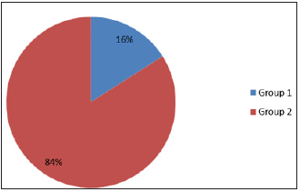
Figure 2:
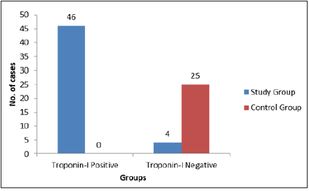
Figure 3:
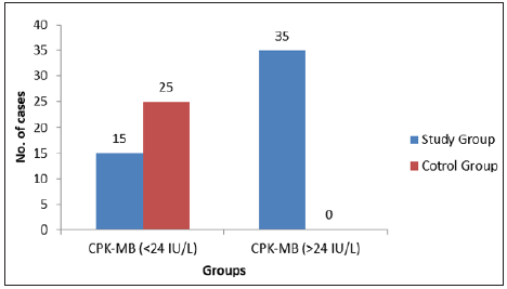
Figure 4:
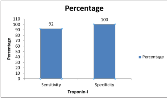
Figure 5:
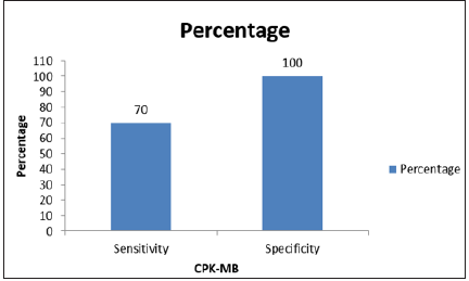
Table 1:

Table 2:

Table 3:

Table 4:

Table 5:

Table 6:

Table 7:

Table 8:

Table 9:

Table 10:

Table 11:

Table 12:

Discussion
AMI is one of the leading causes of death worldwide in both men
and women. Heart tissue death occurs within 6-12 hours of attack.
So, to diagnose AMI in early stage, several markers were used [7]. In
the present study we used the reliability of cardiac Troponin-I and
CPK-MB in these patients. In the past many studies also compare
cardiac Troponin-I and CPK-MB. Thompson et al in 1988 studied 50
patients of AMI and concluded that >95% patients had abnormally
raised CPK-MB (>24IU/L) and <5% patients had CPK-MB within
normal range (<24IU/L) which was highly significant. They also
concluded that 100% of the patients had Troponin-I positive and
0% had Troponin-I negative cases [8].
Hamm et al. [9] in 1997 studied 47 patients of AMI and revealed
that 91% patients had abnormally increased levels of CPK-MB
(>24IU/L) and 9% patients had CPK-MB levels within normal
range (<24IU/L) and was highly significant. They also concluded that 100% of the patients had Troponin-I positive and 0% had
Troponin-I negative cases [9] like Thomson et al. In the present
study, 70% cases had raised CPK-MB (>24IU/L) and 30% cases had
(<24IU/L) levels of CPK-MB, and 92% cases had Troponin-I positive
and 8% cases had Troponin-I negative while in control group 0%
individuals had CPK-MB (>24IU/L) and 100% individuals had CPKMB
(<24IU/L) and 0% individuals had Troponin-I positive and
100% had Troponin-I negative. Statistical analyses comparing CPKMB
between study and control groups was significant. Our results
comply with the results of the above-mentioned studies that CPKMB
and Troponin-I are highly sensitive (70% and 92%) and highly
specific (100% and 100%) in patients of AMI.
We also compared the sensitivity and specificity of CPK-MB
and Troponin-I in the present study with other studies in the study
group. Sawhney et al in 2004 conducted a study in 18 patients and
concluded that sensitivity and specificity of Troponin-I and CPKMB
was 83%, 87% and 93%, 100% respectively [9]. Meraz et al.
[10] in 2006 also conducted a study on 40 patients of AMI and
demonstrated that sensitivity and specificity of Troponin-I and CPKMB
was 95%, 95% and 40%, 50% respectively [10]. In the present
study, we recruited 50 patients and concluded that sensitivity and
specificity of Troponin-I and CPK-MB was 92%, 100% and 70%,
100% respectively from comparison of sensitivity and specificity
of cardiac Troponin-I in the present study and other studies, it is
concluded that our study contradicts the study done by Sawhney et
al and but consistent with Meraz et al. [10]. But in the case of CPKMB,
our study consistent with the study done by Meraz et al. [10]
and contradicts the study done by Sawhney et al.
Conclusion
In conclusion, whenever we consider the role of cardiac Troponin-I and CPK-MB in the diagnosis of AMI, we required both CPK-MB and Troponin-I for the diagnosis of AMI. Troponin-I rises within 6 hours of chest pain, remains raised for 10 days and CPKMB is raised within 6-12 hours of chest pain and comes back to normal levels within 48 hours. So, we can say that fresh attack will be assayed by serial CPK-MB whereas Troponin-I will not be able to indicate the fresh infarction, but Troponin-I is a better indicator of AMI. The limitation of the study was small sample size, so I suggest a larger sample size to conclude the further result.
References
- Mintz E (1997) Emergency department management of acute myocardial infarction. Mt Sinai J Med 64(4-5): 258-274.
- Garg VK (2018) CPK-MBW or troponin i-marker for acute myocardium infarction: Which is Better? EC Cardiol 10(5): 5-6.
- Nicholson C (2004) A systematic review of the effectiveness of oxygen in reducing acute myocardial ischaemia. Journal of Clinical Nursing. Wiley/Blackwell (10.1111): 13: 996-1007.
- Ryan TJ, Anderson JL, Antman EM, Braniff BA, Brooks NH, et al. (1996) ACC/AHA guidelines for the management of patients with acute myocardial infarction. A report of the American College of Cardiology/American Heart Association Task Force on Practice Guidelines (Committee on Management of Acute Myocardial Infarction). J Am Coll Cardiol 28(5): 1328-1428.
- Anand SS, Islam S, Rosengren A, Franzosi MG, Steyn K, et al. (2008) Risk factors for myocardial infarction in women and men: Insights from the INTERHEART study. Eur Heart J 29(7): 932-940.
- Kutsal A, Saydam GS, Yucel D, Balk M (1991) Changes in the serum levels of CK-MB, LDH, LDH1, SGOT and myoglobin due to cardiac surgery. J Cardiovasc Surg (Torino) 32(4): 516-522.
- Bruyninckx R, Aertgeerts B, Bruyninckx P, Buntinx F (2008) Signs and symptoms in diagnosing acute myocardial infarction and acute coronary syndrome: A diagnostic meta-analysis, British Journal of General Practice. Royal College of General Practitioners 58: 105-111.
- Thompson WG, Mahr RG, Yohannan WS, Pincus MR (1988) Use of creatine kinase MB isoenzyme for diagnosing myocardial infarction when total creatine kinase activity is high. Clin Chem 34(11): 2208-2210.
- Hamm CW, Goldmann BU, Heeschen C, Kreymann G, Berger J, et al. (1997) Emergency room triage of patients with acute chest pain by means of rapid testing for cardiac Troponin T or Troponin I. N Engl J Med 337(23): 1648-1653.
- Meraz Soria CA, Camarena Alejo G, Elizalde González JJ, Aguirre Sánchez J, Martínez Sánchez J, et al. (2006) Usefulness of rapid bedside assay of cardiac troponin I and creatine phosphokinase-MB in acute ischemic coronary syndromes. Arch Cardiol Mex 76(1): 37-46.
© 2020 Vivek Kumar Garg. This is an open access article distributed under the terms of the Creative Commons Attribution License , which permits unrestricted use, distribution, and build upon your work non-commercially.
 a Creative Commons Attribution 4.0 International License. Based on a work at www.crimsonpublishers.com.
Best viewed in


.jpg)


 Editorial Board Registrations
 Submit your Article
 Refer a Friend
 Advertise With Us
.jpg)


.jpg)


.bmp)
.jpg)
.png)
.jpg)


.jpg)


.png)

.png)


.png)


