- Submissions

Full Text
Open Access Research in Anatomy
CT Diagnosis of Rare Case of Morgagni Hernia in an Adult
Antonio Gligorievski*
University Clinic of Surgery, North Macedonia
*Corresponding author: Antonio Gligorievski, University Clinic of Surgery, Skopje, North Macedonia
Submission: March 10, 2020 Published: July 23, 2021
ISSN: 2577-1922
Volume2 Issue4
Abstract
Introduction: Morgagni’s hernia is the rarest of the congenital diaphragmatic defects with a reported frequency of 1% to 5.1% Morgagni hernias are more commonly seen on the right side and it occurs more commonly in females. However, most patients remain asymptomatic, and the majority of cases are discovered incidentally while investigating unrelated problems. Aim: The aim of this paper is to present a rare case of Morgagni hernia in adults and to emphasize the significant role of CT imaging in reaching the exact diagnosis of this condition. Case Report: We present a rare case of 46 years old female with an incidentally detected congenital diaphragmatic hernia on the right side. CT scan is the investigation of choice for diagnosis of Morgagni’s hernia as it allows clear visualization of the defect, and its contents. The diagnosis is established by the presence of a retrosternal mass of herniated omentum or by a transverse colon. Surgical treatment options include a transabdominal or transthoracic repair, but in the case of larger defects, a mesh may be necessary. Conclusion: Most asymptomatic cases were found in adults by chest radiography for unrelated problems. The diagnosis in the suspected case can be confirmed by a CT scan.
Keywords: Adult; Morgagni; Computed Tomography; Congenital; Diaphragmatic; Hernia; repair
Introduction
Morgagni hernia was first described by Giovanni Battista Morgagni, an Italian anatomist, and pathologist in 1769 [1]. The foramen of Morgagni, also known as the sternocostal hiatus is a triangular space caused by a congenital defect in the fusion of septum transverses of the diaphragm and the costal arches [2]. Herniation of abdominal contents through this foramen is known as the Morgagni hernia. The majority of these hernias occur on the right, but leftsided hernias have been reported. Morgagni hernia is the rarest form of congenital hernia, representing 2 to 3% of all case [3,4]. The majority of Morgagni hernias are diagnosed late because most adults can be asymptomatic or present with non-specific respiratory and gastrointestinal symptoms [5]. Diagnosis can be difficult, and a missed diagnosis can lead to life-threatening complications. In most adult cases these hernias are noted incidentally on chest X-ray. Depending on the contents of the hernia (omentum, stomach, small intestine, colon or liver) it can appear differently on chest radiography and the diagnosis can be missed. For example, if omentum is present in the sac, a solid pericardiac shadow will appear on the chest radiographs [6]. Another option would be a barium enema for adults. CT with oral contrast is the investigation of choice as it allows clear visualization of the defect, and its contents [7]. When investigations are non-diagnostic, confirmation by laparoscopy may be needed. Although the majority of these hernias are asymptomatic, the repair is recommended to avoid future complications. If the hernia is small or if it contains omentum only, the operation is indicated when symptoms are recurrent. Operation is indicated when the colon is in the sac, as there is a high risk of obstruction. Treatment options include transabdominal or transthoracic repair [8,9].
Presentation of Case (Case Report)
The aim of this report is to highlights the importance of early CT scanning in reaching the exact and early diagnosis of congenital diaphragmatic hernia, particularly since a delay in diagnosis can result in significant morbidity and mortality. Herein, we present a rare case of 46 years old female with an incidentally detected congenital diaphragmatic hernia on the right side and to emphasize the significant role of imaging for the diagnosis of this condition. She complained about epigastric and abdominal pain for one week and a moderate form of dyspnea. There was no history of any previous abdominal or thoracic trauma. She had no other underlying disease. Her physical examination revealed pain in the epigastric area but did not reveal any significant abnormality. She was sent to the Radiology Department for further evaluation. Posteroanterior (PA) and latero-lateral (LL) chest X-ray revealed that the left diaphragm was unexceptionally high. It was decided to perform a CT scan of the chest and abdomen to clarify the changes seen in the initial chest radiograph. CT scan of the chest and abdomen revealed the presence of a left-sided anterolateral diaphragmatic defect with herniation of the transverse colon including mesenteric fat and omentum into the left hemithorax (Figures 1-3).
Figure 1:Noncontrast CT scans of the thorax and abdomen.
A. Mediastinal window;
B. Lung window: At the level of Th7 axial CT images showed a large Morgagni hernia containing omental fat
(white arrow) and transverse colon loop (black arrow). There were no signs of obstruction or strangulation.
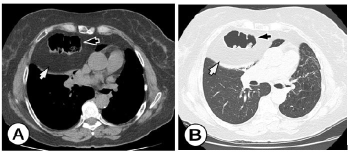
Figure 2:Noncontrast axial CT scans of the thorax and abdomen.
A. Mediastinal window;
B. Lung window: At the level of Th10 axial CT images showed an anterior defect (foramen of Morgagni) in the
right and left hemidiaphragm, with a large Morgagni hernia containing omental fat (white arrow) and transverse
colon loop (black arrow). There were no signs of obstruction or strangulation.
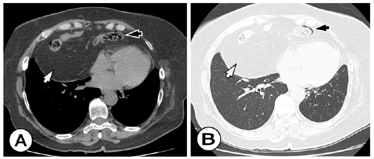
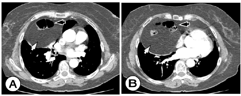
Figure 3:Contrast-enhanced axial CT scans of the thorax and abdomen at the level of aorta ascenders
A. at the level of the right pulmonary artery
B. Contrast-enhanced axial CT images show a large Morgagni hernia containing omental fat (white arrow) and
transverse colon loop (black arrow). There were no signs of obstruction or strangulation.
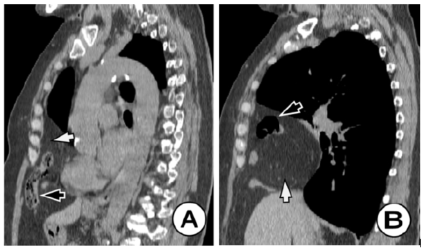
Intraperitoneal fat was responsible for the opacity seen in the x-ray. The herniated omentum and transverse colon passes through the defect, filling the chest cavity and causing the compression of the lower lobe of the lung, and a shift of mediastinal structures to the left side (Figure 4). CT scan confirmed the diagnosis of left-sided Morgagni hernia (Figure 5). The patient was counseled about the possible complications of diaphragmatic hernia that might occur if left untreated. The patient was operated on, and the diagnosis of left-sided hernia was confirmed. Transabdominal operative intervention for correction of the Morganic hernia has been performed and the diaphragm defect is closed with a mesh. The postoperative has no complications and the patient is discharged in good general condition.
Figure 4:Noncontrast Coronal reformatted CT image of the thorax and abdomen (A) and (B): Coronal reformatted CT image in 46 years old woman showed an anterior defect (foramen of Morgagni) in the right and left cardiophrenic angle, with a large Morgagni hernia containing omental fat (white arrow) and transverse colon loop (black arrow). There were no signs of obstruction or strangulation.
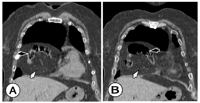
Figure 5:Noncontrast Sagittal reformatted CT image of the thorax and abdomen (A) and (B): Sagital reformatted CT image shows anterior diaphragmatic defect (foramen of Morgagni) and a large Morgagni hernia containing colon (black arrow) and omental fat (white arrow) There were no signs of obstruction or strangulation.
Discussion
Diaphragmatic hernias of Morgagni were first described in 1769 as anatomical defects in the anterior diaphragm that allow herniation of abdominal viscera into the thorax [1]. The presence of this hernia in adulthood is very rare and, when symptomatic, it is usually associated with chronic respiratory symptoms (dyspnea, fatigue, pain) or gastrointestinal involvement with occlusive symptoms (constipation, occlusion) [10]. However, most patients remain asymptomatic, and the majority of cases are discovered incidentally while investigating unrelated problems [11]. Despite being a congenital condition, many cases present only later in life. It is believed this is due to the presence of a sac that limits herniation. Morgagni hernias are more commonly seen on the right side despite protection from the liver. It has been suggested that this happens because the presence of extensive pericardial attachments on the left provides extra support [10]. It occurs more commonly in females [9,12]. Usually, the hernia sac contains the transverse colon followed by omentum, stomach, and small intestine but occasionally the liver may also protrude into the sac [13]. They are the rarest of congenital diaphragmatic hernias, making up 2-3% of cases [3,4]. Incidental findings of this condition in adults are less common with only 81 asymptomatic cases reported in a recent review [3]. Symptomatic adult cases of Morgagni hernias are even rarer with only 12 cases described [2]. Almost 90% of Morgagni hernias are reported to be on the right side, with 2% located on the left and 8% bilateral [14].
Depending on the contents of the hernia, the images in the chest radiography may appear differently [10]. On a chest radiograph, Morgagni hernias is frequently diagnosed incidentally as homogeneous masses in the right cardio phrenic angle [6]. Most frequently, it will show a solid pericardiac shadow due to the presence of omentum and the differential diagnosis should include a pleuropericardial cyst, pericardial fat mass, lipoma or liposarcoma, mesothelioma, diaphragmatic cyst, intrathoracic tumors, and eventration of the diaphragm [3,7,15]. However, it must be noted that plain radiography may be normal especially in intermittent herniation [5]. Another diagnostic option would be a barium enema, having the advantage of being less expensive. Ultrasonography has been shown to be useful in assessing diaphragmatic hernias, but CT is the most sensitive an accurate diagnostic method as it gives excellent anatomical detail on the contents of the hernia and its complications such as strangulation [16,17]. CT scan is the investigation of choice for diagnosis of Morgagni hernia. The diagnosis is established by the presence of a retrosternal mass of fat density representing herniated omentum or by a combination of omentum and a colon [12]. Magnetic resonance imaging can distinguish Morgagni hernia from other mediastinal masses. In our case, the diagnosis was made by CT.
Although the majority of patients are asymptomatic, the repair is almost always recommended to avoid future complications, especially when the colon is herniated due to the risk of obstruction [8]. On the other hand, if it contains the only omentum, surgery is only indicated in the presence of symptoms [18]. Surgical treatment options include a transabdominal or transthoracic repair [19]. For a smaller defect, primary closure is usually done, but in the case of larger defects, a mesh may be necessary [19,20].
Conclusion
In conclusion, Morgagni hernia is a rare type of congenital diaphragmatic hernia in both adults and children and is more common on the right side. A missed diagnosis can lead to life threatening complications such as obstruction or strangulation which warrants early surgical intervention. Most asymptomatic cases were found in adults by chest radiography for unrelated problems. The diagnosis in the suspected case can be confirmed by a CT scan. Surgery by abdominal approach is the management of choice.
References
- Morgagni GB (1769) The seats and causes of diseases investigated by anatomy. Millar and Cadell, London, UK, Volume 3: 205.
- Eren S, Ciri F (2005) Diaphragmatic hernia: Diagnostic approaches with review of the literature. Eur J Radiol 54(3): 448-459.
- Comer TP, Clagett OT (1966) Surgical treatment of hernia of the foramen of Morgagni. J Thorac Cardiovasc Surg 52(4): 461-468.
- Loong TP, Kocher HM (2005) Clinical presentation and operative repair of hernia of Morgagni. Postgrad Med J 81(951): 41-44.
- Marinceu D, Loubani M, O Grady H (2014) Late presentation of a large Morgagni hernia in an adult. BMJ Case Rep
- Daneshvar S, Shriki J, Sohn H, Rahimtoola SH (2010) Morgagni-type diaphragmatic hernia presenting as an abnormal cardiac silhouette. Am J Med 123(10): e11-e12.
- Arora S, Haji A (2008) Adult Morgagni hernia: The need for clinical awareness, early diagnosis and prompt surgical intervention. Ann R Coll Surg Eng 90(8): 694-695.
- Daneshvar S, Shriki J, Sohn H, Rahimtoola SH (2010) Morgagni-type diaphragmatic hernia presenting as an abnormal cardiac silhouette. Am J Med 123(10): e11-12.
- Nasr A, Fecteau A (2009) Foramen of Morgagni hernia: Presentation and treatment. Thorac Surg Clin 19(4): 463-468.
- Horton JD, Hofmann LJ, Hetz SP (2008) Presentation and management of Morgagni hernias in adults: A review of 298 cases. Surg Endosc 22(6): 1413-1420.
- Pfannschmidt J, Hoffmann H, Dienemann H (2004) Morgagni hernia in adults: results in 7 patients. Scand J of Surg 93(1): 77-81.
- Colakoğlu O, Haciyanli M, Soytürk M, Colakoğlu G, Simşek I (2005) Morgagni hernia in an adult: A typical presentation and diagnostic difficulties. Turk J Gastroenterol 16(2): 114-116.
- Abraham V, Myla Y, Verghese S, Chandran BS (2012) Morgagni-larrey hernia, a review of 20 cases. Indian J Surg 74(5): 391-395.
- Aghajanzadeh M, Khadem S, Khajeh S, Gorabi HE, Ebrahimi H, et al. (2012) Clinical presentation and operative repair of Morgagni hernia. Interact Cardiovasc Thorac Surg 15(4): 608-611.
- Komatsu T, Takahashi Y (2013) Is this a mediastinal tumor? A case of Morgagni hernia complicated with intestinal incarceration mistaken for the mediastinal lipoma previously. Int J Surg Case Rep 4(3): 302-304.
- Malav M, Amit Kumar D, Ajay M, Samir R (2016) Strangulated Morgagni’s hernia: A rare diagnosis and management. Case Rep Surg 2016: 2621383.
- Kelly MD (2007) Laparoscopic repair of strangulated Morgagni hernia. World Journal of Emergency Surgery 2(1): 27.
- Durak E, Gur S, Cokmez A, Atahan K, Zahtz E, et al. (2007) Laparoscopic repair of a Morgagni hernia. Hernia 11(3): 265-270.
- Harrington SW (1951) Clinical manifestations and surgical treatment of congenital types of diaphragmatic hernia. Rev Gastroenterol 18(4): 243-256.
- Loong TP, Kocher HM (2005) Clinical presentation and operative repair of hernia of Morgagni. Postgrad Med J 81(951): 41-44.
© 2021 Antonio Gligorievski. This is an open access article distributed under the terms of the Creative Commons Attribution License , which permits unrestricted use, distribution, and build upon your work non-commercially.
 a Creative Commons Attribution 4.0 International License. Based on a work at www.crimsonpublishers.com.
Best viewed in


.jpg)


 Editorial Board Registrations
 Submit your Article
 Refer a Friend
 Advertise With Us
.jpg)


.jpg)


.bmp)
.jpg)
.png)
.jpg)


.jpg)


.png)

.png)


.png)


