

- About Us
- Information
-

The Author ensures that the research has been conducted responsibly and ethically with adherence to all relevant regulations. read more..
- For Authors
- For Reviewer
- Manuscript Guidelines
- Membership
- Publication Ethics
-
- Journals
- Reprints
- e-Books
- Videos
- Policies
- Contact Us
 COVID-19
- Submissions



Full Text
Journal of Biotechnology & Bioresearch
Generation of Self-Developing Crown Cells (SD-Crown Cells) from Self- Organizing DNA Fraction (SO-DNA)
Shoshi Inooka*
Japan Association of Science Specialists, Japan
*Corresponding author:Shoshi Inooka, Japan Association of Science Specialists, Japan
Submission: September 21, 2026;Published: September 30, 2026

Volume6 Issue 4September 30, 2026
Abstract
Artificial cell-like structures can emerge from minimal molecular components providing a platform for exploring autonomous biological behaviors. In this study, DNA fractions of artificial cells, hereafter termed Gas-DNA crown cells (G-Sph crown cells), were generated from Sphingosine (G-Sph), DNA and adenosine produced by incubation under standard culture condition. SO-DNA, designated as the selforganizing DNA fraction, surprisingly, produced autonomous colony-like structures (Self-developing crown cells; SD- crown cells). SD-crown cells exhibit circular or irregular shapes and show dense internal organization. The properties of SO-DNA varied depending on the DNA source used for the production of DNA crown cells, indicating that DNA origin plays an important role in shaping the self-organizing capacity of the fraction. These findings describe a new research field that is distinct from synthetic autonomous DNA systems in which DNA not only functions as genetic information, but also acts as a driver of autonomous assembly and emergent biological behavior.
Keywords:DNA crown cells; Sphingosine-DNA; G-crown cell seeds; G-Sph crown cells; Gas powder; SODNA; SD-crown cells
Introduction
Synthetic biology aims to construct minimal cell-like systems from defined molecular components, enabling the investigation of the fundamental principles underlying biological organization. The author previously developed artificial cells, termed DNA crown cells, by combining sphingosine, DNA, and adenosine [1-5]. As a result of these experiments, several kinds of artificial cells based on DNA crown cells, such as antibiotic-producing crown cells and crown cells Nema, have been prepared [6-17]. One of these G-Sph crown cells can be prepared using gas-exposed sphingosine, which was prepared using gasses such as dimethyl disulfide [18]. These cells, which were produced with the cultures of gas-powder instead of sphingosine [19], represent a highly minimal synthetic system capable of forming stable crown cell-like structures. When DNA was extracted from G-Sph crown cells, a reproducible and distinct fraction emerged, which the author has designated as a self-organizing DNA fraction (SO-DNA). SO-DNA contains characteristic envelope-forming particles that appear during the extraction process and subsequently generate SD-crown cells. These self-developing crown cell-like structures emerge during culture of SO-DNA, suggesting that SO-DNA retains functional units capable of self-organization, independent of intact cellular structures. Importantly, the properties of SO-DNA differ depending on the source of the DNA used to construct G-crown cells. Fractions derived from Escherichia coli DNA and other DNA sources exhibited distinct autonomous behavior, indicating that the origin of the DNA influences the emergent properties of SO-DNA. This work extends synthetic biology into a new research direction that the author refers to as synthetic autonomous DNA systems. Specifically, this new field investigates the potential application of DNA as a functional material driven by self-organizing and emergent biological phenomena. SO-DNA provides a unique platform for exploring minimal autonomous systems and the potential roles of DNA in non-genetic biological functions.
Materials and Methods
Materials
Escherichia coli DNA (Sigma-Aldrich, USA), adenosine (Sigma- Aldrich; Wako, Japan), and Gas-exposed sphingosine (prepared by gas-powder treatment [20]. Agar plates and agar medium (SMA) (AS ONE, Japan). DNAs-icil-F (Rizo Inc., Tsukuba, Japan).
Methods
Preparation of SO-DNA from G-Sph crown cells: The procedure begins with the preparation of G-Sph crown cells, which were prepared using cultures of G-crown cell seeds [19]. G-E. coli crown cell seeds were prepared in a mixture containing G-Sph, E. coli DNA and adenosine, as described previously [20].
The DNA fraction was extracted using the following methods:
A. G-Sph crown cells were collected from two culture plates
(Figure 1).
B. G-Sph crown cells were centrifuged (15,000rpm, 10min), the
supernatant was removed and 1ml of DNAs-icil-F was added.
C. The cell lysate was mixed with 500μL of chloroform.
D. After centrifugation (15,000rpm for 10min at 4 °C), the
aqueous phase containing nucleic acids was collected.
E. The supernatant was then mixed with 800μL of isopropanol.
F. The mixture was centrifuged at 15,000rpm for 10min and the
aqueous phase was collected.
G. DNA was precipitated by adding 500μL of ethanol and collected
by centrifugation at 15,000rpm for 10min at 4 °C.
H. After removing the supernatant, the precipitate was recovered
and dried for 10min.
I. The precipitate was then used as the SO-DNA for subsequent
culture experiments and stored at about -20 °C until use.
Culture of SO-DNA: First, 50μL of SO-DNA (approximately 12, 5μg) was poured onto an agar plate and spread with a culture spreader, followed by incubation at 37 °C for 18h.
Secondary culture: After 18h of culture, objects (OD-cells) were collected by washing with 6mL of distilled water. Then, 200μL of this mixture was poured onto a new agar plate, spread using a spreader, and incubated for 1 or 2 days at 37 °C.
Results and Discussion
General observations
Objects on the plates could be observed directly with the naked eye and under a light microscope. Figure 1 shows a photograph of G-Sph crown cells used for the collection of SO-DNA. (Left) Image of a colony, (right) image of collected objects. A total of 104, 5μg of DNA was obtained from these objects. The DNA concentration of the SO-DNA was estimated by measuring the absorbance of 260nm. Because the fraction contained envelope-forming particles, the absorbance value at 260nm reflects both the dissolved DNA and the particle DNA, and therefore represents approximate rather than precise measurements of DNA. Figure 2 shows a microscopic image of SO-DNA isolated from G-Sph crown cells. The SO-DNA contained numerous envelope-forming particles, which were comprised of assemblies or rounded units surrounded by thin boundaries. These particles resemble the envelope structures observed previously in intact G-Sph crown cells [21] and are considered potential selforganizing seeds within the fraction. Figure 3 shows SD-crown cells that were formed during agar medium culture of SO-DNA. Figure 4 shows the growth behavior of SD-crown cells on agar medium and the microscopic appearance of SD-crown cells after 18h of primary culture. Figure 5 shows SD-crown cells at the time of secondary culture.
Figure 1: Photograph of G-Sph crown cells used to collect DNA fractions (SO-DNA). (Left) Colony image, (right) image of collected objects.
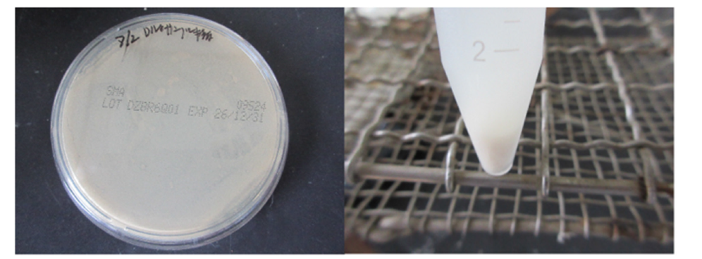
Figure 2: Microscopy image of SO-DNA showing a round object (Figure 2a), with an envelope-like object (Figure 2b). Small round objects (Figure 2c) were observed within the envelope-like object (approximate size of the objects shown in Figure 2d is 6-7μm; actual size of Figure 2d is approximately 2-2.5μm because Figure 2 is enlarged 3 times).

Figure 3: Colony formation on agar medium, showing cultures of SO-DNA. Microorganism-like objects were observed.
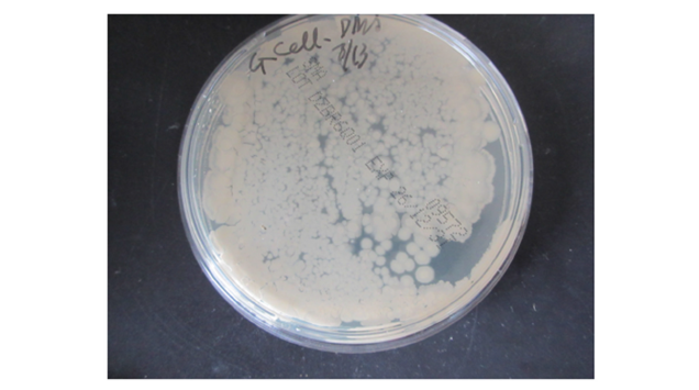
Figure 4: Growth behavior of OD-crown cells on agar medium, showing the microscopic appearance of the edge of objects similar in appearance to colonies after 1 day of primary culture. Numerous small objects with variable morphology were observed. Approximate size of ← is 7-8μm
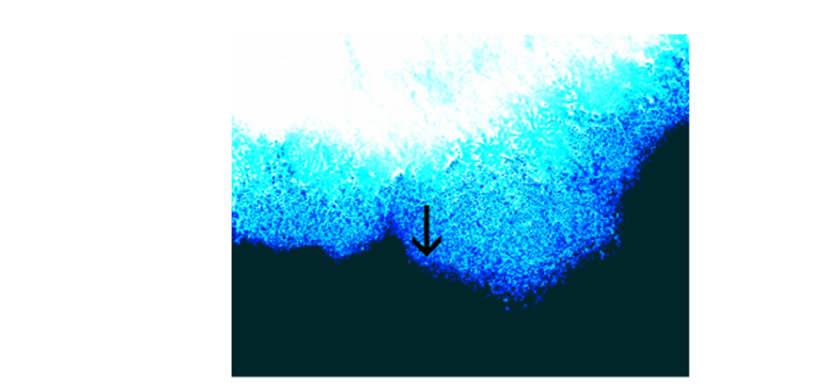
Figure 5: Colony formation of secondary culture. Microorganism-like objects were observed across the plate.
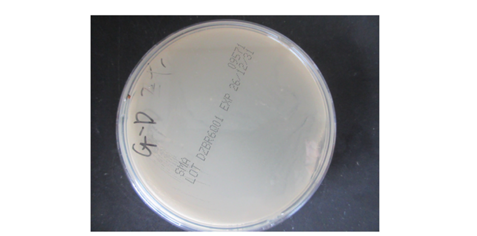
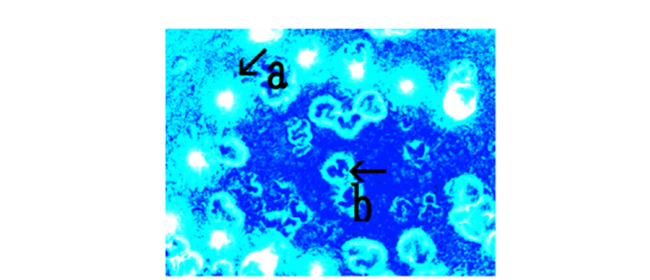
Figure 6 shows the growth behavior of SD-crown cells after 1 day of secondary culture on agar medium. SD-crown cells exhibited circular or irregular shapes and dense internal organization (Figure 6b). Figure 7 shows the growth behavior of SD-crown cells after 2 days of secondary culture. The image shows both aggregation and structural integration (Figure 7b), suggesting that the condensed wavy aggregates contribute to colony formation. The findings of the present study demonstrate that G-Sph crown cells produced from G-Sph, DNA, and adenosine give rise to unique SO-DNA that exhibits autonomous behavior. Because G-Sph crown cells can be cultured serially [17], cultivable objects may be contained within the original G-Sph crown cells. It is considered that most of these envelope-forming particles were collected in SO-DNA, while the remainder may be collected in another fraction. Further, the observation of these objects (envelope-particles) recovered from G-Sph crown cells suggests that structural or functional units remain as envelope-forming particles (Figure 2), and that these retain the capacity for autonomy in SO-DNA (Figures 4,6&7). The ability of SO-DNA to generate SD-crown cells indicates that DNA-associated components can act as autonomous assembly units outside intact cells. A notable finding is that the autonomous properties of SO-DNA depend on the DNA source used for constructing the G-Sph crown cells, suggesting that the DNA origin influences the molecular composition and the structural characteristics of the resulting fraction. Such differences may reflect variations in the DNA sequence, or interactions with G-Sph and adenosine during cell generation. This observation opens the possibility that SO-DNA represents a tunable autonomous system whose behavior can be modulated through the selection of different DNA sources.
Figure 6: Growth behavior of SD-crown cells at 1 day of secondary culture on agar medium. An object containing a dense ring-like object (Figure 6b) was observed (Figure 6a). Approximate size of the ring in Figure 6b is 300μm in diameter.
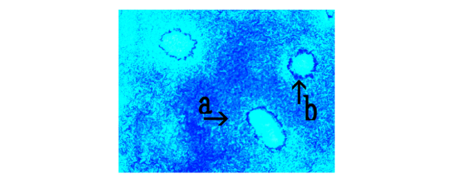
Figure 7: Growth behavior of SD-crown cells at 2 days of secondary culture on agar medium. Soft objects containing colorless, round objects were observed (Figure 7a). The objects contained condensed wavy structures (Figure 7b). Approximate size of the object shown in Figure 7a is 300μm.
The concept of SO-DNA expands the functional role of DNA beyond its conventional genetic capacity. In this system, DNA potentially acts as a driver of autonomous assembly, contributing to emergent biological behavior without requiring intact cellar machinery. This positions SO-DNA within the emerging field of systematic autonomous DNA systems, which explores DNA based self-organization and the development of minimal autonomous units. Overall, SO-DNA provides a new platform for investigating autonomous molecular systems and may contribute to understanding how minimal components can generate lifelike behavior. Little is known about the mechanisms underlying colony formation and the extent to which DNA origin can modulate autonomous properties. Future studies will therefore examine whether G-Sph crown cells and SD-crown cells emerge when synthetic DNA, instead of native DNA, is used.
Acknowledgements
The author thanks Lisa Monna (Rizo Inc., Tsukuba, Japan) for assistance with DNA extraction and useful discussions.
References
- Inooka S (2012) Preparation and cultivation of artificial cells. App Cell Biol 25: 15-18.
- Inooka S (2016) Investigation of the chemical composition of artificial cell seeds-Sphingosine-DNA bound components from extracts of the meat from adult Ascidiara. LICRLS, pp. 534-540.
- Inooka S (2016) Preparation of artificial cells using eggs with Sphingosine-DNA. J Chem Eng Process Technol 17: 277.
- Inooka S (2016) Aggregation of sphingosine-DNA and cell construction using components from egg white. Integrative Molecular Medicine 3(6): 1-5.
- Inooka S (2017) Systematic preparation of artificial cells (DNA crown cells). J Chem Eng Process Technol 8: 2.
- Inooka S (2025) Separation of antibiotic-producing cells from beer produced in co-cultures of DNA (streptomyces) crown cells with yeast. Annals of Reviews and Research 12(3).
- Inooka S (2025) Isolation of antibiotic producing cells from plate culture of egg powder enclosing DNA (bovine meat) crown cells and beef extract. Annals of Reviews and Research 12(5).
- Inooka S (2025) Separation of antibiotic producing cells from culture fluids of egg white powder enclosed DNA (streptomyces) crown cells and yeast. International Journal of Bioprocess & Biotechnological Advancements 10(1): 499-504.
- Inooka S (2025) Separation of antibiotic-producing cells from agar cultures of egg powder-enclosed DNA (hepato-blastoma cell Line: HepG2) crown cell and yeast. American Journal of Biomedical Science & Research 26(2): 2642-1747.
- Inooka S (2025) Isolation of antibiotic-producing cells from cultures of egg cultures of egg powder enclosing DNA (Hepg2) crown cells and salmon roe. Trends in General Medicine 3(1): 1-5.
- Inooka S (2025) Antibiotic-producing cells produced from a plate culture of egg powder enclosing DNA (Nannochloropsis Species) crown cells and yeast. Scientific Journal Biology & Life Sciences 4(2).
- Inooka S (2025) Antibiotic producing cells from a plate culture of egg white powder enclosed DNA (bovine meat) crown cells and Iponala Nil-seed extract. Stem Cells Regen Med 9(3).
- Inooka S (2025) Antibiotic-producing cells in milk cultures of egg powder-enclosed DNA (ascidians sea squirt) crown cells cultured with peptide (Glu-Glu) Partners. In J Biopro Biotechnol Advance 10(1): 505-511.
- Inooka S (2026) Microscopic appearance and cultivation of objects (Crown Asci gluglu cells nema) from agar culture of antibiotic crown Ascci gluglu cells stimulated with gluglu as a partner. Archive in Biomedical Engineering & Biotechnology 6(3):
- Inooka S (2026) Microscopic appearance of objects (Crown coli-glu.glu cells nema) from agar culture of antibiotic crown E. coli-gluglu cells stiulated with GluGlu. Chem Pharm Re 8(1): 1-7.
- Inooka S (2026) Microscopic appearance of objects (crown coli-amino acid cells nema) from agar culture of amino-acid stimulated antibiotic crown E. coli amino acid cells. In J Biopro Biotechnol Advance 10(1).
- Inooka S (2026) Serial cultivation of crown cells Nema in agar medium. Biomed J Sci Tech Res 65(2): 57521-57532.
- Inooka S (2026) Gas generation by egg-white-enclosed DNA crown cells in agar culture. In J Biopro Biotechnol Advance 10(1): 530-539.
- Inooka S (2026) Gas associated emergence of living cells from non-living DNA crown cell seeds. Trends Gen Med 4(2): 1-9.
- Inooka S (2026) Emergence of living cells by gas-exposed sphingosine, DNA and adenosine. Stem Cell & Regenerative Medicine 10(3): 1-8.
- Inooka S (2026) Order-dependent formation of self-organizing, microscopic objects from gas-exposed sphingosine, DNA, and adenosine. Nanotechnology & Applications 9(3): 1-4.
© 2026 Shoshi Inooka. This is an open access article distributed under the terms of the Creative Commons Attribution License , which permits unrestricted use, distribution, and build upon your work non-commercially.
Editor In Chief





.jpg)




























Signup for Newsletter

Quick Links
 Editorial Board Registrations
 Submit your Article
 Best Paper of the Volume
 Reprints
 Refer a Friend
 Advertise With Us
Our Recent Edition



Top Editors



.jpg)

















.bmp)
.jpg)
.png)
.jpg)













.jpg)






.png)

.png)




.png)




Financial Support

Sponsors

Latest e-Books

Latest Video

 a Creative Commons Attribution 4.0 International License. Based on a work at www.crimsonpublishers.com.
Best viewed in

 | Above IE 9.0 version