

- About Us
- Information
-

The Author ensures that the research has been conducted responsibly and ethically with adherence to all relevant regulations. read more..
- For Authors
- For Reviewer
- Manuscript Guidelines
- Membership
- Publication Ethics
-
- Journals
- Reprints
- e-Books
- Videos
- Policies
- Contact Us
 COVID-19
- Submissions



Full Text
Journal of Biotechnology & Bioresearch
Immobilization and Optimization of Trichoderma Harzianum β-Glucosidase for In Vitro Inhibition of Oil Palm Fungal Pathogens
Muhammad Muhammad2,3*, Roswanira Abdul Wahab1,3, Fahrul Huyop3, Mohd Hefni Rusli4, Umar Aliyu Abdullahi2, Muhammad Ali Dikwa2, Ali Ahmad5, Saminu Ali Kofar Kwaru2, Rabiu Salihu2, Hamisu Shuaibu Idris2, Usman Lawan Ubani2, Sa’adatu Abba Yusuf2 and Aisha Auwal Umar2
1 Department of Chemistry, Faculty of Science, Universiti Teknologi Malaysia (UTM), Malaysia
2 Department of Microbiology and Biotechnology, Federal University Dutse (F.U.D), Nigeria
3 Enzyme Technology and Green Synthesis Group, Faculty of Science, Universiti Teknologi Malaysia (UTM), Malaysia
4 Plant Pathology and Biosecurity Unit, Biology and Sustainability Research Division, Malaysia Palm Oil Board, Malaysia
5 Department of Biochemistry and Molecular Biology, Federal University Dutsinma, Nigeria
*Corresponding author:Muhammad Muhammad, Department of Microbiology and Biotechnology, Faculty of Life Sciences, Federal University Dutse P.M.B. 7156, Dutse, Jigawa State, Nigeria
Submission: June 16, 2026;Published: July 20, 2026

Volume6 Issue 3July 20, 2026
Abstract
This study optimized the physico-chemical parameters for immobilized cellulolytic β-glucosidase produced by Trichoderma harzianum Rifai strain Th12 using oil palm frond leaves, an agro-industrial waste substrate, via solid state fermentation. The enzyme was evaluated as an eco-friendly alternative to synthetic chemical fungicides for managing Ganoderma boninense, Fomitopsis meliae, Bipolaris sorokiniana and Phoma herbarum, the causal agents of basal stem rot, brown rot, root rot and leaf spot diseases in oil palm, respectively. Optimization of cultivation conditions was performed using Taguchi orthogonal array design based on five factors: pH, incubation period, temperature, surfactant concentration, and enzyme loading, each at four levels. The maximum activity of free β-glucosidase was 30.50U/g. Under optimized conditions-pH 7.0, 6 days incubation, 30 ℃, 2.0mg/mL enzyme concentration, and 0.05%v/v surfactant the immobilized β-glucosidase exhibited a peak activity of 92.40U/g. The linear model generated by Taguchi analysis accurately predicted the optimal cultivation parameters for T. harzianum Rifai Th12. Antifungal efficacy assays revealed that the immobilized β-glucosidase produced inhibition zones of 92.59%, 82.39%, 81.35%, and 86.21% against G. boninense, F. meliae, B. Sorokiniana, and P. herbarum, respectively, under in vitro conditions. The crude immobilized enzyme demonstrated superior antifungal activity compared to synthetic fungicide tested. These findings demonstrate that immobilized cellulolytic β-glucosidase from T. harzianum Rifai Th12 is a viable, sustainable biocontrol agent. It offers a promising alternative to synthetic fungicides for protecting oil palm against major fungal diseases..
Keywords:Immobilization; Biological agent; β-glucosidase; Taguchi orthogonal array design; Enzyme loading; Optimization; Solid state fermentation; Eco-friendly alternative
Introduction
Fungal pathogens such as Ganoderma boninense, Fomitopsis sp., Bipolaris sorokiniana and Phoma herbarum are among the causal agents that cause devastating disease in crops especially in oil palm. They can cause enormous losses in both quantity and quality of crop yields in more than 500 plant species of monocots and dicots viz. legumes plant, cereals plant, vegetables, fruits, and oil seed plant, and this is a major economic issue in the global agricultural sector [1-4]. To eliminate these devastating plant diseases, various agriculturalists use different synthetic chemicals, but the technique is ineffective due to its poisonous or toxicity effect to the living system present in the environment as well as the economic infeasibility. Even though, active pesticide ingredients may be effective as a means of preventive measures of fungal pathogens especially in oil palm, the soil born nature of the fungal pathogens renders the effort futile. Apart from being costly, the continuous application of synthetic chemicals pesticides could hinder the nature of the delicate balance of beneficial soil born microorganisms, leading to pesticide resistance microorganism [2-5]. The antagonist (T. harzianum sp.) has been widely used against the G. boninense and other fungal pathogens of oil palm as biocides [6]. T. harzianum is among the various potential antagonistic fungal species which is potent for improving the systemic resistance and act as plant growth promoters [2,5,7,8]. Aside to the familiar microbial control mechanisms such as competition for nutrient or shelter and ability to produce microbial metabolites [9], T. harzianum also produces hyphae that penetrate into the G. boninense, B. sorokiniana, F. meliae and P. herbarum fungal pathogens and which later secrete extracellular enzymes such as β-glucosidase to digest the cell walls and eventually kills the fungal pathogens [2,9,10].
It is well known that the cellulolytic β-glucosidase secreted by T. harzianum sp. is evolutionary adapted to specifically hydrolyze the cell wall components of G. boninense, B. sorokiniana, F. meliae and P. herbarum [11-16]. This β-specific cellulase enzyme targets and explicitly hydrolyzes the β-linkage of amorphic β-1,3:1,6-β-glucan filling material of the chitin-based cell wall of G. boninense [12- 14]. The cellulase secreted by T. harzianum hydrolyze the cell wall components of B. sorokiniana targets and explicitly hydrolyzes the β- linkage of amorphic β-1,4-chitin-based filling material of the cell wall of B. sorokiniana [11,3,17], and also target and hydrolyze the β-linkage of endo-1,4-glucan in F. meliae [16] as well as β-1,3:1,6- glucan in P. herbarum [15]. By such excellent behavior of T. harzianum, it was hypothesized that direct application of immobilized cellulolytic enzymes especially β-glucosidase to suppress the growth of these pathogens could be likely feasible. The technique suggested here may be a promising and environmentally friendly. The technique proposed here could increase the cellulolytic β-glucosidase production and reduces the initial costs of raw materials using the agro-industrial waste oil palm frond leaves substrate [2]. Another important aspect is the optimization of the cultivation media of the immobilized cellulolytic β-glucosidase produced. Due to the incapability of conventional approach in optimization processes [18- 21]. Therefore, one of the best statistical method Taguchi was used. In fact, it has been proved importance and successful in optimizing the cultivation conditions of immobilized enzymes [22-26]. The present research aimed at immobilization and optimization of cellulolytic β-glucosidase produced in batch cultures of T. harzianum Rifai using agro-industrial waste oil palm frond leaves as suitable substrate. The optimization process uses 4-level-5-factors Taguchi design of experiment to evaluate five parameters which includes pH, incubation time, temperature, surfactant and enzyme concentrations to provide the maximum cellulolytic β-glucosidase activity. Additionally, plate inhibition assays were used to confirm the effectiveness of the immobilized cellulolytic β-glucosidase produced to suppress the G. boninense, B. sorokiniana, F. meliae and P. herbarum fungal pathogens of oil palm.
Materials and Methods
Sample collections of oil palm frond leaves substrate, T. harzianum Rifai and fungal pathogens
Fresh oil palm frond leaves were collected from oil palm plantation province of University Teknologi Malaysia in Johor, Malaysia. Then, air dried, grounds using a table grinder (Wellmac RT-08, Taiwan) follow by sieved through 1.18-3mm Endecott’s test sieves (London, UK). The T. harzianum Rifai utilized in the present study were graciously collected from [5]. Following the method described by [2], the axenic culture of T. harzianum Rifai strain Th12 was sub cultured on potato dextrose agar (PDA; Oxoid Ltd, Basingstoke, England). The mycelia of fungus grown on potato dextrose agar (PDA; Ltd, Basingstoke, England) slants were kept in an incubator at 30 ℃ for 7 days and then kept at 4 ℃ before use. The fungal pathogens viz; G. boninense, B. sorokiniana, P. herbarum and F. meliae were obtained from Malaysia Palm Oil Board (MPOB) as well as Federal Land Development Authority (FELDA) Kulai Johor Malaysia. Then, sub cultured on potato dextrose agar (PDA; Oxoid Ltd, Basingstoke, England). The pure mycelia of the fungi grown on potato dextrose agar (PDA; Ltd, Basingstoke, England) slants were kept in an incubator at 30 ℃ for 7 days and then kept at 4 ℃ before use. The in vitro antagonistic efficacy of crude extract of T. harzianum Rifai utilized in this study was reported in our previous work Muhammad et al. [27].
Cellulolytic β-glucosidase production by Solid State Fermentation (SSF) using oil palm frond leaves substrate
In this study, β-glucosidase was produced using the grounded OPFL as the suitable substrate, following the method described by [2] with slight modifications, the SSF was conducted in 250ml Erlenmeyer flasks at 30 ℃ containing 5 grams of raw OPFL to stimulate the production of crude cellulolytic β-glucosidase from the cultures of T. harzianum Rifai Th12. The sieved 5g of raw OPFL was moistened with 20ml of a modified Mandel production medium containing 1.4g/L (NH4)2 SO4, 2g/L KH2 PO4, 0.3g/L urea, 0.3g/L CaCl2, MgSO4 0.005g/L, FeSO4 0.0016g/L, MnSO4 H2 O 0.0014g/L, ZnSO4 7H2 O 0.002g/L, CoCl2 0.75g/L, Peptone 1.0g/L, and 1ml/L Tween 80 (pH 5.2) [28-30] to achieve a final moisture level of 80% with the help of commercial moisture analyzer (MX50, A&D Weighing Co., Ltd., Japan). All flask was autoclaved (5 minutes at 121 ℃ 20psi) and allowed them to cool at room temperature. 5ml (104- 105 spores/mL) spore suspension was inoculated and incubated at 30 ℃ for 7 days followed by extraction of cellulolytic β-glucosidase cocktail.
Extraction of cellulolytic β-glucosidase enzyme cocktail
To extract the cellulolytic β-glucosidase enzyme cocktail after 7 days of incubation, cold sodium citrate buffer (0.05M, pH 4.8) in a 1:10 ratio was suspended into the culture medium containing the fermented substrate in the Erlenmeyer flask and shake at 120r/ min for maximum 30 minutes. Then the mixture was centrifuged for 30 minutes at 4000rpm, the pellet was discarded, and supernatant was used as the crude cellulolytic β-glucosidase enzyme and checked for its activity [2].
Enzyme assay for crude cellulolytic β-glucosidase activity
Cellulolytic activity of β-glucosidase was assayed as described by [2] using p-nitrophenyl-β-D-glucopyranoside (pNPG, Sigma Plot) as a substrate. In which equal amount of enzyme and substrate (25μL of enzyme and 25μL of pNPG) 10mmol/L was suspended in 50μL of phosphate buffer (pH 7.0) and incubated at 45 ℃ for 30 minutes. Then the reaction was stopped by the addition of 100μL of NaOH-glycine buffer (0.4mol/L, pH10.8). The developed color was read at 430nm, by using a spectrophotometer (T60 V; S/N:21-1610- 1-0091; AC230V [31-41] HZ 150W). The amount of sugar released was quantified using glucose standard calibration curve. One unit of cellulolytic β-glucosidase activity was defined as the amount of enzyme needed to liberate 1μmol/L of pNPG per minute under the assay conditions. The specific activity was expressed in μmol min- 1mg-1 protein. The dilution series of pNPG standard curve for determining the β-glucosidase activity was expressed in Table 1.
Table 1:Standard curve of pNPG for determining the β-glucosidase activities Dilution series of pNPG (430nm).

Determination of cellulolytic β-glucosidase protein content and total reducing sugar assay
The concentration of the protein in the cell extracts of crude cellulolytic β-glucosidase was measured using the Bradford assay method [42]. 1.0mL of the crude β-glucosidase was mixed with 1.5mL of Bradford reagent and the optical density of the mixture was measured immediately at wavelength of 430nm. A standard curve of protein concentration was constructed using Bovine Serum Albumin (BSA), BSA of 0.2% (w/v) and used as a stock solution. The preparation of BSA a series of diluted standard solutions was summarized in Table 2. The concentrations of protein in enzyme sample were determined using the standard curve for protein concentration. Then, the optical densities were plotted on a graph against their respective concentrations using Microsoft Excel software. Following the method described by Miller GL [43] the total reducing sugar in the crude enzyme cocktail was determined through the 3,5-dinitrosalysalic acid method, using the glucose as the standard. 1mL of 3,5-dinitrosalysalic acid and 2 drops of 0.1 M NaOH were mixed with an aliquot 1mL of the sample. The mixture was then boiled at 100 ℃ for 5 mins and cooled at room temperature prior to read the absorbance at 430nm. The hydrolysis efficiency of total reducing sugar concentrations was expressed in mg/g of the substrate (oil palm frond leaves) and calculated as in equation (1) [44]:

Table 2:Standard BSA dilution series for the determination of the crude cellulolytic β-glucosidase protein content (430nm).

Where “Є” represents the percentage (%) of the hydrolysis efficiency and the concentration of sugar released represents the total reducing sugar released in the crude enzyme cocktail. Cellulose and hemicellulose concentrations are expressed in g/g oil palm frond leaves substrate and values 1.11 and 1.12 are the standard conversion factors for calculating the levels of liberated sugar monomers from the hydrolyses of their respective sugar polymers.
Sodium dodecyl sulphate polyacrylamide gel electrophoresis for determination of T. harzianum Rifai cellulolytic β-glucosidase molecular weight
To determine the molecular weight of the T. harzianum Rifai cellulolytic β-glucosidase, an SDS-PAGE (TGXTMFastcast, Bio-Rad, Sigma-Aldrich, United States) 5% stacking and a 12% separating gel, respectively, was used as described by [45]. A 50μL buffer sample (0.05% bromophenol blue, 5% β-mercaptoethanol, 10% glycerol, and 1% SDS in 0.25 M Tris-HCl buffer; pH 6.8) was added into a 100μL of protein sample in an Eppendorf tube, boiled in hot water bath for 5mins, cooled to room temperature and electrophoresed. The protein bands were visualized by staining with Coomassie Brilliant Blue G (Sigma-Aldrich, United States) and distained in diluted acetic acid overnight. The molecular weight of the β-glucosidase was determined in comparison to marker protein (standard protein marker, 15-250kDa; Sigma, USA).
Optimization of cultivation parameters for the cellulolytic β-glucosidase produced by T. harzianum Rifai using One Variable at A Time (OVAT) method
Optimization of cultivation conditions for the activity of the crude β-glucosidase was performed and the influence of fermentation production parameters pH (3.0-9.0), incubation temperature (25-55 ℃), incubation time (1-7 days), enzyme concentration (2, 4, 6, 8 and 10mg/mL) and surfactant concentration (0.1-0.5) was study.
Effect of pH
The effect of cellulolytic β-glucosidase optimum pH activities was determined by incubating the crude enzyme cocktails (produced under optimal SSF conditions) by T. harzianum Th12 in various buffers solution (sodium citrate buffer 100mM pH 3.0,4.0,5.0,6.0,7.0,8.0 and 9.0) sodium phosphate buffer 100Mm pH 7-10, glycine- NaOH buffer 100Mm pH 9.0, and sodium hydroxide buffer 100mM pH10.0) at a ratio of 1:1 and incubated for 12h, in dissolved pNPG. For the assessment of pH stability, the crude enzyme was incubated at optimum temperature in buffers of different pH (3.0-9.0) with incubation time of 30-300 minutes.
Effect of incubation temperature
The effects of optimum incubation temperature on enzyme activity were determined by incubating the enzyme-substrate mixture for 30 minutes at varying temperature (25, 30, 35, 40, 45, 50, and 55 ℃) in sodium acetate buffer (20mM) at pH 7.0. Aliquots were withdrawn at regular intervals, cooled in an ice bath and the residual activity was calculated using pNPG assay. The thermal stability study was carried out by pre- incubating the enzyme solution at 25 ℃ to 55 ℃ at regular periods of 12 hours intervals. The activity and stability of the crude enzymes produced by the T. harzianum Th12 fungi was assessed between 25-55 °C. For stability, crude enzyme was incubated at the various temperatures (25-55 °C) for 30-300mins, respectively. Result was expressed as the % of residual activity, assayed at regular time intervals [30].
Effect of incubation time
For the evaluation of the effect of incubation time, the crude enzyme was incubated at optimum temperature in buffers of different pH (3.0-9.0) with incubation time of 24-168 hours (7days). (24, 48, 72, 96, 120, 144, and 168hrs).
Effect of enzyme concentrations (mg/mL)
The effect of the amount of enzyme loaded for the optimum inoculum level that would give the best enzyme activity were determined, by inoculating varying amount (1-5mL) of freshly prepared crude cellulolytic β-glucosidase in (50mL) of the fermentation production media in duplicates flasks and incubate for 1-7 days under optimum pH and temperature conditions.
Effect of surfactant concentrations
Surfactant (Tween 80) at different concentrations (0.1, 0.2, 0.3, 0.4, and 0.5) (% v/v) were used. For the effect of surfactants, 50μL of crude β-glucosidase was incubated in 50μL of the surfactants for I hour under optimum conditions of pH and temperature. Then, the residual activity was determined. The activities of assayed in the absence surfactants were designated as the negative control, expressed as 100%.
Screening of immobilization process by OFAT and optimization of immobilized cellulolytic βeta-glucosidase using Taguchi method
Screening of immobilization process by OFAT was conducted by the addition of dried composite (5.0g) in 25mL cellulolytic β-glucosidase in a shaker for 18h at 4 ℃. After washing with deionized water three times, the immobilized beads were stored in a vessel containing 0.01M of acetate buffer (pH 3.5) at 4 ℃. To develop a stable support based on the maximum yield of β-glucosidase produced from the T. harzianum Rifai Th12. The beads were functionalized in aqueous solution of surfactant (0.01, 0.02, 0.03, and 0.04 %) for 7h at room temperature. The beads were removed from the surfactant solution, and wash with ultra-pure water then keep in a desiccator overnight to dry. The beads were then bathed in 25mL of filtered β-glucosidase solution (0.25, 0.50, 0.750, and 1.0mg/ mL) and monitored at different immobilization times (2h, 6h, 15h, and 24h) and temperatures (25, 30, 35 and, 40 ℃). The influence of pH (pH 3-9) on the immobilization method was monitored using different buffers system (50mM) which includes acetate buffer (pH 3-5), potassium phosphate buffer (pH 6-7), Glycine-NaOH buffer (pH 8-9). The immobilized β-glucosidase obtained was detached from the solution followed by washing with buffer to remove the remaining un-immobilized β-glucosidase (if any) from the surfaces of the beads. Then, the beads were kept overnight in a desiccator prior further use. The parameter that yielded a maximum percentage of specific activity of β-glucosidase was considered the optimum and then used in the optimization of β-glucosidase immobilization using Taguchi design from Design Expert 7.1.6 robust statistical software using L16 orthogonal array. In the present study, the production factors optimized were; immobilization temperature (30, 40, 50, and 60 ℃), immobilization time (2, 4, 6, and 8 days), pH (5, 7, 9, and 11), enzyme concentration (0.5, 1.0, 1.5, and 2.0mg/mL), and surfactant concentration (0.025, 0.050, 0.075, and 0.1). The experiment was conducted in triplicate.
Quantification of immobilized β-glucosidase protein content and activity
The Bradford method at 595nm was use for the quantification of immobilized β-glucosidase and BSA was used as the protein standard. The Immobilization Protein (IP) and immobilization yield was determined by using the equations below:
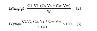
Where: IP refers to the amount of β-glucosidase loaded on the bentonite composite support (mg protein/g support); IY is the immobilization yield (%); C1, Cs and Cw are the concentration of β-glucosidase (mg/mL) of initial, final and for the wash solution respectively; V1, Vs, and Vw are the volumes of β-glucosidase (mL) of initial, final and for the wash solution and W refers to as the weight of bentonite support used in immobilization of β-glucosidase.
Recovery and esterification activities of immobilized β-glucosidase
The recovery activity of the immobilized β-glucosidase was conducted with slight modifications from that of what previously reported by 35 and 36. Here, the levulinic acid and 1-butanol (ratio 2:1) esterification reaction was used for determining the recovery activity. This was achieved by comparing the activity of immobilized β-glucosidase with that of free β-glucosidase activity. The esterification reaction of β-glucosidase was conducted in a 25mL of screw-capped vial which contained a mixture of 1-butanol and levulinic acid (340mM and 180mM) with an appropriate amount of immobilized β-glucosidase. The mixture was vortexed at 200rpm in heptane at 40 ℃ for 2h and a 300μl aliquot was taken at zero hour (0h) and 45min intervals. Acetone: Ethanol (50%v/v) was used to terminate the reaction prior titration with NaOH (0.1M) to estimate the total acid content using phenolphthalein solution as indicator [46]. The experiment was conducted in triplicate and calculation of β-glucosidase esterification was done using equation 4 and a recovery activity of immobilized β-glucosidase was determined using equation 5.

Where: V0 and V1=Volumes (mL) of NaOH used to neutralize unreacted levulinic acid in the reaction mixture with and without β-glucosidase respectively.
M = Concentration (mol/L) of NaOH
E = Weight of protein in immobilized β-glucosidase
T= Time (mins) of incubation

Fourier Transform Infrared (FTIR) and X-Ray Diffraction (XRD) patterns of neat bentonite, free and immobilized β-glucosidase
In the present study, the Fourier Transform Infrared (FTIR) spectra of bentonite support material, immobilized and free β-glucosidase from the region of 4000 to 1000cm-1 were obtained by using a Frontier FT-IR spectrometer (Perkin-Elmer Company, USA) serial number 96064. The experiment was carried out at C19, UTM Johor Bahru Malaysia. Whereas the X-Ray Diffraction (XRD) patterns of bentonite, immobilized and free β-glucosidase were obtained using X-Ray Diffractor meter (XRD) model RIGAKU SMARTLAB (2018) serial number BD680001165-01. The experiment was performed at T05, UTM Johor Bahru Malaysia.
Experimental design and optimization of cellulolytic β-glucosidase produced by T. harzianum Rifai strain Th12 using Taguchi statistical analysis method
The present study used a robust Taguchi statistical analysis method to evaluate the parametric interactions and determine the set of optimal parameters to yield the highest production of β-glucosidase produced by the T. harzianum Rifai fungi via solid state fermentation process. In this study, a five-factor, four level of orthogonal array Taguchi design from the Design Expert 7.1.6 statistical software was used. The L16 array has 16 experimental runs that evaluate the effect of five parameters and their levels viz. pH (3-9), incubation time (2-8 days), incubation temperature 30-60 ℃, surfactant concentration (0.025-0.1% v/v) and enzyme concentration (0.5-2.0mg/mL).
Analysis of Variance (ANOVA) for optimization using Taguchi
To evaluate the suitability of the model for defining the response and optimized conditions, analysis of variance is important. The Sum of squares and Fisher’s value would suggest the effect of optimum response and significance of the model as well as the effect or influence of individual factors contributed to the high production yield of β-glucosidase. The p-value determine the probability of getting the values by noise where; <0.05 p-value considered to be significant [47]. The regression coefficient R-squared (R2) values describe the model fit quality and express the optimization conditions variability. The predicted and adjusted R2 experimental data express the nature of the model. The final stage of optimization using the Taguchi software is to validate the result, which was done by selecting the triplicated verification runs at maximum conditions. Followed by calculating the percentage (%) contribution of individual factors in the process using the equation below:

Where; SSF=The sum of squares for the fth variable and SST=- Sum of squares of all variables
Biocontrol activity of immobilized β-glucosidase in the inhibition of fungal pathogens in a plate
The inhibition of growth of fungal pathogens caused by immobilized cellulolytic β-glucosidase was performed in a petri dish as described by [2,48,49], with slight modification using PDA as substrate. A 5mm disc from the active 5-days old of fungal pathogens mycelia margins was placed on a PDA plate at the center of the plate. Wells were made on the fungal mycelia then filled with 15μL of immobilized cellulolytic β-glucosidase and synthetic fungicide (benomyl) as control followed by incubation at 30 ℃ for 72h before examination. The diameters of the halo Zones of Inhibition (ZOI) indicating complete inhibition were measured by comparing with that of control plates without the cellulolytic immobilized β-glucosidase as described by [49,48,2]. The percentage of inhibition was calculated using the (Eqn. 7).

R1 refers to the radius of the radial growth of the pathogen towards the opposite side in the control plate. The term R2 describes the pathogens’ radius of radial growth towards the cellulolytic β-glucosidase in the test plate.
Statistical analysis
Statistical analysis was performed using IBM SPSS version 22.0 software. Statistical inference for the normality of the data of percentage in plate inhibition of immobilized β-glucosidase experiments in the inhibition of fungal pathogens tested was analyzed using non parametric test (Mann-Whitney) due to the fact that, the data was not normally distributed at significance level of 0.05% by considering the Shapiro-Wilk as described by [50,51].
Results and Discussion
Cellulolytic β-glucosidase production through solid state fermentation using oil palm frond leaves substrate
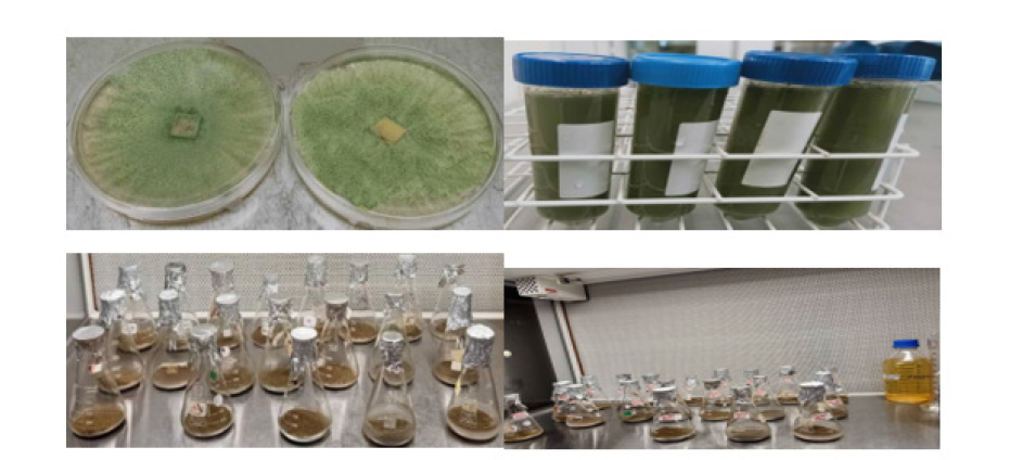
Production and extraction of extra-cellular β-glucosidase by T. harzianum Rifai was successful using the oil palm frond leaves as suitable substrate. The schematic representation of the preparatory steps involved in the production of β-glucosidase by T. harzianum was depicted in Figure 1(a-d).
Figure 1:Preparation of extra-cellular β-glucosidase production via solid state fermentation. a. Trichoderma harzianum Rifai (Th12) culture, b. Trichoderma harzianum Rifai harvested spores c. Dried and grinded OPFL unsterilized d. Sterilized and grinded OPFL (1.18 and 3mm particle size) with fermentation production media.
Enzyme assay for determining the crude cellulolytic β-glucosidase activity and protein content
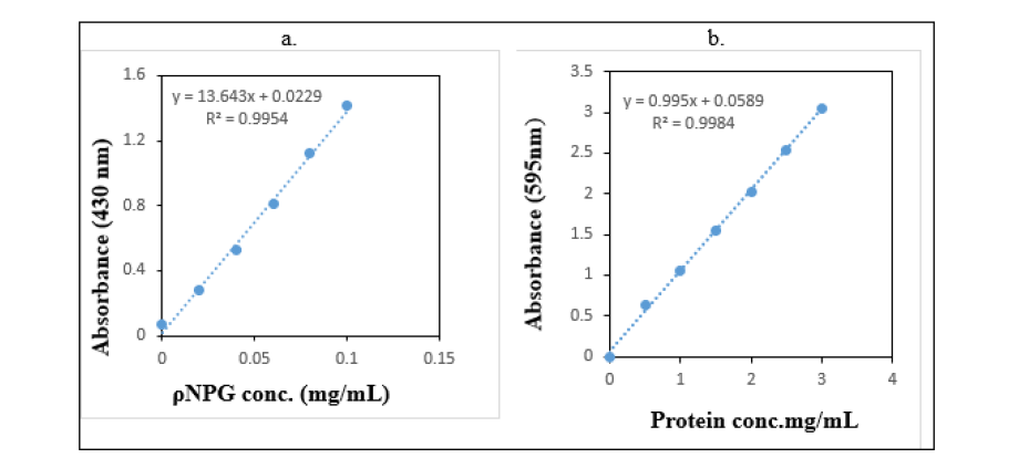
In this study, the activity of free cellulolytic β-glucosidase was determined by considering the ρNPG (4-Nitrophenyl-β-D-glucopyranoside) and Bovine Serum Albumin (BSA) standard curve for the protein content. High R-square value (0.9954 and 0.9984) observed in the standard curves of ρNPG and BSA respectively showed a correlation between the absorbance (enzymatic activity) and protein content concentration. This is clear indication of suitable measurement of β-glucosidase activity and protein content of the β-glucosidase produced. Based on the outcomes of the standard curve seen in this study (Figure 2a, 2b), the β-glucosidase activities were 10.42U/mL. Whereas, 30.50mg/mL protein content in the stock solution of β-glucosidase was obtained.
Figure 2:Standard curve of a. ρNPG and b. Bovine Serum Albumin (BSA) for determining the activity of β-glucosidase and protein content.
Determination of cellulolytic β-glucosidase total soluble protein and total reducing sugar assay
In this study, the total soluble protein content reached the higher level of 4.40±0.01 on day 4 of incubation (Table 3). This lead to have higher concentration of total reducing sugar of 10.53±0.05 (Table 3). The positive growth observed on fungi in SSF is related to the nature and structural components such as cellulose, hemicellulose and lignin present in the oil palm frond leaves substrate. This is in consistent to findings reported by [52]. The concentrations of total reducing sugar and total soluble protein content was depicted in Table 3.
Table 3:Concentrations of Total Reducing Sugar (TRS)
and Total Soluble Protein (TSP).
Data are presented as mean ± standard deviation and the
experiment was conducted in triplicate.

Determination of T. harzianum Rifai cellulolytic β-glucosidase molecular weight by SDS
Figure 3:SDS-PAGE analysis of β-glucosidase produced
from T. harzianum Rifai.
Note: The arrow showed the estimated location for
β-glucosidase on lane 2 (L2).
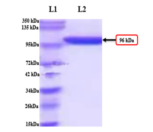
The SDS-PAGE of β-glucosidase produced by the T. harzianum Rifai revealed a single band with estimated 96kDa in the present study. This is in consistent with the findings reported by the previous researchers [53-56]. Lane 1 (L1) is the DNA molecular weight marker. Based on the molecular weight, the estimated location for β-glucosidase was labelled on lane 2 (L2) in this study (Figure 3).
Optimization of SSF parameters for the production of β-glucosidase by T. harzianum Rifai using One Variable at A Time (OVAT) method
Effect of initial fermentation pH: Initial fermentation pH is very important parameter that affects the β-glucosidase production. The effect of initial fermentation pH for the activity of β-glucosidase produced by T. harzianum Rifai was investigated at pH range 3-9 (Figure 4a). In this study, there was an increase of β-glucosidase activity at pH 7.0 after which the activity declined at pH 8 to pH 9 (Figure 4a). In the present study, the β-glucosidase activity was increased from pH 3.0 (5.9U/mL) to pH 7.0 (93.6U/ mL). A serious reduction of activity was seen at pH 8 and pH 9. The lower activity observed at pH 3 is attributed to the slight loss of β-glucosidase catalytic active structure. The low pH tends to produce negatively charge enzyme whereas, the high pH tends to cause reversible protonation of the amino acid functional groups leading to produce positively charge enzyme. The outcome for the pH stability of the β-glucosidase produced by T. harzianum Rifai strain Th12was investigated by pre incubating the β-glucosidase solution at pH 3-9 for 12h (30, 60, 120, 180, 240, 300mins) at 30 ℃ and the outcomes are shown in (Figure 4b). The result of the pH in the present study, showed that the structure of β-glucosidase remained unaffected at its optimum pH level 7.0 with (93.6U/mL) activity which is in consistent with findings previously reported by [57,55]. Therefore, the β-glucosidase produced in the present study was found to be effective in the wide range of pH 3-9 and the result infer good management of oil palm plant in the field against fungal pathogens affecting oil palm.
Effect of incubation temperature: Temperature is very essential factor that influence the activity and stability of the β-glucosidase produced. In this study, Figure 4c showed that β-glucosidase activities increase with increasing temperature. It was observed that as the temperature increased the activity of cellulolytic β-glucosidase is increased (55 ℃ 90.03U/mL) (Figure 4c) and the lower the temperature, the lower the activity (25 ℃ 15.9U/ mL) (Figure 4c). This is because of the rigidity of the β-glucosidase structure and its ability in unfolding enzyme protein are not achieve to its catalytic active form [20]. So, in vivo approach for the control of fungal pathogens affecting the economic crops especially oil palm in our concern may be economically ineffective. Therefore, formulation of β-glucosidase from the potential T. harzianum Rifai strain Th12 as an active ingredient of biocides for the management of various fungal diseases affecting oil palm is important. Thermal stability investigated in the present study (Figure 4d) showed that β-glucosidase was able to resist high temperature (55 ℃) and long incubation period (300mins). This corresponds to findings reported by [57]. It has been reported that, different β-glucosidase from thermophilic fungi range between 40 ℃ and 50 ℃ [57]. Therefore, β-glucosidase produced by T. harzianum Rifai Th12 in this study is stable, recommending its utilization for industrial and agricultural applications. Such thermal stability observed in the present study may be advantageous for in vitro enzymatic hydrolysis of lignocellulose biomasses which may also be useful for controlling the pathogenic fungi such as G. boninense, F. meliae, B. sorokiniana, and P. herbarum by degrading their protective cell walls. This was reported in our study somewhere else [58].
Figure 4:Effect of various fermentation production parameters for the activity of free β-glucosidase produced by T. harzianum Rifai (Th12) through SSF. a. effect of different pH on the activity of β-glucosidase b. pH stability c. effect of incubation temperature on the activity of β-glucosidase d. Thermostability e. effect of incubation time on the activity of β-glucosidase f. effect of enzyme concentration on the activity of β-glucosidase and, g. effect of surfactant concentration on the activity of β-glucosidase.
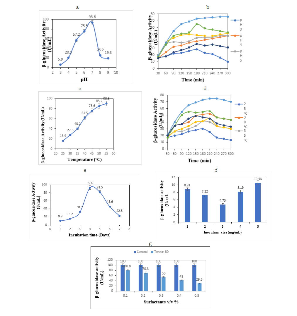
Effect of incubation time: Incubation time is another vital factor in production of β-glucosidase by T. harzianum Rifai strain Th12. In this study, the cellulolytic β-glucosidase production via SSF was conducted for seven (7) days of incubation, where the optimum level of β-glucosidase activities was established at day 4 of incubation time (Figure 4e) with the highest β-glucosidase activity 92.4U/mL and lower activity (9.8U/mL) which was observed at day 1 (Figure 4e). The result of the optimum activity of free β-glucosidase produced by T. harzianum Rifai strain Th12 on day four (4) of incubation is consistent with the findings reported by [59]. In this study, the activity starts to decrease when it reaches the maximum activity at 4 days. It has been previously reported that, the longer the incubation times in SSF process, the higher the yields of cellulolytic enzymes produced. However, β-glucosidase were produced in large quantity at 7 days as reported by [60]. Considering the different reports on cellulolytic β-glucosidase produced by T. harzianum Rifai, it is clear indication of its excellent potential activities.
Effect of enzyme concentration: Enzyme concentration in the production of β-glucosidase by T. harzianum Rifai strain Th12 is another important factor that influence the activity of β-glucosidase. In this research, the optimum enzyme concentrations for β-glucosidase production were 5mg/mL with highest activity of 10.53 and lowest activity 4.73 (U/mL) (Figure 4f). Insufficient inoculum size would adversely affect the entire microbial growth when cultivating conditions for the growth of fungus in the production of cellulolytic β-glucosidase. It has been reported that large amount of inoculum size tends to cause reduction in nutrient for the fungal growth which leads to poor enzyme yield produced. Similarly, little amount of inoculum size in SSF would tends to bring very minute mycelial biomass which resulted into low enzyme production. The optimum β-glucosidase of T. harzianum activity of (10.53U/mL) at 5mg/mL enzyme concentration (Figure 4f) corresponds to the previous findings reported by [10].
Effect of surfactant concentration: Surfactant is another crucial factor in the production of β-glucosidase by T. harzianum Rifai strain Th12. The outcomes of the present study revealed that the best surfactant concentration for β-glucosidase production was 0.1v/v % of Tween 80 (Figure 4g). Most of the surfactants are able to increase the enzymatic reactions and increase the availability of nutrients in the active site [60]. Due to the polar heads present on non-ionic group of surfactants they cannot easily pass-through water- soluble protein and they have little protein denaturing potential [41]. Tween 80 has the larger hydrophobic surface that can easily to attached to protein and form some structural conformations to the proteins [31]. In this study, the higher the concentration of the surfactant the lower the activity. Here, the activity (80.8U/mL) was higher at the concentration of 0.1%v/v than 29.5 at the concentration of 0.5 % v/v surfactant concentration (Figure 4g). The activity of cellulolytic β-glucosidase in SSF at different concentrations of surfactant (Tween 80) in comparison to control was shown in Figure 4g.
Screening of immobilization process by One Factor at A Time (OFAT) method
Effect of immobilization pH: For determining the effect of immobilization pH on β-glucosidase onto the bentonite composite, the support activation and the subsequent immobilization stage (25 ℃, 3h) were conducted at different pH values (3, 5, 7, and 9). The result revealed that immobilization pH 7 in phosphate buffer reveals the optimum protein content with highest amount of 16.64 ± 7.2mg/g (Figure 5a) with activity 98.66U/mL (Figure 5b) p<0.05. Whereas the lowest amount of protein content (4.75mg/g) with less activity of 84.66U/mL (<0.05) was observed (Figure 5b). The optimum activity of immobilized β-glucosidase seen on pH 7 (98.66U/mL) (Figure 5b) could be attributed to the ionization of the acidic and basic amino acid residues in the active site’s micro-environment. The lower activity 84. 66U/mL (Figure 5b) observed could be due to acidic buffer solution rich in hydrogen ions (H+ ions). This corresponds to the previous studies on lipase immobilization reported by [18,23,32,33,47,34,35].
Effect of immobilization temperature: The effect of immobilization temperature was determined using a various immobilization temperatures ranging from 25 ℃-40 ℃ (25, 30, 35, and 40 ℃). The results are expressed in Figure 5c. The results reveals that there were significance activities of immobilized β-glucosidase assessed at various temperature (<0.05). The highest activities of immobilized β-glucosidase were observed at 30 ℃ with optimum protein content 37.21±0.2mg/g (Figure 5c) and immobilization specific activity of 98.96±4.2U/mL (Figure 5d). The observed result is consistent with findings reported by [23]. The lower protein content 25.42mg/g observed at 40 ℃ (Figure 5c) was due to the thermal deactivation occurred on the β-glucosidase. The obtained result seen here showed that the amount of immobilized β-glucosidase formulated onto the bentonite composite at 30 ℃ temperature is adequate to catalyze the optimum β-glucosidase production.
Effect of immobilization time: The effect of immobilization time onto the bentonite immobilization support of β-glucosidase was assessed. The outcome of the protein content and specific activity of immobilized β-glucosidase was determined and the results are expressed in (Figures 5e & 5f). The result of the optimum protein content revealed 25.76mg/g of support with specific activity of 90.90±4.8U/mL (Figure 5e & 5f) when the immobilized β-glucosidase incubated at different incubation time ranging from 2h, 6h, 15h, and 24h (Figure 5e). Here, the results revealed that the least protein content of 14.21mg/g of support at immobilization time of 24h revealed the lower immobilized β-glucosidase specific activity of 81.9±4.8 (p<0.05). The longer the immobilization time (24h) the lesser the specific activity. This indicated that, the 2h immobilization time of β-glucosidase is sufficient for the production of β-glucosidase. The lower specific activity observed in this study is attributed to the gradual denaturation of β-glucosidase. Our findings are in consistence with the findings reported on lipase by [23,47].
Figure 5:Efficacy of immobilized β-glucosidase on bentonite composite support on the enzymatic production of cellulolytic β-glucosidase catalyzed by immobilized β-glucosidase within 2h of reaction at optimum pH 7, temperature 30 ℃, 1.0mg/mL concentration of β-glucosidase, and 0.04 % v/v surfactant concentration. Note: a & b immobilization pH, c & d temperature of immobilization, e & f time of immobilization of β-glucosidase, g & h concentration of immobilized β-glucosidase, and i & j concentration of surfactant.
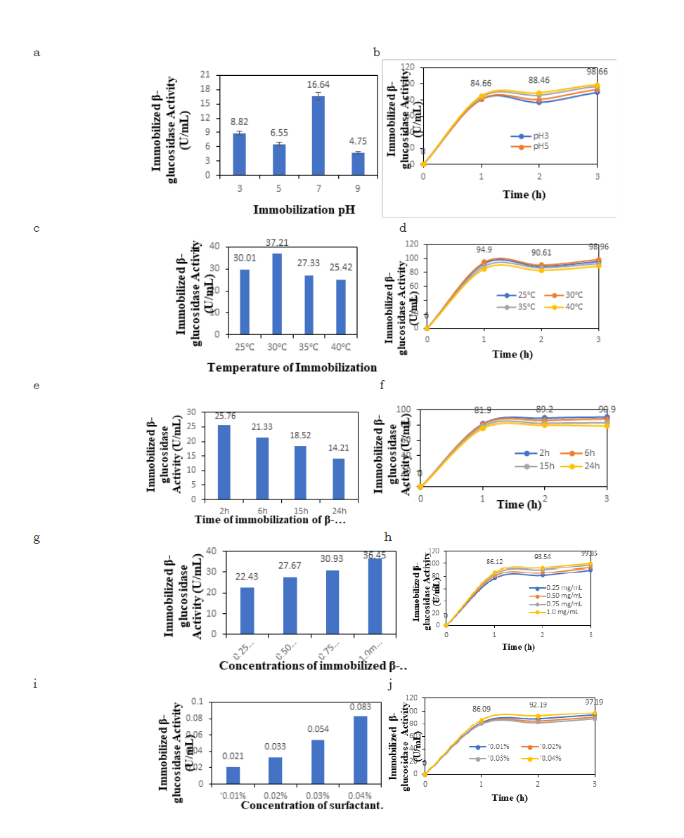
Effect of concentration of β-glucosidase: The effect of concentration of β-glucosidase onto the bentonite composite support was carried out at various concentrations ranging from 0.25 mg/ mL to 1.0mg/mL (0.25, 0.50, 0.75, and 1.0mg/mL of freeze dried β-glucosidase in 10mL phosphate buffer). The total amount of optimum β-glucosidase protein content bound to the support (bentonite composite) was (36.45mg/g) at 1.0mg/mL concentration in which the specific activity (99.85±6.9 U/mL) was observed and the result was expressed in Figure 5g. The results showed the optimum protein content is 36.45mg/g (Figure 5g) with optimum specific activity 99.85±6.9U/mL (Figure 5h). The result showed that as the β-glucosidase concentration increases, the number of enzymes loaded rises (Figure 5g). Therefore, 1.0mg/mL of β-glucosidase concentration is sufficient for immobilization process. This result corresponds to the findings on lipase reported by [23,36].
Effect of surfactant (Tween 80): The Figure 5i shows the protein contents of immobilized β-glucosidase on bentonite conducted at various concentrations of surfactant (Tween 80). In which the result revealed a highest protein content of 0.083mg/g for 0.04% concentration. This revealed the highest activity of 97.19±5.6U/mL (Figure 5j). The result is consistent with findings reported by [23]. Therefore, this study discovered that a 0.04%(v/v) of surfactant (Tween 80) was optimal to produce the highest β-glucosidase activity of (97.19U/mL, p\0.05) (Figure 5j).
Quantification of immobilized β-glucosidase protein content and activity
Table 4:a. Factors for estimating the protein content of
immobilized β-glucosidase b. Specific and residual activities
of free and immobilized betaglucosidase c. recovery
activity of free and immobilized β-glucosidase.
Note: Specific activity = number of enzyme units per mL
divided by protein concentration.

For the determination of β-glucosidase immobilization protein content and immobilization yield percentage, equation 2 and 3 were use and equation 4 and 5 were used for the determination of immobilized β-glucosidase esterification activity and recovery activity respectively. The evaluation on the immobilized protein revealed a maximum enzyme loading of 5mg/g. Therefore, immobilization of β-glucosidase on to the bentonite composite support had a great effect on the specific activity of the immobilized β-glucosidase. The β-glucosidase reached its optimum specific activity of 0.60±0.02 (Table 4) U/mg and relative activity of 199 (%) (Table 4), here the relative activity of the obtained results was higher than that of free β-glucosidase 110 (%) and lower specific activity was observed in free β-glucosidase (Table 4). These results revealed that immobilized β-glucosidase has great impact when activated onto the support. The esterification result in this study did not occur in the negative control. This is clear that an esterification assay of the activated bentonite composite without β-glucosidase has no any impact of the catalytic activity. Therefore, the catalytic activity seen here is attributed to the availability of β-glucosidase absorbed by the support after immobilization process.
Fourier Transform Infrared (FTIR) of bentonite, immobilized and free β-glucosidase
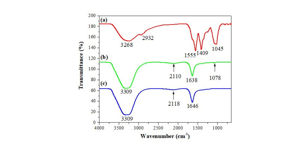
The FTIR spectra for (a) bentonite, (b) immobilized β-glucosidase and (c) free β-glucosidase are expressed in Figure 4 (a-c). The bentonite spectrum (Figure 4a) revealed a broad adsorption band at 3268 cm-1 could be associated with the stretching vibration of -OH group caused by H2 O in the hydrogen bond [37- 39]. Whereas, other bands observed at 2932, 1555, and 1409 cm-1 in bentonite were asymmetric and symmetric stretching vibrations peak of -COOH [38-40]. Another broad adsorption bands were observed in immobilized and free β-glucosidase at 3309 cm-1 (Figure 4b & 4c). The bands spectra of immobilize and free β-glucosidase revealed a shifted prominent band at 2110, 1638, 2118, and (1646 cm-1) as shown using arrow (Figure 4b & 4c) which give the C-O vibration. Another band 1078 cm-1 was also observed in immobilized β-glucosidase. These outcomes are clear indication of the presence of Amide I (C-O), II (N-H binding) and Amide III (C-N, C-C) stretching vibrations from peptide chains in β-glucosidase which corresponds to findings on immobilization of candida rugosa lipase onto a montmorillonite composite support reported by [24]. Here, crosslinking of β-glucosidase and bentonite is successful and the aromatic - CH, vibrations of the hydrophilic interactions of the matrix rings with β-glucosidase forms the peak spectra observed. The migration of -OH vibration is a clear indication of larger hydrogen bond that form the OH of carbohydrate with OH of β-glucosidase (Figure 6). The outcomes of our research are consistence with findings reported by [38-40].
Figure 6:FTIR spectra of a. neat bentonite b. immobilized β-glucosidase c. free β-glucosidase.
X-Ray Diffraction pattern (XRD) of bentonite, immobilized and free β-glucosidase
Figure 7:X-Ray Diffraction pattern (XRD) of a. bentonite b. immobilized βeta-glucosidase and c. free βeta-glucosidase.
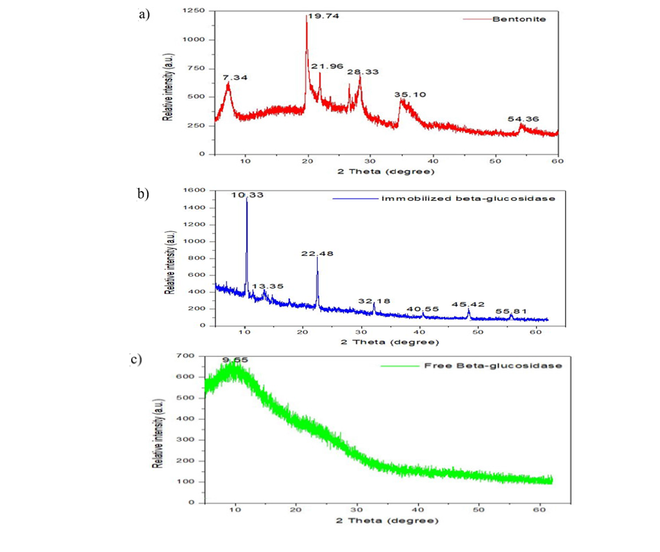
In this research, the crystallinity of the bentonite which contain the montmorillonite, beidellite, nontronite as well as hectorite clay minerals, free β-glucosidase and immobilized β-glucosidase were evaluated by x-Ray Diffraction (XRD) method (Figure 7(a-c)). The crystal structure of bentonite was found to have the relative intensities peaks of 7.34, 19.74, 21.96, 28.33, 35.10 and 54.36 respectively at 2 Theta degrees (Figure 7a). The relative intensities peaks of immobilized β-glucosidase were found to have 10.33, 13.35, 22.48, 32.18, 40.55, and 45.42 respectively at 2 Theta degrees (Figure 7b). For the free β-glucosidase, the peak intensities of 9.55 (Figure 7c) was observed in the present study. This corresponds with the standard information of International Center for Diffraction Data (ICDD). The crystalline structure of the peaks obtained in immobilized β-glucosidase (Figure 7b), is a clear indication of the support (bentonite) proved to be a successful polymer material for β-glucosidase immobilization. The XRD patterns of the obtained result revealed the existence of pure peaks of bentonite crustal structure, immobilized and free β-glucosidase which is in consistent with findings reported by [48,51-63].
Optimization of immobilization process of cellulolytic βeta-glucosidase using L16 orthogonal array of Taguchi
Table 5:Taguchi L16 orthogonal array experimental design with the actual and predicted values of immobilized β-glucosidase.

The optimization of β-glucosidase immobilization process onto the bentonite composite support using the L16 orthogonal array for optimizing the activity of the parametric conditions was successful in this study (Table 5). The actual and predicted values are expressed in Table 5 expressing the combination of best parameters and optimum activity (run 11) (Table 5). The result of the statistical analysis of variance revealed that all the five parameters were significance at (p<0.05) significance level (Table 6). The linear model and effect of individual process parameters on response were clearly observed in this study. The sum of squares (1232.64) and f-value 102.66 signify the linear of the model at 95% confidence level (Table 6). Also, the result of statistical analysis showed the model was significant in optimization process in the selected levels of process factors. In the present study, the contribution of incubation time with 47.93 % is higher than all other factors followed by enzyme concentration (28.65 %), surfactant concentration (8.15), temperature (7.79 %) and pH (5.58 %) (Table 6). The standard deviation (1.55), the residual sum of square (24.01), R-square value (0.9809), adjusted R-square (0.9713) and predicted R-square (0.9502) (Table 6) reveal the fitting model and identify the best β-glucosidase immobilization conditions.
The regression model equation obtained from ANOVA signify the significance factors with 95% confidence in the present study. Equation in terms of actual factor activity in this study is presented below:
Actual activity (U/g)= +77.61+2.81*A+8.23*B- 3.32*C+3.39*D+6.35*E.
Where A=is pH, B=incubation time (day), C=incubation temperature (℃), D=surfactant (% v/v) and E=enzyme concentration (mg/mL).
Here, the regression model equation by which actual activity of the parametric conditions is compared with that of predicted (Figure 8). In this study, the experimental data was closely similar with the predicted values. Therefore, the model is absolutely effective in optimization of immobilization process of β-glucosidase. In the present study, the effect of each individual significant parameters of β-glucosidase immobilization process was assessed for the optimum β-glucosidase immobilization activity in which the contribution of each parameter was found to be significance (p>0.05) (Table 6). The effect of immobilization pH in the present study was evaluated between pH 3-9 and the result s was presented in Figure 9a. The maximum pH was seen at pH 7 with highest activity of (88.73U/mL). Increasing the pH level to pH 9 lower the activity of immobilized β-glucosidase to (75.65U/mL) (Figure 9a). Increasing the incubation time of immobilization to 8 days in this study decreased the activity of the immobilized β-glucosidase from (95.69U/mL) to (71.47U/mL) (Figure 9b). Increasing the immobilization time of the β-glucosidase leads to the formation of multilayer of β-glucosidase molecules overlaying the immobilization support and disrupt the products and substrate. Our results are consistent with findings reported by [23].
Table 6:(a) ANOVA of factors affecting the process of immobilization and (b) statistical variables analyzed by ANOVA.

Figure 8:Actual and predicted response on the activity of immobilized β-glucosidase.
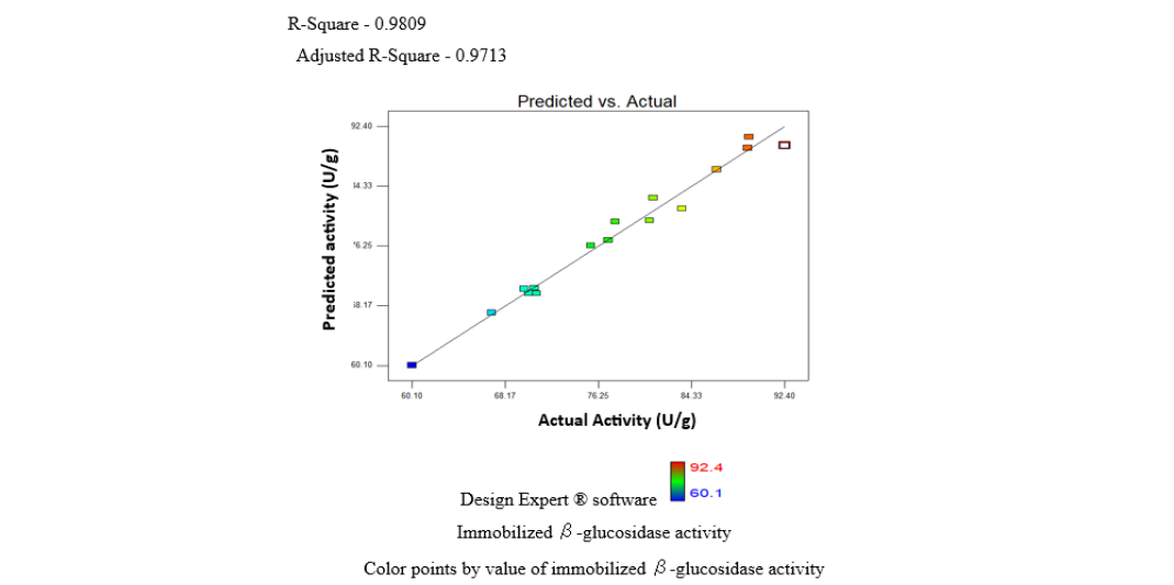
Figure 9:The effect of parameters a. pH, b. incubation temperature c. incubation time, d. enzyme concentrations and e. surfactant concentrations.
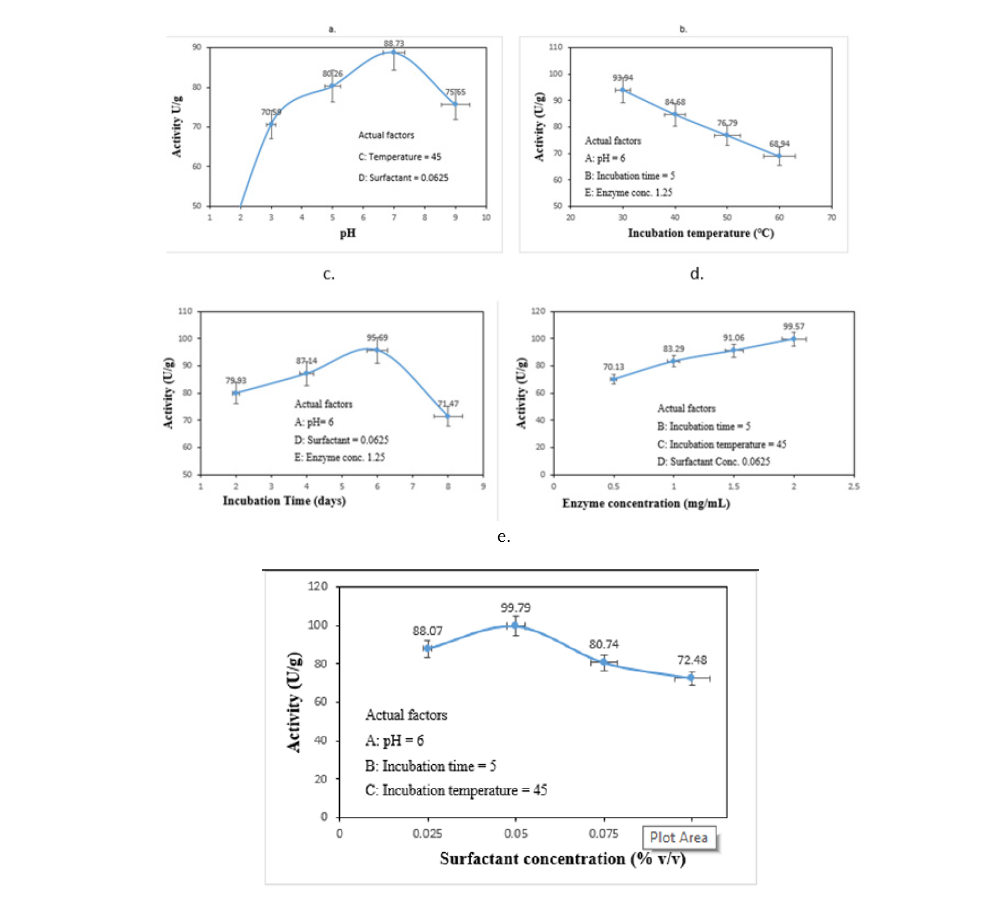
The effect of immobilization incubation temperature ranging from 30-60 ℃ in the present study was evaluated and the result is clearly seen and depicted in Figure 9c. The optimum activity of immobilized β-glucosidase was observed at 30 ℃ temperature with the optimum activity of immobilized β-glucosidase (93.94U/mL) (Figure 9c). Further increased in immobilization temperature above 30 ℃ leads to lower the activity of the enzyme (Figure 9c). The effect of surfactant (Tween 80) in the optimization of β-glucosidase immobilization process was evaluated here (Figure 9d). The activity of immobilized β-glucosidase was found to be higher (99.79U/ mL) at the concentration of 0.050%v/v. High amount of surfactant in the optimization of β-glucosidase immobilization process may lower the activity of the enzyme. In this research, addition of surfactant beyond 0.05% v/v concentration decreased the activity of immobilized enzyme from (99.79U/mL) to (72.48U/mL) (Figure 9d). The reduction in the activity of immobilized β-glucosidase is attributed to the ability of surfactant in promoting the complexity of the enzymatic reactions and equilibrium or suppress the enzyme itself [47]. In this research, the influence of various concentration of immobilized β-glucosidase ranging from 0.5-2.0mg/mL for optimization process was evaluated, in which the maximum activity was observed at 2.0mg/mL (Figure 9e) with maximum activity of 99.57U/mL (Figure 9e). The activity of immobilized β-glucosidase is increasing as the concentration rises. This is associated with the effective collision between the amino groups of β-glucosidase with carbonyl groups of activated immobilization support. Moreover, this study discovered a significance interaction between the pH and immobilization incubation time, incubation time and immobilization incubation temperature, surfactant concentration and enzyme concentration as well as enzyme concentration and pH. The results are depicted in Figure 10(a-e) respectively. The result was validated in Table 7.
Figure 10:Interactive effect and contour plots of a pH and b incubation time, b incubation time and c incubation temperature, d surfactant concentration and e enzyme concentration, e enzyme concentration and a pH to influence the activity of immobilization process of β-glucosidase.
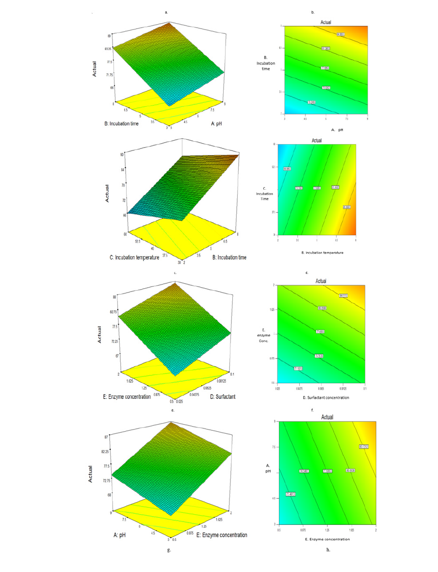
Table 7:Validation of the optimum conditions to immobilize β-glucosidase onto bentonite composite support.

Biocontrol activity of immobilized β-glucosidase in the inhibition of fungal pathogens in a plate
Figure 11:Efficacy of immobilized β-glucosidase in the inhibition of fungal pathogens tested in a plate a. Immobilized β-glucosidase against G. boninense, b. Benomyl against G. boninense c. G. boninense control d. Immobilized β-glucosidase against F. meliae, e. Benomyl against F. meliae f. F. meliae control g. Immobilized β-glucosidase against B. sorokiniana h. Benomyl against B. sorokiniana i. B. sorokiniana control, and J. Immobilized β-glucosidase against P. herbarum, k. Benomyl against P. herbarum and l. P. herbarum control.
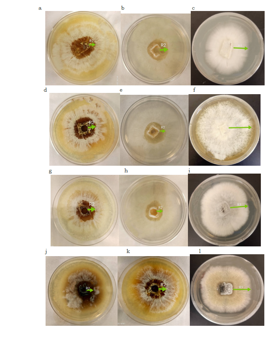
Figure 12:Percentage (%) of inhibition caused by a. immobilized β-glucosidase and b. Benomyl synthetic fungicide against G. boninense, F. meliae, B. sorokiniana and P. herbarum fungal pathogens.
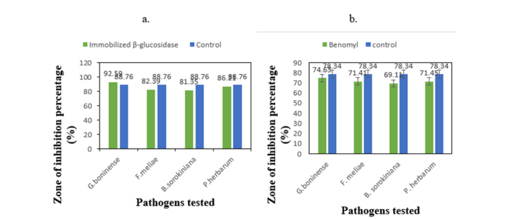
The results of the inhibition caused by immobilized β-glucosidase in the inhibition of fungal pathogens in a plate (Figure 11(a,d,g,j)) revealed 92.59%, 82.39%, 81.35% and 86.21% (Figure 12a) inhibited G. boninense, F. meliae, B. sorokiniana and P. herbarum fungal pathogens respectively compared to control (78.34%) (Figure 12a). Whereas, in benomyl (Figure 11(b,e,h,k)), 74.63, 71.41, 69.11,71.11 percentage (%) of inhibition with 88.76 (%) in control was observed (Figure 12b). Our study is consistent with findings of inhibitory effect of immobilized enzyme on the growth of Fusarium oxysporum and penicillium digitatum reported by [37]. Similarly, our findings correspond to the recent findings reported by [2] on inhibition caused by free β-glucosidase on the management of M. phaseolina causative agent of charcoal rot in soybean. Due to the abnormality of the data, the Mann Whitney test of statistic was performed for comparison between the effect of immobilized β-glucosidase and that of synthetic chemical (benomyl) in a plate inhibition assay. Using the Mann-Whitney U-test, the data were analyzed, and the effects of both immobilized β-glucosidase and benomyl were determined compared to controls. In this study, the effect of immobilized cellulolytic β-glucosidase and benomyl were separately recorded for the inhibition of fungal pathogens followed by comparison between the two. The data of the effects and that of controls were recorded and tabulated in Table 8.
Table 8:The effect of immobilized β-glucosidase and benomyl in plate inhibition assay on fungal pathogens tested.

The effect of immobilized cellulolytic β-glucosidase and benomyl synthetic fungicide on G. boninense, F. meliae, B. sorokiniana and P. herbarum fungal pathogens. The data of the actual effects are recorded and presented as median (range). Mann-Whitney U test was used for comparing the pathogens tested with either immobilized β-glucosidase or benomyl with the level of significance of 0.05. S and NS represent significance (p<0.05) and not significant differences (p>0.05) for comparisons between individual treatment, respectively. In the present study, significance difference (<0.05) was seen in G. boninense, F. meliae, B. sorokiniana and P. herbarum with median 92.54, 81.06, 81.86 and 86.04, having range (85.24- 98.53), (73.76-89.26), (70.62-90.35) and (75.02-99.10) respectively (Table 8) using immobilized β-glucosidase (Table 7). Similarly, another significance difference (<0.05) was observed in the pathogens tested using a benomyl in plate inhibition assay with median 74.54, 71.27, 69.15 and 68.07 range (68.65- 83.33), (65.32-80.15), (62.16- 79.08), and (61.09-78.00) in G. boninense, F. meliae, B. sorokiniana and P. herbarum respectively (Table 8). In comparison to the outcomes observed among the group (immobilized β-glucosidase and benomyl) after tested on fungal pathogens revealed a large median difference in pathogens tested with immobilized β-glucosidase rather than that of synthetic fungicide (benomyl) (Table 7). The outcomes clearly showed the advantage of immobilized β-glucosidase on the inhibition of fungal pathogens over the synthetic fungicide (benomyl). This is because β-glucosidase immobilized on to support material bentonite in our own case by physical absorption method can easily absorb and bind with substrates due to the present of its large surface area. Therefore, the influence of immobilized β-glucosidase in the inhibition of fungal pathogens appears to be effective, reliable and recommendable.
Conclusion
Enzymes are commonly applied in production of several substances that need excessive cost for their production by chemical processes. So, the optimization of different culture conditions for improving β-glucosidase production is of immense interest but the enzymatic processes have unfavorable drawbacks including sensitivity to harsh environmental conditions, studies are demanded to find out the mechanism of fugal degradation of cellulosic materials as well as the mechanism of antifungal activity. It was thought to use fungal enzyme to convert them to useful material for production of β-glucosidase. Hence, the novelty of this work is presented by production of β-glucosidase and biocontrol of phytopathogenic fungi using purified fungal β-glucosidase. The potent antagonistic potency of culture filtrates against fungal pathogens of oil palm highlights the ability to apply novel and safe biofungicides. In this study, Trichoderma harzianum Rifai strain Th12 was utilized for cellulolytic β-glucosidase production via solid-state fermentation using oil palm frond leaves as substrate. Process parameters were optimized using one-variable-at-a-time approach. The crude enzyme was successfully immobilized onto a bentonite-composite polymer support, which demonstrated high efficiency for enzyme binding. Immobilized β-glucosidase exhibited superior catalytic activity compared to the free enzyme and effectively hydrolyzed substrate to release reducing sugars. In vitro assays revealed that immobilized β-glucosidase from T. harzianum Th12 showed stronger inhibitory effects against Ganoderma boninense, Fomitopsis meliae, Bipolaris sorokiniana, and Phoma herbarum than the synthetic fungicide Benomyl. These results position immobilized β-glucosidase from T. harzianum Th12 as a promising biotechnological candidate for plant protection. Its enhanced stability and antifungal potency offer broad prospects for sustainable management of fungal diseases. Further research is warranted to elucidate the mechanisms of cell wall degradation and the antifungal mode of action of this enzyme against phytopathogenic fungi.
Acknowledgement
The authors would like to acknowledge Malaysia Palm Oil Board (MPOB) for the provision of already identified and characterized pathogenic fungi for this research.
References
- Aich S, Singh RK, Kundu P, Pandey SP, Datta S (2017) Genome-wide characterization of cellulases from the hemi-biotrophic plant pathogen, Bipolaris sorokiniana, reveals the presence of a highly stable GH7 endoglucanase. Biotechnology for Biofuels 10: 135.
- Khalili E, Huyop F, Myra Abd Manan F, Wahab RA (2017) Optimization of cultivation conditions in banana wastes for production of extracellular β-glucosidase by Trichoderma harzianum Rifai efficient for in vitro inhibition of Macrophomina phaseolina. Biotechnology & Biotechnological Equipment 31(5): 921-934.
- Kumar J, Schäfer P, Hückelhoven R, Langen G, Baltruschat H, et al. (2002) Bipolaris sorokiniana, a cereal pathogen of global concern: Cytological and molecular approaches towards better control. Molecular Plant Pathology 3(4): 185-195.
- Rebitanim NA, Hanafi MM, Idris AS, Abdullah SNA, Mohidin H, et al. (2020) GanoCare® improves oil palm growth and resistance against Ganoderma basal stem rot disease in nursery and field trials. BioMed research international 2020: 3063710.
- Khalili E, Javed MA, Huyop F, Rayatpanah S, Jamshidi S, et al. (2016) Evaluation of Trichoderma isolates as potential biological control agent against soybean charcoal rot disease caused by Macrophomina phaseolina. Biotechnology & Biotechnological Equipment 30(3): 479-488.
- De Silva NI, Brooks S, Lumyong S, Hyde KD (2019) Use of endophytes as biocontrol agents. Fungal Biology Reviews 33(2): 133-148.
- Manera AP, Da Costa Ores J, Amaral Ribeiro V, Veiga Burkert CA, Kalil SJ (2008) Optimization of the culture medium for the production of β-galactosidase from Kluyveromyces marxianus CCT 7082. Food Technology and Biotechnology 46(1): 66-72.
- Monteiro VN, Do Nascimento Silva R, Steindorff AS, Costa FT, Noronha EF, et al. (2010) New insights in Trichoderma harzianum antagonism of fungal plant pathogens by secreted protein analysis. Current microbiology 61(4): 298-305.
- Strakowska J, Błaszczyk L, Chełkowski J (2014) The significance of cellulolytic enzymes produced by Trichoderma in opportunistic lifestyle of this fungus. Journal of Basic Microbiology 54(S1): S2-S13.
- Zhang Z, Liu JL, Lan JY, Duan CJ, Ma QS, et al. (2014) Predominance of Trichoderma and Penicillium in cellulolytic aerobic filamentous fungi from subtropical and tropical forests in China, and their use in finding highly efficient β-glucosidase. Biotechnology for Biofuels 7(1): 1-14.
- Geimba MP, Riffel A, Agostini V, Brandelli A (1999) Characterisation of cellulose‐hydrolysing enzymes from the fungus Bipolaris sorokiniana. Journal of the Science of Food and Agriculture 79(13): 1849-1854.
- Himmel ME, Ding SY, Johnson DK, Adney WS, Nimlos MR, et al. (2007) Biomass recalcitrance: Engineering plants and enzymes for biofuels production. Science 315(5813): 804-807.
- Hu Y, Lian L, Xia J, Hu S, Xu W, et al. (2020) Influence of PacC on the environmental stress adaptability and cell wall components of Ganoderma lucidum. Microbiological research 230: 126348.
- McCleary BV, Draga A (2016) Measurement of β-glucan in mushrooms and mycelial products. Journal of AOAC International 99(2): 364-373.
- Philippini RR, Martiniano SE, Dos Santos JC, Da Silva SS, Chandel AK (2019) Fermentative production of Beta‐Glucan: Properties and potential applications. Bioprocessing for Biomolecules Production, pp. 303-320.
- Shah F, Mali T, Lundell TK (2018) Polyporales brown rot species Fomitopsis pinicola: Enzyme activity profiles, oxalic acid production, and Fe3+-reducing metabolite secretion. Applied and environmental microbiology 84(8): e02662-02617.
- Peltonen S (1995) Comparison of xylanase production by fungal pathogens of barley with special reference to Bipolaris sorokiniana. Mycological Research 99(6): 717-723.
- Abd Manan FM, Abd Rahman IN, Marzuki NHC, Mahat NA, Huyop F, et al. (2016) Statistical modelling of eugenol benzoate synthesis using Rhizomucor miehei lipase reinforced nanobioconjugates. Process Biochemistry 51(2): 249-262.
- Isah AA, Mahat NA, Jamalis J, Attan N, Zakaria II, et al. (2017) Synthesis of geranyl propionate in a solvent-free medium using Rhizomucor miehei lipase covalently immobilized on chitosan-graphene oxide beads. Preparative Biochemistry and Biotechnology, 47(2): 199-210.
- Wahab RA, Basri M, Rahman RNZRA, Salleh AB, Rahman MBA, et al. (2014) Enzymatic production of a solvent-free menthyl butyrate via response surface methodology catalyzed by a novel thermostable lipase from Geobacillus zalihae. Biotechnology & Biotechnological Equipment 28(6): 1065-1072.
- Wang YH, Feng JT, Zhang Q, Zhang X (2008) Optimization of fermentation condition for antibiotic production by Xenorhabdus nematophila with response surface methodology. Journal of Applied Microbiology 104(3): 735-744.
- Elias N, Wahab RA, Chandren S, Jamalis J, Mahat NA, et al. (2020) Structure and properties of lipase activated by cellulose-silica polyethersulfone membrane for production of pentyl valerate. Carbohydrate polymers 245: 116549.
- Elias N, Wahab RA, Jye LW, Mahat NA, Chandren S, et al. (2021) Taguchi orthogonal design assisted immobilization of Candida rugosa lipase onto nanocellulose-silica reinforced polyethersulfone membrane: Physicochemical characterization and operational stability. Cellulose 28(9): 5669-5691.
- Hussin FNNM, Attan N, Wahab RA (2020) Taguchi design-assisted immobilization of Candida rugosa lipase onto a ternary alginate/nanocellulose/montmorillonite composite: Physicochemical characterization, thermal stability and reusability studies. Enzyme and microbial technology 136: 109506.
- Jacob AG, Wahab RA, Mahat NA (2021) Ternary biogenic silica/magnetite/graphene oxide composite for the hyperactivation of Candida rugosa lipase in the esterification production of ethyl valerate. Enzyme and Microbial Technology 148: 109807.
- Shunmugam S, Teo HL, Elias N, Wahab RA (2021) Taguchi orthogonal design for optimizing a unified ternary process to valorize oil palm leaves for nanocellulose isolation. Jurnal Teknologi 83(2): 95-106.
- Muhammad M, Roswanira A Wahab, Fahrul H, Mohd HR, Naji A M, et al, (2026) Antagonistic activity of crude cellulase (β-glucosidase) produced by Trichoderma harzianum against some fungal pathogens of oil palm. J Biotech Biores 6(2): 867-881.
- Ang S, Shaza E, Adibah Y, Suraini A, Madihah M (2013) Production of cellulases and xylanase by Aspergillus fumigatus SK1 using untreated oil palm trunk through solid state fermentation. Process Biochemistry 48(9): 1293-1302.
- Mandels M, Weber J (1969) The production of cellulases. In: ACS Publications, Washing DC, USA.
- Xu X, Lin M, Zang Q, Shi S (2018) Solid state bioconversion of lignocellulosic residues by Inonotus obliquus for production of cellulolytic enzymes and saccharification. Bioresource Technology 247: 88-95.
- Lai JQ, Li Z, Lü YH, Yang Z (2011) Specific ion effects of ionic liquids on enzyme activity and stability. Green chemistry 13(7): 1860-1868.
- Mohamad N, Buang NA, Mahat NA, Jamalis J, Huyop F, et al. (2015) Simple adsorption of Candida rugosa lipase onto multi-walled carbon nanotubes for sustainable production of the flavor ester geranyl propionate. Journal of Industrial and Engineering Chemistry 32: 99-108.
- Onoja E, Wahab RA (2020) Robust magnetized oil palm leaves ash nanosilica composite as lipase support: Immobilization protocol and efficacy study. Applied biochemistry and biotechnology 192(2): 585-599.
- Osuna Y, Sandoval J, Saade H, López RG, Martinez JL, et al. (2015) Immobilization of Aspergillus Niger lipase on chitosan-coated magnetic nanoparticles using two covalent-binding methods. Bioprocess and Biosystems Engineering 38(8): 1437-1445.
- Guauque Torres MdP, Foresti ML, Ferreira ML (2013) Cross-Linked Enzyme Aggregates (CLEAs) of selected lipases: A procedure for the proper calculation of their recovered activity. AMB Express 3(1): 1-11.
- Naidja A, Huang P (1996) Deamination of aspartic acid by aspartase-Ca-montmorillonite complex. Journal of Molecular Catalysis A: Chemical 106(3): 255-265.
- Öztürk N, Tabak A, Akgöl S, Denizli A (2008) Reversible immobilization of catalase by using a novel bentonite-cysteine (Bent-Cys) micro composite affinity sorbent. Colloids and Surfaces a Physicochemical and Engineering Aspects 322(1-3): 148-154.
- Wen X, Zeng Z, Du C, Huang D, Zeng G, et al. (2019) Immobilized laccase on bentonite-derived mesoporous materials for removal of tetracycline. Chemosphere 222: 865-871.
- Brijwani K, Vadlani PV (2011) Cellulolytic enzymes production via solid-state fermentation: Effect of pretreatment methods on physicochemical characteristics of substrate. Enzyme research 2011: 860134.
- Cao SL, Huang YM, Li XH, Xu P, Wu H, et al. (2016) Preparation and characterization of immobilized lipase from Pseudomonas cepacia onto magnetic cellulose nanocrystals. Scientific reports 6: 20420.
- Cengiz S, Çavaş L, Yurdakoç K (2012) Bentonite and sepiolite as supporting media: Immobilization of catalase. Applied Clay Science 65-66: 114-120.
- Bradford MM (1976) A rapid and sensitive method for the quantitation of microgram quantities of protein utilizing the principle of protein-dye binding. Analytical biochemistry 72: 248-254.
- Miller GL (1959) Use of dinitrosalicylic acid reagent for determination of reducing sugar. Analytical chemistry 31(3): 426-428.
- Ezeilo UR, Wahab RA, Mahat NA (2020) Optimization studies on cellulase and xylanase production by Rhizopus oryzae UC2 using raw oil palm frond leaves as substrate under solid state fermentation. Renewable Energy 156: 1301-1312.
- Laemmli U (1970) Most commonly used discontinuous buffer system for SDS electrophoresis. Nature 227: 680-686.
- Onoja E, Chandren S, Razak FIA, Wahab RA (2018) Extraction of nanosilica from oil palm leaves and its application as support for lipase immobilization. Journal of biotechnology 283: 81-96.
- El-Shora HM, El-Sharkawy RM, Khateb AM, Darwish DB (2021) Production and immobilization of β-glucanase from Aspergillus Niger with its applications in bioethanol production and biocontrol of phytopathogenic fungi. Scientific reports 11(1): 1-11.
- Asril M, Mubarik NR, Wahyudi AT (2014) Blight disease on oil palm. Research Journal of Microbiology 9(3): 265-277.
- Mahat N, Zainol-Abidin N, Nordin N, Abdul-Wahab R, Jayaprakash P (2016) Patterns of oviposition and development of Chrysomya megacephala (Fabricius) (Diptera: Calliphoridae) and Chrysomya rufifacies (Macquart) (Diptera: Calliphoridae) on burned rabbit carcasses. Forensic Science International 260: 9-13.
- Mahat NA, Yin CL, Jayaprakash PT (2014) Influence of paraquat on Chrysomya megacephala (Fabricius) (Diptera: Calliphoridae) infesting minced‐beef substrates in Kelantan, Malaysia. Journal of forensic sciences 59(2): 529-532.
- Yoon LW, Ngoh GC, Chua ASM (2013) Simultaneous production of cellulase and reducing sugar through modification of compositional and structural characteristic of sugarcane bagasse. Enzyme and microbial technology 53(4): 250-256.
- Elham K, Fahrul H, Muhammad AJ, Naji AM, Kalaivani B, et al. (2018) Assessments on the catalytic and kinetic properties of beta-glucosidase isolated from a highly efficient antagonistic fungus Trichoderma harzianum. Bioscience Journal 34(4): 830-847.
- Jeya M, Joo AR, Lee KM, Tiwari MK, Lee KM, et al. (2010) Characterization of β-glucosidase from a strain of Penicillium purpurogenum Applied Microbiology and Biotechnology 86(5): 1473-1484.
- Kaur J, Chadha BS, Kumar BA, Kaur G, Saini HS (2007) Purification and characterization of ß-glucosidase from Melanocarpus sp. MTCC 3922. Electronic Journal of Biotechnology 10(2): 260-270.
- Mateos SE, Cervantes CAM, Zenteno E, Slomianny MC, Alpuche J, et al. (2015) Purification and partial characterization of β-Glucosidase in Chayote (Sechium edule). Molecules 20(10): 19372-19392.
- Garcia NFL, Santos FRdS, Gonçalves FA, Paz MFd, Fonseca GG, et al. (2015) Production of β-glucosidase on solid-state fermentation by Lichtheimia ramosa in agroindustrial residues: Characterization and catalytic properties of the enzymatic extract. Electronic Journal of Biotechnology 18(4): 314-319.
- Marzuki NHC, Mahat NA, Huyop F, Aboul-Enein HY, Wahab RA (2015) Sustainable production of the emulsifier methyl oleate by Candida rugosa lipase nanoconjugates. Food and Bioproducts Processing 96: 211-220.
- Garcia NFL, Da Silva Santos FR, Bocchini DA, Da Paz MF, Fonseca GG, et al. (2018) Catalytic properties of cellulases and hemicellulases produced by Lichtheimia ramosa: Potential for sugarcane bagasse saccharification. Industrial Crops and Products 122: 49-56.
- Zimbardi AL, Sehn C, Meleiro LP, Souza FH, Masui DC, et al. (2013) Optimization of β-glucosidase, β-xylosidase and xylanase production by Colletotrichum graminicola under solid-state fermentation and application in raw sugarcane trash saccharification. International Journal of Molecular Sciences 14(2): 2875-2902.
- Hermoso J, Pignol D, Kerfelec B, Crenon I, Chapus C, et al. (1996) Lipase activation by nonionic detergents: The crystal structure of the porcine lipase-colipase-tetraethylene glycol monooctyl ether complex. Journal of Biological Chemistry 271(30): 18007-18016.
- Poorakbar E, Shafiee A, Saboury AA, Rad BL, Khoshnevisan K, et al. (2018) Synthesis of magnetic gold mesoporous silica nanoparticles core shell for cellulase enzyme immobilization: Improvement of enzymatic activity and thermal stability. Process Biochemistry 71: 92-100.
- Santos CA, Morais MA, Terrett OM, Lyczakowski JJ, Zanphorlin LM, et al. (2019) An engineered GH1 β-glucosidase displays enhanced glucose tolerance and increased sugar release from lignocellulosic materials. Scientific Reports 9(1): 4903.
- Wu L, Lu X, Wu ZS, Dong Y, Wang X, et al. (2018) 2D transition metal carbide MXene as a robust biosensing platform for enzyme immobilization and ultrasensitive detection of phenol. Biosensors and Bioelectronics 107: 69-75.
© 2026 Muhammad Muhammad. This is an open access article distributed under the terms of the Creative Commons Attribution License , which permits unrestricted use, distribution, and build upon your work non-commercially.
Editor In Chief






.jpg)































Signup for Newsletter

Quick Links
 Editorial Board Registrations
 Submit your Article
 Best Paper of the Volume
 Reprints
 Refer a Friend
 Advertise With Us
Our Recent Edition

.jpg)



Top Editors




.jpg)

















.bmp)
.jpg)
.png)
.jpg)













.jpg)






.png)

.png)




.png)




Financial Support

Sponsors

Latest e-Books

Latest Video

 a Creative Commons Attribution 4.0 International License. Based on a work at www.crimsonpublishers.com.
Best viewed in
a Creative Commons Attribution 4.0 International License. Based on a work at www.crimsonpublishers.com.
Best viewed in 
 | Above IE 9.0 version