

- About Us
- Information
-

The Author ensures that the research has been conducted responsibly and ethically with adherence to all relevant regulations. read more..
- For Authors
- For Reviewer
- Manuscript Guidelines
- Membership
- Publication Ethics
-
- Journals
- Reprints
- e-Books
- Videos
- Policies
- Contact Us
 COVID-19
- Submissions



Full Text
Investigations in Gynecology Research & Womens Health
Gynaecological and Obstetric Causes of Iron Deficiency in Women
Emmanuel Andrès*, Xavier Jannot, Jean-Edouard Terrade and Noël Lorenzo-Villalba
Service de Médecine Interne, Clinic of Anemia, Hôpitaux Universitaires de Strasbourg (HUS), France
*Corresponding author:Emmanuel Andrès, Service de Médecine Interne, Clinic of Anemia, Hôpitaux Universitaires de Strasbourg (HUS), Strasbourg, Frances
Submission:September 26, 2026;Published: October 28, 2026
ISSN: 2577-2015 Volume6 Issue2
Abstract
Iron deficiency, with or without anaemia, is one of the most common and most persistently under-recognised medical conditions affecting women of reproductive age. Its two leading mechanistic pathways are gynaecological - chronic or acute menstrual and uterine blood loss - and obstetric - the greatly increased iron requirement of pregnancy compounded by peripartum blood loss. This Seminar reviews the principal gynaecological causes of iron deficiency, structured around the FIGO PALM-COEIN classification of abnormal uterine bleeding, with particular attention to uterine leiomyoma, adenomyosis, endometrial polyps, endometriosis, and bleeding disorders such as von Willebrand disease. It then reviews the obstetric causes, including the physiological iron cost of pregnancy, multiple gestation and short interpregnancy intervals, placental pathology, and postpartum haemorrhage, which together make pregnancy and the puerperium a period of maximal vulnerability to iron deficiency. We discuss current diagnostic thresholds and screening recommendations from FIGO, the American College of Obstetricians and Gynecologists, the World Health Organization, and the US Preventive Services Task Force, and we summarise the evidence base for oral iron, intravenous iron formulations, and hormonal and surgical treatments of the underlying gynaecological cause. Despite a high prevalence and well-documented adverse effects on quality of life, cognition, cardiovascular tolerance, and pregnancy outcome, iron deficiency in women remains frequently unrecognised because heavy menstrual bleeding is normalised by patients and under-elicited by clinicians. Systematic questioning about menstrual and obstetric history, coupled with routine ferritin measurement in at-risk women, is a simple intervention with the potential to substantially reduce the burden of this largely preventable and treatable condition.
Introduction
Iron deficiency is the most common nutritional deficiency worldwide and the leading cause of anaemia, affecting an estimated 1·2 billion people globally [1]. Women of reproductive age bear a disproportionate share of this burden: They enter each menstrual cycle with iron stores roughly a third the size of those in men (approximately 300mg versus 1000mg), and every episode of menstrual, gynaecological, or obstetric blood loss draws directly on this already limited reserve [2]. Unlike most other populations in whom iron deficiency triggers investigation for occult gastrointestinal blood loss or malabsorption, the leading cause of iron deficiency in women of reproductive age is physiological or pathological blood loss from the female genital tract - through menstruation, structural or functional uterine disease, or pregnancy and childbirth [1,3].
Despite this, the relationship between women’s reproductive health and iron status is persistently under-recognised, both by the public and by clinicians. Heavy menstrual bleeding is common - affecting between a tenth and a third of menstruating women by conservative estimates, and considerably more by some population surveys - yet it is often normalised by women themselves, dismissed as an inconvenience rather than raised as a medical symptom, and infrequently and inconsistently asked about even when patients present with unexplained iron deficiency [3-7]. The World Health Organization only formally recognised heavy menstrual bleeding as a risk factor for anaemia in its 2023 anaemia-reduction framework, decades after the pathophysiological link was first established [6,8]. A parallel pattern of under-recognition affects pregnancy, where progressive expansion of maternal iron requirement across gestation is compounded, for a substantial minority of women, by antenatal bleeding, multiple gestation, or postpartum haemorrhage, yet screening practices and treatment thresholds continue to vary widely between national and international guidelines [7,9].
This Seminar reviews the gynaecological and obstetric causes of iron deficiency in women, their underlying mechanisms, the current evidence for screening and diagnosis, and the therapeutic options available for both the underlying cause and the resulting iron deficit. We use the term iron deficiency to denote depleted iron stores irrespective of haemoglobin concentration, and iron deficiency anaemia to denote the subset of cases in which erythropoiesis has become iron-restricted; the distinction matters clinically because symptoms, quality-of-life impairment, and some adverse pregnancy outcomes occur well before anaemia is biochemically apparent [10-12].
Search Strategy and Selection Criteria
We searched PubMed/MEDLINE for articles published in English between January 2000 and September 2026, using combinations of the terms “iron deficiency”, “iron deficiency anaemia”, “heavy menstrual bleeding”, “abnormal uterine bleeding”, “uterine fibroids”, “leiomyoma”, “adenomyosis”, “endometriosis”, “von Willebrand disease”, “pregnancy”, “postpartum haemorrhage”, “placenta praevia”, “placenta accreta”, and “intravenous iron”. We prioritised systematic reviews, meta-analyses, and guideline documents from major professional societies (FIGO, ACOG, WHO, NICE, USPSTF, SOGC), supplemented by key primary studies. Reference lists of identified articles were hand-searched for additional relevant reports. The final reference list was selected on the basis of relevance, methodological quality, and, where available, recency.
Epidemiology and Burden
Population-based data consistently place non-pregnant women among the groups at highest risk of iron deficiency worldwide, alongside young children and pregnant women [1,13]. In crosssectional cohorts of women presenting with heavy menstrual bleeding, low ferritin (<15-30μg/L, depending on the threshold used) has been reported in 40-45% of participants, and frank anaemia in a third to over 40% [4,14]. Among adolescents, a Swedish school-based survey using the validated SAMANTA questionnaire found a heavy-menstrual-bleeding prevalence of 53% and an iron-deficiency prevalence of 40% in a cohort of 394 post-menarchal students, with meat-restricted diets compounding menstrual losses [15]. In women with an underlying bleeding disorder such as von Willebrand disease, the burden is still greater: A Swedish national cohort found low ferritin in 45% and anaemia in 18% of participants, findings that likely under-estimate the true prevalence given loss to follow-up in registry-based designs [16].
Beyond menstruation, uterine structural disease adds substantially to this burden. In large retrospective and real-world cohorts, anaemia is documented in a third or more of women undergoing surgery for uterine fibroids, and preoperative anaemia is itself an independent predictor of adverse perioperative cardiovascular and thromboembolic outcomes, with an associated near-doubling of 12-month mortality after fibroid surgery in one large propensity-matched analysis of the TriNetX research network [17,18]. In endometriosis, cross-sectional data show a prevalence of iron deficiency of over 50%, with iron deficiency present in nearly half of patients who did not report heavy menstrual bleeding on direct questioning - underscoring that reliance on a single symptom to trigger screening will systematically miss a substantial fraction of at-risk women [19].
Pregnancy compounds this baseline vulnerability. The World Health Organization estimates that around 40% of pregnant women worldwide are anaemic, and iron deficiency accounts for approximately 50-60% of these cases [20]. In low- and middleincome settings, prevalence estimates for iron deficiency anaemia in pregnancy approach or exceed 50%, and even in high-income settings, two-centre UK data found that 46% of women were anaemic at some point during pregnancy [20]. Postpartum anaemia, most commonly reflecting unresolved antenatal iron deficiency combined with peripartum blood loss, affects an estimated 50-80% of women worldwide in the weeks after childbirth [21].
Physiology of Iron Balance across Reproductive Life/h4>
Menstrual blood loss is, for most women, the dominant physiological driver of iron turnover across reproductive life. Average menstrual blood loss is on the order of 30-40ml per cycle, translating into an average additional iron requirement of roughly 0·5-1·0mg per day when averaged across the cycle; heavy menstrual bleeding, conventionally defined as total menstrual blood loss exceeding 80mL per cycle (though contemporary guidance increasingly favours patient-reported impact over objective volume measurement), can multiply this iron cost several-fold [3,22,23]. Because duodenal iron absorption is capped at only a few milligrams per day even under conditions of maximal upregulation, sustained heavy menstrual loss readily outstrips dietary compensation, particularly in women who additionally restrict dietary iron intake (for example through vegetarian or vegan diets) or who have coexisting causes of reduced absorption or increased demand [2,7].
Pregnancy multiplies this iron requirement several-fold further. Total iron requirements across a singleton pregnancy are estimated at 800-1000mg, reflecting expansion of maternal redcell mass, transfer to the fetus and placenta, and anticipated blood loss at delivery; the daily requirement for absorbed iron rises from approximately 0·8mg in early pregnancy to approximately 7·5mg in the third trimester, a demand that the great majority of women cannot meet from diet and pre-pregnancy stores alone [20]. Maternal hepcidin, the master regulator of systemic iron availability, is physiologically suppressed as pregnancy advances, favouring both intestinal iron absorption and placental iron transfer; experimental data indicate that maternal hepcidin status is itself a determinant of fetal iron endowment, providing a mechanistic link between maternal iron sufficiency and neonatal iron status at birth [24]. Lactation adds a further, though comparatively modest, iron cost through breast-milk iron content, partly offset by lactational amenorrhoea [20].
Gynaecological Causes of Iron Deficiency
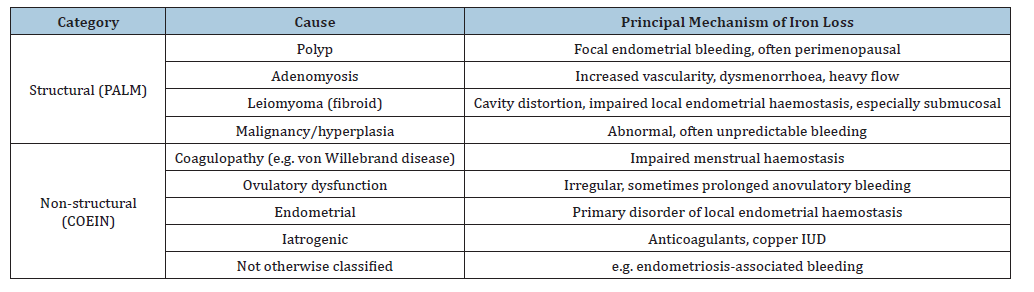
The FIGO PALM-COEIN classification provides a structured framework for the causes of abnormal uterine bleeding, dividing structural causes - polyp, adenomyosis, leiomyoma, malignancy and hyperplasia from non-structural causes - coagulopathy, ovulatory dysfunction, endometrial, iatrogenic, and not-otherwiseclassified [25-27]. Heavy menstrual bleeding, a common final pathway for many of these conditions, is the single most important gynaecological driver of iron deficiency in reproductive-aged women, but the relative contribution of specific structural and nonstructural causes differs by age, parity, and underlying pathology, and more than one cause frequently coexists in the same patient [27].
Uterine fibroids (leiomyoma)
Uterine leiomyomas (fibroids) are the most common benign gynaecological tumours, with a prevalence estimated between 8% and 18% in reproductive-aged women depending on diagnostic method and population studied, rising considerably higher by the time of menopause [15,28]. Fibroids are the leading structural cause of heavy menstrual bleeding severe enough to produce iron deficiency anaemia, particularly when submucosal location increases contact with the endometrial cavity; submucosal fibroids carry a disproportionately high risk of anaemia compared with intramural or subserosal fibroids [27,29-31]. Proposed mechanisms extend beyond simple mechanical distortion of the endometrial cavity: fibroid-conditioned media have been shown to raise local transforming growth factor beta-3, which suppresses bone morphogenetic protein-2 receptor signalling and impairs normal endometrial haemostatic response, while abnormal uterine vascular architecture and altered myometrial contractility around fibroids further compromise haemostasis at the endometrialmyometrial interface [27]. The clinical consequences of fibroidassociated iron deficiency anaemia can be severe. Case reports and case series document profound, occasionally life-threatening anaemia - including reported haemoglobin concentrations below 2g/dL - resulting from chronic or acute fibroid-related haemorrhage, sometimes complicated by reversible anaemic cardiomyopathy [32,33]. At a population level, real-world cohort data from a large multinational research network show that preoperative anaemia in women undergoing fibroid surgery independently predicts adverse 12-month cardiovascular and thromboembolic outcomes, including a near-doubling of all-cause mortality, reinforcing that anaemia associated with fibroids is not merely a laboratory finding but a marker of, and probable contributor to, broader physiological vulnerability that merits correction before elective surgery wherever possible [17].
Adenomyosis
Adenomyosis, defined by the presence of ectopic endometrial glands and stroma within the myometrium, is increasingly recognised - aided by improved ultrasound and MRI diagnostic criteria - as a frequent and previously under-diagnosed cause of heavy menstrual bleeding and secondary iron deficiency [27]. Its pathogenesis remains incompletely understood, with the dominant hypothesis favouring direct invasion of the myometrium by basal endometrium, although alternative theories invoking Müllerian remnants or metaplasia of myometrial stem cells have not been excluded [27]. Mechanistically, adenomyotic myometrium shows increased local vascularity, macrophage infiltration, and elevated tissue factor and inflammatory cytokine expression, plausibly amplifying both menstrual blood loss and dysmenorrhoea severity [27]. Because adenomyosis frequently coexists with fibroids and endometriosis, its independent contribution to iron deficiency can be difficult to isolate in clinical cohorts, but the National Institute for Health and Care Excellence guideline on heavy menstrual bleeding explicitly lists suspected or diagnosed adenomyosis as an indication for first-line consideration of the levonorgestrelreleasing intrauterine system [34].
Endometrial polyps and ovulatory dysfunction
Endometrial polyps are a further structural cause within the PALM-COEIN framework and are a recognised, though generally less severe, contributor to abnormal uterine bleeding and consequent iron loss, particularly in perimenopausal women; symptom severity has been linked in exploratory work to polyp proliferative activity [27]. Ovulatory dysfunction (the “O” of COEIN) - including the irregular, sometimes prolonged bleeding of polycystic ovary syndrome and other causes of anovulation - and endometrial causes of locally dysregulated haemostasis represent important non-structural contributors, particularly at the extremes of reproductive life (adolescence and the menopausal transition), when anovulatory cycles are most frequent [26,27].
Endometriosis
The relationship between endometriosis and iron status is mechanistically distinctive and, in important respects, still incompletely resolved. Endometriosis is characterised locally by iron overload: Recurrent cyclical bleeding into ectopic endometriotic implants and endometriomas generates a markedly pro-oxidant, iron-rich peritoneal environment, with iron accumulation implicated in local inflammation, fibrosis, oxidative DNA damage, and impaired clearance of senescent cells - mechanisms now considered central to disease pathogenesis and progression [33,35]. Paradoxically, despite this local iron excess, systemic iron status in women with endometriosis is frequently normal or reduced, a pattern also observed in a non-human primate model of spontaneous endometriosis, in which iron loss through heavy or irregular menses, compounded by chronic systemic inflammation and hepcidin-mediated sequestration of iron in macrophages, was identified as the principal mechanism underlying reduced hepatic and bone-marrow iron stores [36].
Clinically, this translates into a high prevalence of unrecognised iron deficiency among women with endometriosis. In a crosssectional cohort of 251 patients, iron deficiency was present in 53% and iron deficiency anaemia in 14%; critically, of the 142 patients who did not report heavy menstrual bleeding, 47% were nonetheless iron-deficient, and transferrin saturation identified deficiency in over a third of patients whose ferritin alone was within the normal range [19]. Because fatigue, dizziness, and restless-legs-type symptoms overlap substantially between iron deficiency and endometriosis itself, iron status is easily overlooked as a contributor to symptom burden in this population, and reliance on menstrual-bleeding history alone as a trigger for iron testing will miss a substantial proportion of affected women [19,35].
Coagulopathy and iatrogenic causes
Congenital and acquired bleeding disorders are an important, frequently under-diagnosed, non-structural cause of heavy menstrual bleeding and associated iron deficiency. Von Willebrand disease is the most prevalent inherited bleeding disorder and disproportionately affects women because of the additional haemostatic challenge of menstruation; systematic coagulation screening in large cohorts of women presenting with heavy menstrual bleeding identifies von Willebrand factor deficiency in approximately 9-10% of previously undiagnosed patients [5]. In women with confirmed von Willebrand disease, the combination of heavy menstrual bleeding, iron deficiency, and iron deficiency anaemia is the rule rather than the exception, and cohort data suggest that a meaningful proportion of these women may require specific factor concentrate therapy in addition to iron repletion and hormonal management [16]. Iatrogenic causes, the “I” of PALMCOEIN, include anticoagulant and antiplatelet therapy, which can unmask or aggravate otherwise subclinical menstrual blood loss, and the copper intrauterine device, which characteristically increases menstrual blood volume and is a recognised, dose-related contributor to iron deficiency in users [26,27] (Table 1).
Table 1:FIGO PALM-COEIN causes of abnormal uterine bleeding relevant to iron deficiency.
Obstetric Causes of Iron Deficiency
Pregnancy and the peripartum period constitute the second major axis of iron deficiency risk in women, arising from the interaction of a substantially increased physiological iron requirement (discussed above) with a range of obstetric complications that add acute or chronic blood loss on top of this baseline demand. Because maternal iron deficiency and anaemia are each independently associated with adverse maternal and neonatal outcomes, the causes discussed below are not only common but clinically consequential [10,20].
Multiple gestation, grand multiparity, and short interpregnancy interval
Multiple gestation multiplies fetal and placental iron demand without a proportionate increase in maternal iron absorptive capacity, and is consistently identified as a risk factor for both antenatal and postpartum iron deficiency anaemia [9,37]. Grand multiparity, short interpregnancy intervals that do not allow full repletion of iron stores depleted by a preceding pregnancy, and adolescent pregnancy — in which the mother’s own growth-related iron requirements compete with those of the fetus — are similarly recognised risk factors incorporated into national risk-stratification guidance for antenatal iron screening [9,38]. Guidance from the British Committee for Standards in Haematology, for example, explicitly identifies multiparity greater than three, consecutive pregnancies, adolescent pregnancy, and a history of heavy menstrual bleeding or prior anaemia as indications for proactive ferritin testing rather than reliance on haemoglobin alone [38].
Placental pathology
Placental pathology contributes to obstetric iron deficiency through mechanisms ranging from chronic low-volume antenatal bleeding to acute, life-threatening haemorrhage. Placenta praevia and placental abruption are each associated with variable degrees of antepartum bleeding that can precede and compound the iron deficit accumulated across gestation, while placenta accreta spectrum disorders carry a substantial risk of massive intrapartum and postpartum haemorrhage requiring transfusion [37,39]. Antenatal iron deficiency and anaemia are themselves associated, in observational data, with an increased risk of placenta praevia and with increased rates of caesarean delivery, findings that have been interpreted as evidence of impaired placental implantation and uteroplacental function under conditions of iron insufficiency, although the direction and mechanisms of this association require further clarification [10,40].
Postpartum haemorrhage and caesarean delivery
Postpartum haemorrhage is both a cause and a consequence of iron deficiency: Antepartum anaemia and iron deficiency increase the risk of postpartum haemorrhage, and postpartum haemorrhage, in turn, is the leading acute cause of severe postpartum anaemia [10,40,41]. A large United States cohort analysis of more than 76 million delivery hospitalisations documented a substantial and sustained rise in postpartum haemorrhage rates over the period 2000-2019, from 2·7% to 4·3% of deliveries, with an even steeper rise among women with two or more concurrent risk factors, and antepartum anaemia has since been highlighted as a risk factor meriting explicit inclusion in postpartum-haemorrhage risk-assessment and action-plan tools, a recommendation not yet incorporated into some existing national protocols at the time they were published [41,42]. Because postpartum anaemia affects an estimated 50-80% of women worldwide and is linked to postpartum depression, fatigue, impaired cognition, and premature cessation of breastfeeding, its prevention through antenatal correction of iron deficiency, and its prompt treatment when peripartum blood loss has occurred, are considered a standard-of-care priority in contemporary patient-blood-management pathways for obstetrics [21,40].
Caesarean delivery, whether elective or emergency, is independently associated with greater blood loss than vaginal delivery and, in cohort data, with patterns of more severe postpartum anaemia; a 2024 meta-analysis further demonstrated an association between maternal anaemia and increased rates of caesarean delivery itself, suggesting a bidirectional relationship in which iron deficiency may both predispose to, and result from, operative delivery [10,37].
Diagnostic Approach and Screening Recommendations
Serum ferritin remains the most sensitive and specific first-line test for iron deficiency in the absence of concurrent inflammation, but interpretation differs by clinical context [10,24]. In nonpregnant women, physiologically based analyses of US NHANES data support a ferritin threshold in the region of 25μg/L (rather than the traditional 15μg/L) as more accurately reflecting the point of irondeficient erythropoiesis, though many guidelines and laboratories continue to use lower cut-offs [43]. In pregnancy, ferritin declines physiologically as gestation advances, and international guidance increasingly favours a threshold of approximately 30μg/L (rather than the lower non-pregnant cut-off) to trigger treatment, alongside recognition that ferritin’s acute-phase reactant properties can mask true deficiency in the presence of inflammation, in which case transferrin saturation provides complementary diagnostic information — an approach that identified deficiency in over a third of endometriosis patients whose ferritin alone was normal [19,44].
Screening recommendations vary across professional bodies but converge on the principle that ferritin, not haemoglobin alone, should be measured at least once in pregnancy, and ideally preconceptionally, in women with recognised risk factors. Since 2023, FIGO has recommended ferritin-based screening for iron deficiency pre-conceptionally where possible, and at multiple points during pregnancy, a position now endorsed by several national societies including the Society of Obstetricians and Gynaecologists of Canada [39,41]. The US Preventive Services Task Force, following its 2024 evidence review, recommends screening all pregnant individuals for iron deficiency anaemia, while stopping short of a formal recommendation on non-anaemic iron deficiency screening pending further trial evidence [44,45]. In the non-pregnant gynaecological setting, guideline appraisal work using the AGREE II framework has highlighted substantial heterogeneity between national guidelines in recommended screening triggers, ferritin thresholds, and iron dosing regimens, underscoring a persistent evidence gap that limits harmonised practice [9,46].
Management
Gynaecological causes
Management of gynaecological iron deficiency proceeds along two parallel tracks: Correction of the iron deficit itself, and treatment of the underlying cause of abnormal bleeding. For the latter, the levonorgestrel-releasing intrauterine system is the most consistently recommended first-line hormonal option across international guidelines, including as first-line therapy in women with heavy menstrual bleeding of no identified structural cause, fibroids under 3cm, or suspected adenomyosis; randomised comparisons show that it produces significantly greater improvements in both haemoglobin and serum ferritin than cyclic oral progestin [34,47,48]. Tranexamic acid, an antifibrinolytic agent taken during the days of heaviest flow, and non-steroidal anti-inflammatory drugs are recommended nonhormonal alternatives, particularly where hormonal treatment is undesirable or contraindicated, and can reduce menstrual blood loss by approximately a third to a half [49,50]. For fibroid- or adenomyosis-related bleeding refractory to medical management, options span gonadotrophin-releasing hormone agonists and antagonists, selective progesterone-receptor modulators, and a range of fertility-sparing procedural and surgical interventions (myomectomy, uterine artery embolisation, radiofrequency or focused-ultrasound ablation) through to hysterectomy where fertility preservation is not a priority [27].
For the iron deficit itself, oral iron remains first-line therapy for most non-pregnant women, though tolerability limits adherence, and accumulating evidence supports alternate-day dosing over traditional daily dosing as a means of improving fractional absorption while reducing gastrointestinal side-effects, reflecting hepcidin-mediated diurnal regulation of duodenal iron uptake [20,51]. Intravenous iron - historically reserved for oral-iron intolerance or failure - is increasingly used as first-line therapy in women with more severe iron deficiency anaemia, heavy ongoing losses, or a need for rapid correction (for example before elective fibroid surgery). Modern formulations such as ferric carboxymaltose and ferric derisomaltose allow full-dose replacement in one or two infusions, with a substantially lower risk of hypersensitivity reactions than older high-molecularweight iron dextran preparations, though ferric carboxymaltose carries a recognised risk of transient, usually asymptomatic, hypophosphataemia that should be considered with repeated or high-dose administration [52-54].
Pregnancy and the postpartum period
In pregnancy, most international guidelines recommend oral elemental iron doses in the range of 30-60mg daily for at-risk or anaemic women, though recommended doses vary between 30mg (ACOG, FIGO, CDC) and 60-100mg (WHO, FOGSI), and there is active debate about whether lower, alternate-day dosing regimens might achieve equivalent haemoglobin response with better tolerability and adherence [20,55]. A 2024 Cochrane review of daily oral iron supplementation in pregnancy, alongside a parallel meta-analysis specifically examining supplementation in iron-replete women, together support supplementation’s benefit in deficient women while highlighting continued uncertainty about the optimal strategy in women who are iron-sufficient at baseline [56,57].
Where oral iron is poorly tolerated, ineffective, or insufficient time remains before delivery, intravenous iron has an increasingly well-supported role in pregnancy. Randomised trials and systematic reviews comparing intravenous ferric carboxymaltose with oral iron in pregnant women with anaemia show significantly greater increases in haemoglobin and ferritin with the intravenous route, and a pragmatic randomised trial in late pregnancy found ferric carboxymaltose safe and effective for rapid correction of anaemia close to term [52,53,58]. In the postpartum period specifically, randomised and real-world comparative data support single high-dose intravenous iron (for example ferric derisomaltose or ferric carboxymaltose) over combined oral-and-intravenous or oral-only regimens for more rapid and complete correction of postpartum-haemorrhage-related anaemia: in a retrospective cohort of 91 women with postpartum haemoglobin below 8g/dL, a single 1000mg dose of ferric derisomaltose produced significantly higher haemoglobin, serum iron, and ferritin than conventional intravenous-plus-oral therapy as early as two weeks post-partum, with earlier resolution of anaemia in the single-dose group (53 women) than the conventional-treatment group (38 women), with parallel evidence that intravenous iron reduces postpartum fatigue after significant peripartum blood loss [54,59,60]. Because antenatal anaemia is itself a modifiable risk factor for postpartum haemorrhage, proactive antenatal identification and correction of iron deficiency — rather than reactive treatment after bleeding has already occurred - is now regarded by FIGO and allied bodies as a core preventive strategy, one that intersects directly with the broader patient-blood-management approach to reducing postpartum-haemorrhage-related maternal morbidity [41,45].
Clinical Consequences of Untreated Iron Deficiency
The consequences of unrecognised or inadequately treated iron deficiency in women extend well beyond the laboratory diagnosis of anaemia. Even in the absence of frank anaemia, iron deficiency is associated with fatigue, reduced exercise tolerance, impaired cognitive performance and concentration, hair loss, pica, and restless-legs-type symptoms, all of which materially affect quality of life and, in working-age women, occupational functioning [2,10]. In pregnancy, maternal iron deficiency anaemia - particularly when present in the first trimester - has a more adverse impact on fetal growth than anaemia developing later in gestation, and is associated with increased risk of preterm birth, low birth weight, and small-for-gestational-age neonates, together with longerterm, measurable deficits in offspring neurodevelopment [20,37]. Severe maternal anaemia (haemoglobin below approximately 7g/dL) has been reported to double the risk of maternal death, and postpartum anaemia is independently linked to postpartum depression and early cessation of breastfeeding, outcomes with consequences for both mother and infant that extend well beyond the immediate puerperium [20,21]. The bidirectional relationship between anaemia and postpartum haemorrhage, together with anaemia’s association with increased rates of caesarean delivery, further illustrates how a single, readily correctable deficiency can propagate a cascade of avoidable obstetric morbidity if left unaddressed [10,41].
Controversies and Research Gaps
Despite the accumulating evidence reviewed here, several important gaps remain. First, there is no international consensus on the optimal ferritin threshold, dosing regimen, or timing of screening for iron deficiency across the reproductive lifespan, and existing guidelines diverge considerably when formally appraised [49,50]. Second, robust randomised trial data comparing daily versus alternate-day oral iron dosing, and clarifying which women benefit most from first-line intravenous over oral iron, remain limited, particularly in pregnancy [20,61]. Third, the mechanistic paradox of local iron excess coexisting with systemic iron deficiency in endometriosis warrants further mechanistic study, both to refine screening strategies and to explore whether iron-targeted therapies might modify disease progression rather than merely correct systemic deficiency [33,35]. Finally, and perhaps most importantly from a public-health perspective, the persistent normalisation of heavy menstrual bleeding by patients, and its inconsistent elicitation by clinicians across primary care and specialist settings, remains a major and remediable barrier to timely diagnosis that is unlikely to be solved by biochemical or pharmacological advances alone [3,6].
Conclusion
Iron deficiency in women of reproductive age is common, frequently under-recognised, and disproportionately driven by two intersecting causes: Gynaecological blood loss, principally heavy menstrual bleeding related to structural or non-structural uterine disease, and obstetric iron demand, compounded in a substantial minority of pregnancies by antenatal or peripartum blood loss. Systematic menstrual and obstetric history-taking, routine ferritin measurement in women with recognised risk factors, and a structured approach to both the underlying gynaecological or obstetric cause and the resulting iron deficit together offer a straightforward, evidence-based pathway to reducing a largely preventable and treatable source of morbidity across the reproductive lifespan.
References
- GBD (2021) Anaemia Collaborators. Global, regional, and national burden of anaemia and its underlying causes, 1990-2021: A systematic analysis for the Global Burden of Disease Study 2021. Lancet Haematol.
- Percy L, Mansour D, Fraser I (2017) Iron deficiency and iron deficiency anaemia in women. Best Pract Res Clin Obstet Gynaecol 40: 55-67.
- Sheldon WR, Grimes DA (2023) Iron deficiency and menstrual blood loss: A persistently under-recognised clinical relationship. Am J Obstet Gynecol Rev.
- Silva Filho AL, Caetano C, Lahav A, Grandi G, Lamaita RM (2026) Evaluation of coagulation disorders and iron deficiency in women with heavy menstrual bleeding. Int J Gynecol Obstet 172(1): 250-254.
- Wright TS, Cygan PH (2023) Closing the diagnostic gap in adolescents and young adult women with bleeding disorders: missed opportunities. Obstet Gynecol 142(2): 251-256.
- World Health Organization (2023) Accelerating anaemia reduction: A comprehensive framework for action. Switzerland.
- Akpan IJ, Narang M, Zampaglione E, Marshall S, Stefanik D (2025) Iron deficiency anemia in patients with heavy menstrual bleeding: The patients' perspective from diagnosis to treatment. BMC Womens Health 21.
- Critchley HOD, Munro MG, Shakur-Still H, Roberts I (2021) Menstruation should not be overlooked in control of anaemia. Lancet 397(10268): 26.
- Mintsopoulos V, Tannenbaum E, Malinowski AK, Shehata N, Walker M (2024) Identification and treatment of iron-deficiency anaemia in pregnancy and postpartum: A systematic review and quality appraisal of guidelines using AGREE II. Int J Gynecol Obstet 164(2): 460-475.
- Camaschella C (2015) Iron-deficiency anemia. N Engl J Med 372(19): 1832-1843.
- Auerbach M, DeLoughery TG, Tirnauer JS (2025) Iron deficiency in adults: A review. JAMA 333(20): 1813-1823.
- Andrès E, Federici L, Serraj K, Kaltenbach G (2008) Update of nutrient-deficiency anemia in elderly patients. Eur J Intern Med 19(7): 488-493.
- Terrier B, Resche-Rigon M, Andrès E, Bonnet F, Hachulla E, et al. (2012) Prevalence, characteristics and prognostic significance of anemia in daily practice. QJM 105(4): 345-354.
- Patel NN, Bannow BS (2025) Disparities in discussion of heavy menstrual bleeding in patients with iron deficiency. J Womens Health 17.
- Lisa Söderman, Anna Stubbendorff, Ladfors LV, Bolmsjö BB, Peter Nymberg (2025) Exploring the effect of menstrual loss and dietary habits on iron deficiency in teenagers: A cross-sectional study. PLoS One 20(12): e0336688.
- Myrin-Westesson L, Elfvinge P, Zetterberg E, Olsson A (2025) Prevalence of heavy menstrual bleeding, iron deficiency, iron deficiency anemia, and treatment in women with von Willebrand disease-a cohort study. Res Pract Thromb Haemost 9(4): 102949.
- Stewart EA, Cookson CL, Gandolfo RA, Schulze-Rath R (2017) Epidemiology of uterine fibroids: A systematic review. BJOG 124(10): 1501-1512.
- Hani Essa, Hapangama DK, Ingeborg Welters, Lip GYH (2026) Anaemia and long-term outcomes after surgical treatment of uterine fibroids: Evidence from real-world data. J Thromb Thrombolysis 59(6): 1659-1666.
- Goldberg HR, McCaffrey C, Jonathon Solnik, Nucelio Lemos, Mara Sobel, et al. (2025) High prevalence of undiagnosed iron deficiency in endometriosis patients: A cross-sectional study. Fertil Steril 168(3): 1321-1327.
- O'Toole F, Sheane R, Reynaud N, McAuliffe FM, Walsh JM (2024) Screening and treatment of iron deficiency anaemia in pregnancy: A review and appraisal of current international guidelines. Int J Gynecol Obstet 166(1): 214-227.
- Vanessa Neef, Suma Choorapoikayil, Lotta Hof, Patrick Meybohm, Kai Zacharowski (2024) Current concepts in postpartum anemia management. Curr Opin Anaesthesiol 37(3): 234-238.
- Vitale SG, Riemma G, Franciscis P (2023) Uterine fibroids and heavy menstrual bleeding: Current evidence and future directions. Curr Opin Obstet Gynecol.
- Donnez J, Carmona F, Maitrot-Mantelet L, Dolmans MM, Chapron C (2022) Uterine disorders and iron deficiency anemia. Fertil Steril 118(4): 615-624.
- Sangkhae V, Fisher AL, Chua KJ, Ruchala P, Ganz T, et al. (2020) Maternal hepcidin determines embryo iron homeostasis in mice. Blood 136(19): 2206-2216.
- Munro MG, Critchley HOD, Fraser IS, FIGO Menstrual Disorders Working Group (2011) The FIGO classification of causes of abnormal uterine bleeding in the reproductive years. Fertil Steril 95(7): 2204-2208.
- Bourdon M, Oliveira J, Marcellin L, Santulli P, Bordonne C, et al. (2021) Adenomyosis of the inner and outer myometrium are associated with different clinical profiles. Hum Reprod 36(2): 349-357.
- Ikhena DE, Bulun SE (2018) Literature review on the role of uterine fibroids in endometrial function. Reprod Sci 25(5): 635-643.
- Houmed HH, Benani M, Arfaoui M, Nabil E, Tachinante R (2025) Severe anemia (1·4 g/dL) due to uterine fibroid bleeding with reversible myocardial dysfunction: a case report. Cureus 17(12): e100528.
- Ricci G, Scrimin F, Sartore A, Borelli M, Zito G, et al. (2022) Characteristics of submucous myomas and the risk of anemia. Medicina (Kaunas) 58(11): 1652.
- Kawano M, Okamoto M, Yano M, Kawano Y (2022) Life-threatening anemia due to uterine fibroids: a case series. Exp Ther Med 24(5): 669.
- Petraglia F, Dolmans MM (2022) Iron deficiency anemia: Impact on women's reproductive health. Fertil Steril 118(4): 605-606.
- Holzer I, Ott J, Beitl K, Mayrhofer D, Heinzl F, et al. (2023) Iron status in women with infertility and controls: A case-control study. Front Endocrinol 14: 1173100.
- Kobayashi H, Yamada Y, Kanayama S, Hill CJ, Arshad I, et al. (2023) The role of iron in the pathogenesis of endometriosis: a systematic review. Hum Reprod Open 2023(3): hoad033.
- National Institute for Health and Care Excellence (2018) Heavy menstrual bleeding: assessment and management. NICE guideline NG88.
- Richa Patel, Guruprasad Kalthur, Ratul Datta, Swar Shah, Rahul Dutta (2025) The nexus of iron, senescence, and fibrosis in endometriosis: A narrative review. Antioxidants Reprod Sci 32(12): 3783-3806.
- Atkins HM, Appt SE, Taylor RN, Torres-Mendoza Y, Lenk EE (2018) Systemic iron deficiency in a nonhuman primate model of endometriosis. Comp Med 68(4): 283-293.
- Malinowski AK, Murji A (2021) Iron deficiency and iron deficiency anemia in pregnancy. CMAJ 193(29): E1137-E1138.
- Sue Pavord, Bethan Myers, Susan Robinson, Shubha Allard, Jane Strong, et al. (2012) UK guidelines on the management of iron deficiency in pregnancy. British Committee for Standards in Haematology 156(5): 588-600.
- Christine Lett (2026) Iron deficiency and iron deficiency anemia in pregnancy: A narrative review for Canadian obstetrical practice. J Obstet Gynaecol Can 48(8): 103369.
- Corbetta-Rastelli CM, Friedman AM, Sobhani NC, Arditi B, Goffman D, et al. (2023) Postpartum hemorrhage trends and outcomes in the United States, 2000-2019. Obstet Gynecol 141(1): 152-161.
- Ubom AE, Begum F, Ramasauskaite D, Nieto-Calvache AJ, Oguttu M (2026) FIGO good practice recommendations on anemia in pregnancy, to reduce the incidence and impact of postpartum hemorrhage. Int J Gynecol Obstet 171(3): 993-1007.
- Hani Faysal, Tarek Araji, Ahmadzia HK (2022) Recognizing who is at risk for postpartum hemorrhage: Targeting anemic women and scoring systems for clinical use. Am J Obstet Gynecol MFM 5(2S): 100745.
- Mei Z, Addo OY, Jefferds ME, Sharma AJ, Flores-Ayala RC, et al. (2021) Physiologically based serum ferritin thresholds for iron deficiency in children and non-pregnant women: A US NHANES serial cross-sectional study. Lancet Haematol 8(8): e572-e582.
- US Preventive Services Task Force, Nicholson WK, Silverstein M, Wong JB, Coker TR, et al. (2024) Screening and supplementation for iron deficiency and iron deficiency anemia during pregnancy: US Preventive Services Task Force recommendation statement. JAMA 332(11): 906-913.
- Cantor AG, Holmes R, Bougatsos C, Atchison C, DeLoughery T, et al. (2024) Screening and supplementation for iron deficiency and iron deficiency anemia during pregnancy: Updated evidence report and systematic review for the US Preventive Services Task Force. JAMA 332(11): 914-928.
- Mansour D, Hofmann A, Gemzell-Danielsson K (2021) A review of clinical guidelines on the management of iron deficiency and iron-deficiency anemia in women with heavy menstrual bleeding. Adv Ther 38(1): 201-225.
- Kaunitz AM, Meredith S, Inki P, Kubba A, Sanchez-Ramos L (2009) Levonorgestrel-releasing intrauterine system and endometrial ablation in heavy menstrual bleeding: A systematic review and meta-analysis. Obstet Gynecol 113(5): 1104-1116.
- Andrew M Kaunitz, François Bissonnette, Ilza Monteiro, Eeva Lukkari-Lax, Yoriko DeSanctis, et al. (2012) Levonorgestrel-releasing intrauterine system for heavy menstrual bleeding improves hemoglobin and ferritin levels. Contraception 86(5): 452-457.
- (2010) Special feature: Tranexamic acid, nonhormonal treatment for heavy menstrual bleeding. OB/GYN Clin Alert.
- Mariam Barseghyan, Chae-Kim J, Catherino WH (2024) The efficacy of medical management of leiomyoma-associated heavy menstrual bleeding: A mini review. Front Reprod Health 5(1): 4-8.
- Siebenthal HK, Gessler S, Vallelian F, Steinwendner J, Kuenzi U, et al. (2023) Alternate day versus consecutive day oral iron supplementation in iron-depleted women: A randomized double-blind placebo-controlled study. EClinical Medicine 65: 102286.
- Pasricha SR, Moya E, Ataíde R, Glory Mzembe, Rebecca Harding, et al. (2025) Ferric carboxymaltose for anemia in late pregnancy: A randomized controlled trial. Nat Med 31(1): 197-206.
- Barish CF, Koch T, Butcher A, Morris D, Bregman DB (2012) Safety and efficacy of intravenous ferric carboxymaltose (750mg) in the treatment of iron deficiency anemia: Two randomized, controlled trials. Anemia 2012: 172104.
- Misugi T, Nakamoto E, Kurihara Y, Kohei Kitada, Akihiro Hamuro, et al. (2025) Real-world comparative effectiveness of ferric derisomaltose versus conventional iron treatment in postpartum hemorrhage-related anemia. Cureus 17(9): e93490.
- American College of Obstetricians and Gynecologists (2021) Anemia in pregnancy: ACOG practice bulletin no. 233. Obstet Gynecol 138(2): e55-e64.
- Finkelstein JL, Cuthbert A, Weeks J, Sudha Venkatramanan, Larvie DY, et al. (2024) Daily oral iron supplementation during pregnancy. Cochrane Database Syst Rev 8(8): CD004736.
- Hansen R, Sejer EP, Holm C, Schroll JB (2023) Iron supplements in pregnant women with normal iron status: a systematic review and meta-analysis. Acta Obstet Gynecol Scand 102(9): 1147-1158.
- Jose A, Mahey R, Sharma JB, Bhatla N, Saxena R, et al. (2019) Comparison of ferric carboxymaltose and iron sucrose complex for treatment of iron deficiency anemia in pregnancy - randomised controlled trial. BMC Pregnancy Childbirth 19(1): 54.
- Holm C, Thomsen LL, Norgaard A, Langhoff-Roos J (2017) Single-dose intravenous iron infusion or oral iron for treatment of fatigue after postpartum haemorrhage: a randomized controlled trial. Vox Sang 112(3): 219-228.
- Derman RJ, Bellad MB, Somannavar MS, Bhandari S, Mehta S, et al. (2025) Single-dose intravenous iron vs oral iron for treatment of maternal iron deficiency anemia: a randomized clinical trial. Am J Obstet Gynecol 233(2): 120.e1-120.e18.
- Ali MS, Mahmoud Elfiki, Abdalla Elfeky, Amr Ibrahim, Mariem Zayed, et al. (2026) Efficacy and safety of intravenous ferric carboxymaltose versus oral iron in pregnant women with anemia: A systematic review and meta-analysis. Eur J Clin Pharmacol 82(8): 195.
© 2026 Emmanuel Andrès. This is an open access article distributed under the terms of the Creative Commons Attribution License , which permits unrestricted use, distribution, and build upon your work non-commercially.
Editor In Chief







.jpg)






























Signup for Newsletter

Quick Links
 Editorial Board Registrations
 Submit your Article
 Best Paper of the Volume
 Reprints
 Refer a Friend
 Advertise With Us
Our Recent Edition

Top Editors




.jpg)
















.bmp)
.jpg)
.png)
.jpg)













.jpg)





.png)

.png)




.png)




Financial Support

Sponsors

Latest e-Books

Latest Video

 a Creative Commons Attribution 4.0 International License. Based on a work at www.crimsonpublishers.com.
Best viewed in

 | Above IE 9.0 version