- Submissions

Full Text
Gastroenterology Medicine & Research
Sigmoid Volvulus as a Cause of Intestinal Obstruction in Pregnancy: A Case Report
Paul Naseef*, Ayman Abou Elnour and Ahmed Ramy
Department of Obstetrics and Gynecology, Ain Shams University, Cairo, Egypt
*Corresponding author: Paul Naseef, Department of Obstetrics and Gynecology, Ain Shams University, Cairo, Egypt
Submission:September 19, 2022;Published: October 11, 2022

ISSN 2637-7632Volume7 Issue2
Abstract
Sigmoid volvulus is one of the rare causes of intestinal obstruction in pregnancy and can result in significantly increased maternal and fetal mortality rates, especially in the case of delayed diagnosis. In this report, we present a 34-week-pregnant lady presenting with intestinal obstruction due to sigmoid volvulus and her management.
Keywords:Sigmoid volvulus; Intestinal obstruction; Colostomy
Introduction
Intestinal Obstruction (IO) in pregnancy, first reported by Houston in 1830, is a rare but extremely serious condition that has a high maternal and fetal mortality rate owing to delayed diagnosis. The reported incidence of intestinal obstruction complicating pregnancy ranges from one in every 66,431 to one in 1,500 births. A retrospective study of 66 cases of intestinal obstruction complicating pregnancy and the puerperium, found that adhesive IO (58%), volvulus (24%), and intussusception (5%) were the most common causes of mechanical bowel obstruction. 77% of patients with adhesive intestinal obstruction gave history of previous abdominal or pelvic surgery [1]. In a descending order of frequency, volvulus of the colon can occur in the sigmoid colon, caecum, splenic flexure, and transverse colon. Sigmoid volvulus has a diverse geographical and racial distribution, and while it is extremely common in developing countries such as African nations, where it mainly affects young male patients, it is much less common in the West, where it affects the elderly and frail female patients. The Western type of sigmoid volvulus is attributed to persistent constipation; however, a high-fiber diet has been identified as a key factor in the development of sigmoid volvulus in African people [2].
Although Sigmoid Volvulus (SV) is rare, it is one of the most common causes of bowel obstruction that occurs in pregnancy, making up to 24% of reported cases. The prompt diagnosis and timely management of this condition is challenging because its clinical picture is usually masked by the physiological changes of pregnancy which adds to the morbidity and mortality rates associated with this condition. Therefore, early diagnosis and timely surgical intervention are mandatory to improving the overall outcome [3]. In this case report, we present the case of a 34 week-pregnant lady presenting with acute abdominal pain and features of bowel obstruction due to sigmoid volvulus, and her initial, surgical and postoperative management.
Case Report
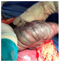
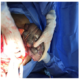
A 24-year-old Egyptian pregnant lady, gravida 1 and para 0, presented to our hospital with 2 days history of intermittent lower abdominal pain that has been exacerbated over the few hours prior to her presentation, severe abdominal distension, and absolute constipation. She had non-contributory past medical or surgical history with no previous abdominal or pelvic surgeries. Her pregnancy had been uneventful until that presentation. She reported taking up to 4grams/day of Acetaminophen at home with minimal pain relief. On examination, she was extremely agitated and uncomfortable, her pulse was 138 beats / minute, her blood pressure was 85/55mmHg, her respiratory rate was 32 breaths/minute and her temperature was 38.2C. Her abdominal examination revealed marked left lower quadrant tenderness, rebound tenderness and rigidity. Her laboratory investigations revealed leukocytosis with left shift (23.2x10^9/L), serum creatinine and BUN were 1.9mg/ dl and 42mg/dl respectively. Her serum lactate was 5.1mg/dl. Bedside ultrasound revealed turbid fluid reaching the hepatorenal angle. Aspiration of this fluid, with aseptic technique, under ultrasound guidance yielded a seropurulent discharge. Decision was taken for exploratory laparotomy hand in hand with IV fluid resuscitation and antibiotics (2gm of Ceftriaxone and 500 mg of Metronidazole). The patient’s Mean Arterial Pressure (MAP) was still 60mmHg despite adequate fluid replacement, so a loading dose of IV norepinephrine infusion was started at a rate of 10mcg/min followed by a maintenance dose of 6mcg/min to optimize patient’s Blood Pressure (BP). General surgery team was called for intraoperative attendance and patient was transferred to the operating room. A midline abdominal incision with supra-umbilical extension was performed. Upon entry into the peritoneal cavity, about 300ml of seropurulent exudates were found and suctioned. The sigmoid and transverse colon was markedly dilated and a picture of sigmoid volvulus was found. The dilated segment of the colon proximal to the site of obstruction friable, and gangrenous as shown in Figure 1 & 2. A lower segment caesarean section was done yielding a live female infant weighing 1950 grams whose APGAR scores were 8 at 1 minute and 9 at 5 minutes. Mobilization and resection of the dilated gangrenous segment of the colon was performed by the general surgery team and diverting colostomy with a Hartman’s procedure was done. 2 wide bore intra-peritoneal were placed and mass closure of the anterior abdominal wall was done. Delayed primary closure of the skin was undertaken 48 hours later after showing no signs of surgical site infection.
Figure 1: Showing dilated gangrenous colon proximal to site of obstruction.

Figure 2: Showing dilated gangrenous colon proximal to site of obstruction.

Post-operatively, the patient was kept on broad on broadspectrum antibiotics and was transferred to the Intensive Care Unit (ICU) for postoperative care. She was gradually weaned off cardiovascular support and had a smooth postoperative course. She was discharged home 10 days later.
Discussion
Intestinal obstruction has a low incidence in pregnancy ranging from 1 in 1500 to 1 in 66431 deliveries. Its causes during pregnancy include congenital or surgical adhesions, volvulus, intussusceptions, hernia, and appendicitis. Sigmoid volvulus is one of the most prevalent causes accounting for up to 24% of cases [4]. Pregnancy itself is thought to be the trigger for sigmoid volvulus where the gravid uterus may cause it by displacing, compressing, and partially obstructing a redundant or abnormally long sigmoid colon. This could explain why sigmoid volvulus occurs more frequently in the third trimester of pregnancy as shown in our case who presented at 34 weeks gestation. Despite the higher risk in the third trimester, reports of this problem occurring in the first trimester as well as the puerperium have been reported [5]. When a pregnant woman experiences a clinical triad of abdominal pain, distension, and absolute constipation, the diagnosis of sigmoid volvulus should be considered. It has been observed in literature, in match with our case, that the mean time from the onset of obstruction to clinical presentation is 48 hours. This is mainly due to the fact that pregnancy can conceal the clinical picture by causing abdominal pain, nausea, and leukocytosis in otherwise healthy women [6].
This delay in diagnosis and surgical intervention in pregnancy has a substantial impact on the maternal and fetal final outcomes. These outcomes have been directly related to the degree of bowel ischemia and subsequent sepsis. Literature has shown that all maternal deaths occured in patients who had a two-day or more delay in presentation and surgical intervention. Individuals who presented after 48 hours of onset of symptoms had 5 fetal deaths, compared to 2 fetal deaths in patients who presented early in the disease’s course. This finding emphasises the importance of a high degree of clinical suspicion in cases of intestinal obstruction in pregnant women. This information should be stressed to general practitioners and community obstetricians who are largely responsible for the treatment of these individuals [7]. Intestinal obstruction in pregnant women is treated in the same way as in nonpregnant women. Early surgical intervention is the cornerstone of treatment preferably via a midline vertical laparotomy. If adequate intestinal exposure cannot be acquired due to a larger gravid uterus in the third trimester, a caesarean section must be performed. The whole bowel should be meticulously explored for other sites of obstruction. Intestinal viability must be carefully checked and segmental resection with or without anastomosis is frequently required [8]
Conclusion
Sigmoid volvulus complicating pregnancy is extremely rare, however, when it occurs, may lead to considerable maternal and fetal morbidity and mortality. In patients who present with abdominal pain, distension, and absolute constipation, a high index of clinical suspicion is required for prompt diagnosis. Delayed diagnosis and treatment results in higher fetal and maternal morbidity and mortality especially after 48 hours. Therefore, early diagnosis and appropriate surgical intervention is crucial to improving maternal and fetal outcomes as shown in our case.
Author Contribution
P. Naseef was involved in data collection, interpretation and drafting the article. A. Abou Elnour and A. Ramy revised and edited the manuscript. The final version to be published was approved by all the authors.
Acknowledgement
None.
Conflict of Interest
The authors declare no conflict of interest.
Ethical Approval
This case report was approved by the Research Ethics Committee (REC) of Faculty of Medicine, Ain Shams University
Consent
Written informed consent was obtained from the patient to publish this report in accordance with the journal’s patient consent policy.
Funding Information
None.
References
- Perdue PW, Johnson HW, Stafford PW (1992) Intestinal obstruction complicating pregnancy. Am J Surg 164(4): 384-388.
- Madiba TE, Thomson SR (2000) The management of sigmoid volvulus. J R Coll Surg of Edinb 45(2): 74-80.
- Aftab Z, Toro A, Abdelaal A, Dasovky M, Gehani S, et al. (2014) Endoscopic reduction of a volvulus of the sigmoid colon in pregnancy: Case report and a comprehensive review of the literature. World Journal of Emergency Surgery 9(1): 1-6.
- Kolusari A, Kurdoglu M, Adali E, Yildizhan R, Sahin HG, et al. (2009) Sigmoid volvulus in pregnancy and puerperium: A case series. Cases J 2: 9275.
- Harer WB JR, Harer WB SR (1958) Volvulus complicating pregnancy and puerperium; report of three cases and review of literature. Obstet Gynecol 12(4): 399-406.
- Keating JP, Jackson DS (1985) Sigmoid volvulus in late pregnancy. J R Army Med Corps 131(2): 72-74.
- Iwamoto I, Miwa K, Fujino T, Douchi T (2007) Perforated colon volvulus coiling around the uterus in a pregnant woman with a history of severe constipation. J Obstet Gynaecol Res 33(5): 731-733.
- Redlich A, Rickes S, Costa SD, Wolff S (2007) Small bowel obstruction in pregnancy. Arch Gynecol Obstet 275(5): 381-383.
© 2022 Paul Naseef. This is an open access article distributed under the terms of the Creative Commons Attribution License , which permits unrestricted use, distribution, and build upon your work non-commercially.
 a Creative Commons Attribution 4.0 International License. Based on a work at www.crimsonpublishers.com.
Best viewed in


.jpg)


 Editorial Board Registrations
 Submit your Article
 Refer a Friend
 Advertise With Us
.jpg)


.jpg)


.bmp)
.jpg)
.png)
.jpg)


.jpg)


.png)

.png)


.png)


