

- About Us
- Information
-

The Author ensures that the research has been conducted responsibly and ethically with adherence to all relevant regulations. read more..
- For Authors
- For Reviewer
- Manuscript Guidelines
- Membership
- Publication Ethics
-
- Journals
- Reprints
- e-Books
- Videos
- Policies
- Contact Us
 COVID-19
- Submissions



Full Text
Advances in Complementary & Alternative medicine
NS2130E03, an Anthocyanin-Rich Purple Carrot Extract, Ameliorates Benzalkonium Chloride-Induced Dry Eye in Mice
Sameer Kamalakar Akolkar*, Mohan Gowda CM, Madhukumar MS and Anirudh Ranga
Natural & Essential Oils Private Limited, Mysuru, Karnataka, India
*Corresponding author:Sameer Kamalakar Akolkar, Natural & Essential Oils Private Limited, Mysuru, Karnataka, India
Submission: August 18, 2026;Published: September 04, 2026

ISSN: 2637-7802 Volume 9 Issue 3
Abstract
Dry Eye Disease (DED) is a multifactorial ocular surface disorder characterized by tear-film instability, hyperosmolarity, inflammation, and epithelial dysfunction. This study evaluated the protective and therapeutic effects of NS2130E03, a standardized anthocyanin-rich purple carrot (Daucus carota subsp. sativus) extract, in a Benzalkonium Chloride (BAC)-induced murine model of DED. Male BALB/c mice (n=36) were randomized into six groups (n=6/group): normal control, BAC-induced pathological control, cyclosporine A (0.05%), and NS2130E03 at 50, 150, or 500mg/kg/day. NS2130E03 was administered orally for 14 days, while 0.2% BAC was applied topically for 7 consecutive days. Tear secretion, tear-film stability, and tear osmolarity were assessed using the Schirmer test, Tear Break-Up Time (TBUT), and TearLab osmometry, respectively.
Serum IL-1β, IL-6, TNF-α, and MUC5AC were quantified by ELISA, and ocular tissues were evaluated histopathologically. BAC significantly impaired tear secretion and tear-film stability and increased tear osmolarity. NS2130E03 significantly improved these parameters dose-dependently (P<0.001). At 500mg/kg, tear secretion and TBUT recovered to approximately 92% and 90% of normal-control values, respectively, while tear osmolarity decreased by 28.6% versus the pathological control. IL-1β, IL-6, and TNF-α decreased by 58.2%, 54.7%, and 57.6%, respectively, while MUC5AC increased by 81.2% (P<0.001). Histopathology demonstrated dose-dependent preservation of ocular tissue architecture. NS2130E03 attenuated BAC-induced DED by improving tear-film parameters, reducing inflammation, enhancing MUC5AC-associated responses, and preserving ocular tissue architecture. These findings support further investigation of anthocyanin-rich purple carrot extract as a potential adjunctive strategy for DED.
Keywords:Dry eye disease; NS2130E03; Purple carrot anthocyanins; Daucus carota; Benzalkonium chloride; Tear-film stability; Ocular inflammation; MUC5AC
Abbreviations: ANOVA: One-way Analysis of Variance; BAC: Benzalkonium Chloride; DED: Dry Eye Disease; H&E: Hematoxylin and eosin; IAEC: Institutional Animal Ethics Committee; IL-1β: Interleukin- 1β; INL: Inner Nuclear Layer; MAPK: Mitogen-Activated Protein Kinase; MUC5AC: Mucin-5 Subtype AC; NF-κB: Nuclear Factor-κB; ONL: Outer Nuclear Layer; PSL: Photoreceptor Segment Layer; RGC: Retinal Ganglion Cell; ROS: Reactive Oxygen Species; TBUT: Tear Break-Up Time; TNF-α: Tumor Necrosis Factor-α
Introduction
Dry Eye Disease (DED) is a multifactorial disorder of the ocular surface characterized by loss of tear-film homeostasis, tear-film instability, hyperosmolarity, ocular surface inflammation, and neurosensory abnormalities. Although DED can arise from multiple interacting etiologies, disruption of the tear film and ocular surface initiates a self-perpetuating cycle in which tear evaporation and hyperosmolarity promote epithelial injury and inflammation, which in turn further destabilize the tear film [1,2]. Clinically, these alterations manifest as reduced tear secretion, shortened Tear Break-Up Time (TBUT), increased tear osmolarity, ocular surface epithelial damage, and discomfort or visual disturbance. Changes in tear-film mucins and inflammatory mediators are also closely associated with disease severity and ocular surface dysfunction [3]. Oxidative stress and inflammation are increasingly recognized as important components of DED pathophysiology. Excessive generation of Reactive Oxygen Species (ROS) can impair cellular antioxidant defenses, damage ocular surface epithelial cells, and promote activation of inflammatory signaling pathways. Inflammatory mediators, including Interleukin (IL)-1β, IL-6, and Tumor Necrosis Factor-α (TNF-α), contribute to epithelial injury and disruption of ocular surface homeostasis.
Persistent inflammation can additionally affect conjunctival goblet cells and mucin production, thereby compromising the protective and lubricating properties of the tear film [2]. Thus, therapeutic strategies capable of simultaneously counteracting oxidative stress and inflammation may provide broader protection of the ocular surface than approaches directed exclusively toward symptomatic tear replacement. Benzalkonium Chloride (BAC), a commonly used preservative in ophthalmic formulations, is known to exert dose-and exposure-dependent toxic effects on the ocular surface. Repeated topical exposure to BAC can induce epithelial damage, inflammatory responses, apoptosis, mucin depletion, and tear-film instability, making it a widely used experimental model for investigating DED and evaluating potential therapeutic interventions [4,5]. In particular, topical administration of 0.2% BAC in mice has been shown to produce characteristic dry-eyelike alterations, including reduced tear volume and tear breakup time, increased ocular surface inflammation, enhanced TNF-α expression, epithelial apoptosis, and reduction of MUC5AC-positive conjunctival cells [4].
These pathological changes reproduce several important features of human DED and provide a relevant platform for evaluating agents with ocular surface protective, anti-inflammatory, and tear-film stabilizing properties. Natural polyphenolic compounds have attracted considerable interest as potential adjunctive strategies for disorders associated with oxidative stress and chronic inflammation. Among these compounds, anthocyanins are flavonoid pigments widely distributed in fruits and vegetables and are recognized for their antioxidant and anti-inflammatory activities. Experimental evidence indicates that anthocyanins can scavenge reactive oxygen species and modulate inflammatory pathways, including Nuclear Factor-κB (NF-κB), Mitogen-Activated Protein Kinase (MAPK), and related signaling cascades involved in the production of pro-inflammatory cytokines [6,7]. Their ability to influence both oxidative and inflammatory processes provides a mechanistic rationale for investigating anthocyanin-rich botanical preparations in diseases in which oxidative stress and inflammation coexist.
Purple carrot (Daucus carota L.) is a particularly relevant source of anthocyanins and other phenolic constituents. Unlike conventional orange carrots, purple and black carrot varieties contain substantial concentrations of anthocyanins, which constitute a major proportion of their total anthocyanin content. Ferulic acid- and p-coumaric acid-acylated cyanidin derivatives are among the characteristic pigments identified in purple carrots [8]. Importantly, anthocyanin-rich purple carrots exhibit considerable antioxidant capacity, and the antioxidant activity of different purple carrot accessions has been positively associated with their anthocyanin and total phenolic contents [9]. The potential relevance of anthocyanins to ocular surface health is supported by emerging experimental and clinical evidence. Anthocyanin-rich preparations have demonstrated anti-inflammatory and antioxidant effects in biological systems, while a randomized, double-blind, placebocontrolled clinical study reported improvement in dry-eye-related outcomes following administration of an anthocyanin oligomer-rich grape skin extract [10]. Although these findings provide encouraging evidence for the potential of anthocyanin-containing preparations in DED, evidence specifically evaluating standardized anthocyaninrich purple carrot extracts in an established experimental dry-eye model remains limited.
Moreover, the effects of such preparations on tear secretion, tear-film stability, tear osmolarity, inflammatory cytokines, mucinassociated responses, and ocular tissue architecture have not been comprehensively characterized. NS2130E03 is a proprietary hydroalcoholic extract prepared from purple carrot (Daucus carota subsp. sativus) tubers and standardized by HPLC to contain ≥4.89% anthocyanins. The present study therefore investigated the protective and therapeutic effects of NS2130E03 in a BACinduced murine model of dry eye. The effects of three oral doses of NS2130E03 were evaluated using complementary functional and biochemical endpoints, including Schirmer tear testing, TBUT, tear osmolarity, serum IL-1β, IL-6 and TNF-α, MUC5AC levels, and histopathological assessment of ocular tissues. Cyclosporine A was included as a reference treatment. By integrating clinical surrogate measures with inflammatory and histopathological endpoints, this study aimed to determine whether standardized anthocyanin-rich purple carrot extract could attenuate BAC-induced ocular surface dysfunction and restore ocular tissue homeostasis
Materials and Methods
Plant material
NS2130E03, a proprietary natural herbal supplement used in this study was prepared from purple carrot (Daucus carota subsp. Sativus) tubers. It’s a hydroalcoholic extract, developed and named by Natural and Essential oils Pvt Ltd, Mysore, India. NS2130E03 is phytochemically standardized to Anthocyanins ≥4.89% by using HPLC method [11].
Animals
Male BALB/c mice (6-8 weeks old; 22-24g) were procured from ATNT Laboratories (Hyderabad, India). The animals were housed in standard polycarbonate cages under controlled environmental conditions (temperature, 23±3 °C; relative humidity, 30-70%; 12h light/12h dark cycle). Mice were provided with a standard laboratory diet (National Institute of Nutrition, India) and water ad libitum throughout the study. All the experimental procedures were conducted with the approval of the Institutional Animal Ethics Committee (IAEC) of Radiant Research Services Pvt. Ltd., Bangalore, India (Approval no.: RR/IEAC/162-2025) and experiments were conducted in accordance with the guidelines of Committee for the Control and Supervision of Experiments on Animals (CCSEA Registration Number-1803/PO/RcBi/S/2015/CCSEA).
Experimental design
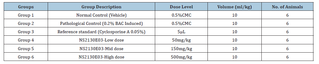
Following an acclimatization period of at least 6 days, thirtysix mice were randomly allocated into six experimental groups (n=6 per group). Group 1 served as the normal control, whereas Group 2 served as the pathological control and received 0.2% Benzalkonium Chloride (BAC) to induce dry eye. Group 3 served as the reference standard and was treated with 0.05% cyclosporine A ophthalmic solution (5μL/eye), administered topically to both eyes twice daily. Groups 4, 5, and 6 served as the treatment groups and received NS2130E03 orally at doses of 50, 150, and 500mg/kg body weight/day, respectively. Following a 7-day pretreatment period, experimental dry eye was induced by topical instillation of 0.2% BAC into the left eye of mice in Groups 2-6 for 7 consecutive days, while mice in the normal control group received an equal volume of physiological saline. Administration of the respective treatments (NS2130E03 at 50, 150, or 500 mg/kg/day and cyclosporine A in the reference standard group) was continued for an additional 7 days following BAC induction. On Day 21, all animals were euthanized. Blood samples were collected via the retro-orbital plexus for biochemical analyses, and both eyes were harvested for histopathological evaluation (Table 1).
Table 1:Experimental design.
Schirmer test
The Schirmer test was used to assess tear production in the mice. Schirmer strips were gently placed in the lateral canthus of the lower eyelid of each eye without anesthesia and left in place for 5 minutes. The length of the wetted area was measured in millimeters to assess tear secretion. The test was performed on day 7 before treatment, on day 14 after dry eye induction, and at the end of the treatment period on day 21 [12]. This allowed evaluation of the impact of Benzalkonium chloride in tear secretion as well as the therapeutic effects of the NS2130E03.
TBUT scoring
Tear Break-Up Time (TBUT) was measured to evaluate tear film stability. A 1-μL drop of 1% fluorescein sodium solution was instilled into the conjunctival sac of each eye using a micropipette. The mice were gently restrained and the tear film was observed using a slitlamp bio microscope equipped with a cobalt blue filter. The time interval between the last complete blink and the appearance of the first dry spot on the corneal surface was recorded in seconds. TBUT was assessed on day 7 before treatment, on day 14 after dry eye induction and at the end of the treatment period on day 21 to monitor the effect of the NS2130E03 on tear film stability [13].
Tear osmolarity
After the treatment period on day 21, tear osmolarity was measured using the Tear Lab osmometer. A single-use Tear Lab test card was carefully removed and taking care not to touch the collection port. The mouse was gently restrained, and the lower eyelid was retracted to expose the lateral tear meniscus. A small tear sample (~50nL) was collected by gently touching the collection port of the test card to the tear meniscus, avoiding direct contact with the eyelid margin or ocular surface. The device automatically analyzed the sample and displayed the tear osmolarity value in mOsm/L on the screen. After measurement, the test card was discarded [14].
Bio-markers analysis
At the end of the experimental period, the animals were anaesthetized with 2.8% isoflurane administered via an induction chamber before terminal blood collection. Following blood collection, the animals were humanely euthanized by Carbon Dioxide (CO₂) inhalation. Serum was separated by centrifugation at 6,000 rpm for 10 minutes and was analysed using a fully automated clinical chemistry analyser (MISPA Ace, Agappe Biomedicals Ltd. India) for the specified parameters. Serum levels of the proinflammatory cytokines Interleukin (IL)-1β, IL-6, Tumour Necrosis Factor-α (TNF-α), and mucin-5 subtype AC (MUC5AC) were quantified using commercially available mouse ELISA kits according to the manufacturers’ instructions. IL-1β (Cat. No. E0192Mo), IL-6 (Cat. No. E0049Mo), TNF-α (Cat. No. E0117Mo), and MUC5AC (Cat. No. E0169Mo) ELISA kits were purchased from BT Labs (China). The absorbance was measured using a BioTek ELISA microplate reader.
Histopathological examination of mouse eyes
Eyes were carefully enucleated from euthanized mice, and a small scleral window was created to facilitate fixative penetration and preserve ocular morphology. The tissues were fixed in Modified Davidson’s (Hartman’s) fixative (20:1 fixative-to-tissue ratio) for 20h at room temperature (20-25 °C) with gentle agitation. After fixation, samples were washed with phosphate-buffered saline (PBS) and dehydrated through a graded ethanol series (30%, 50%, 70%, and 100%). Tissues were then processed in an automatic tissue processor, embedded in paraffin, and sectioned at 5μm using a rotary microtome. Sections were mounted on glass slides, dried overnight, deparaffinized in xylene, and rehydrated through graded ethanol. Hematoxylin and Eosin (H&E) staining was performed using an automated slide stainer by loading the slides and running a pre-set eye staining program that utilized the appropriate reagents and dyes. Finally, slides were mounted with a permanent mounting medium and examined under a light microscope for histopathological evaluation [15].
Statistical analysis
All data, including body weight and biomarker measurements, were statistically analyzed using GraphPad Prism software (version 5.01). Data are presented as the mean±Standard Deviation (SD). Statistical differences between the treatment groups and the pathological control group were evaluated using one-way Analysis of Variance (ANOVA) followed by Dunnett’s multiple comparisons test. The results of the statistical analyses are summarized in the corresponding tables. A value of P<0.05 was considered statistically significant.
Results
Effect of NS2130E03 on tear secretion in the BACinduced dry eye model
Tear secretion was quantified using the Schirmer test to evaluate lacrimal gland function and ocular surface hydration (Figure 1). On Day 7, prior to BAC administration, tear production was comparable among all experimental groups (5.48-5.53mm), indicating the absence of any treatment-related effects on basal tear secretion. Following BAC-induced dry eye (Day 14), the pathological control group exhibited a pronounced reduction in tear production (2.45±0.10mm), representing a 56.1% decrease compared with the normal control group (5.58±0.42mm), confirming successful establishment of the dry eye model. In contrast, pretreatment with NS2130E03 significantly preserved tear secretion relative to the BAC group. Tear production increased by 35.9%, 38.0%, and 43.7% in the low-dose (50mg/kg), mid-dose (150mg/kg), and high-dose (500mg/kg) groups, respectively, while the Cyclosporine A increased tear secretion by 45.7% (all P<0.001 vs. pathological control). Nevertheless, tear secretion in all BAC-treated groups remained below that observed in the normal control animals.
Figure 1:Effect of NS2130E03 on tear secretion in the BAC-induced dry eye model. Schirmer tear test values were measured on Day 7 (before treatment), Day 14 (following BAC-induced dry eye), and Day 21 (following treatment). Data are presented as mean±SD (n=6). Statistical comparisons were performed between the pathological control group (BAC-induced) and treatment groups (NS2130E03-low, mid and high dose, respectively). ***P < 0.001. Higher Schirmer test values indicate increased tear secretion. NS2130E03 treatment progressively restored tear secretion following BAC-induced reduction, with the high-dose group showing the greatest recovery. BAC; Benzalkonium chloride.
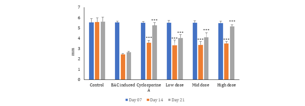
Following seven days of treatment after BAC induction (Day 21), NS2130E03 produced a marked recovery of tear secretion compared with the pathological control group (P<0.001). Tear production increased from 2.65±0.10mm in the pathological control group to 4.05±0.31mm, 4.10±0.45mm, and 5.17±0.16mm in the low, mid, and high-dose NS2130E03 groups, corresponding to 52.8%, 54.7%, and 95.1% improvements, respectively. The Cyclosporine A restored tear secretion to 5.25±0.28mm, representing a 98.1% improvement over the pathological control. Among the NS2130E03-treated groups, the 500mg/kg dose produced the greatest restoration of tear secretion, achieving 91.8% of the tear production observed in the normal control group (5.17 vs. 5.63mm), whereas the 50 and 150mg/kg doses restored approximately 71.9% and 72.8%, respectively, of normal tear secretion. Although the Cyclosporine A exhibited the highest recovery (93.3% of normal control), the highdose NS2130E03 group demonstrated a comparable therapeutic effect. Overall, these findings indicate that NS2130E03 effectively attenuated BAC-induced lacrimal dysfunction and promoted recovery of tear secretion in a dose-dependent manner, with the 500mg/kg dose providing the greatest protection against BACinduced dry eye.
Effect of NS2130E03 on Tear Break-Up Time (TBUT) in the BAC-induced dry eye model
Tear film stability was evaluated by measuring TBUT at baseline (Day 7), following benzalkonium chloride (BAC)-induced dry eye (Day 14), and after the treatment period (Day 21). On Day 7, TBUT values were comparable among all experimental groups (7.50- 7.83s), indicating that pretreatment with NS2130E03 did not alter physiological tear film stability prior to BAC exposure (Figure 2). Following BAC induction, the pathological control group exhibited a marked reduction in TBUT (2.17±0.41s), representing a 72.9% decrease compared with the normal control group (8.00±0.63s), confirming successful induction of tear film instability. In contrast, all NS2130E03-treated groups demonstrated significantly higher TBUT values than the pathological control group (P<0.001), indicating attenuation of BAC-induced tear film disruption. Compared with the pathological control, TBUT increased by 92.2%, 99.5%, and 115.2% in the low-dose (4.17±0.75s), mid-dose (4.33±0.82s), and high-dose (4.67±1.03s) NS2130E03 groups, respectively. The Cyclosporine A group also showed a 115.2% improvement (4.67±0.82s).
Figure 2:Effect of NS2130E03 on Tear Break-Up Time (TBUT) in the BAC-induced dry eye model. TBUT was assessed on Days 7, 14, and 21 to evaluate tear-film stability before induction, following BAC-induced ocular surface damage, and after treatment, respectively. Data are presented as mean±SD (n=6). Statistical comparisons were performed between the pathological control group (BAC-induced) and treatment groups (NS2130E03-low, mid and high dose, respectively). ***P<0.001. Higher TBUT values indicate improved tear-film stability. NS2130E03 treatment increased TBUT compared with the pathological control, indicating restoration of tear-film stability. BAC; Benzalkonium Chloride.
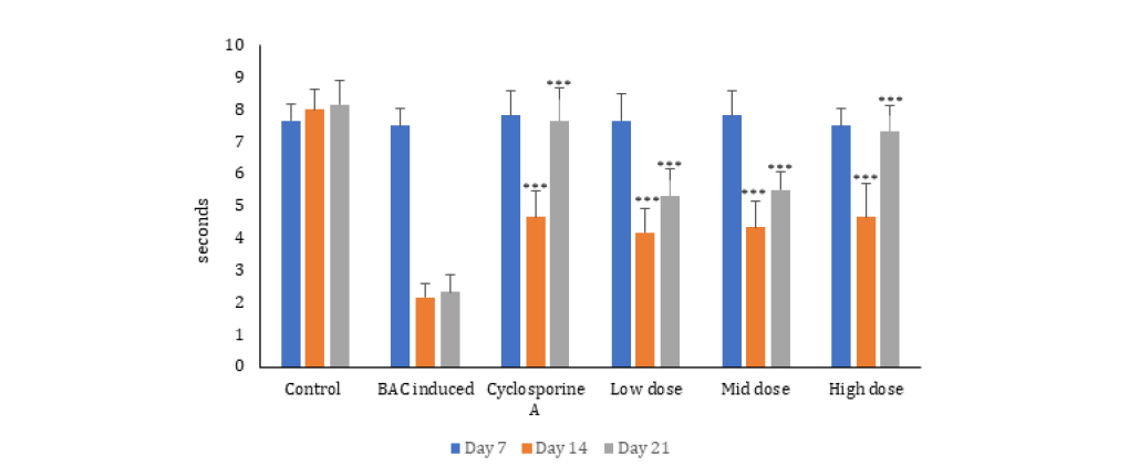
Following continued treatment until Day 21, TBUT remained significantly higher in all NS2130E03-treated groups and the Cyclosporine A group than in the pathological control group (P<0.001). The pathological control group maintained severe tear film instability (2.33±0.52s), whereas NS2130E03 treatment produced a dose-dependent restoration of tear film stability. Relative to the pathological control, TBUT increased by 128.8%, 136.1%, and 214.6% in the low-dose (5.33±0.82 s), mid-dose (5.50±0.55s), and high-dose (7.33±0.82s) groups, respectively. The Cyclosporine A group demonstrated a comparable 229.2% improvement (7.67±1.03s). Furthermore, the high-dose NS2130E03 group restored TBUT to approximately 89.7% of the normal control value (7.33 vs. 8.17s), indicating near-complete recovery of tear film stability. These findings demonstrate that NS2130E03 effectively mitigated BAC-induced tear film instability and promoted a dose-dependent restoration of tear film integrity. The pronounced improvement observed in the high-dose group, which approached the efficacy of the Cyclosporine A, suggests that NS2130E03 preserves tear film homeostasis and protects the ocular surface against dry eye-associated damage.
Effect of NS2130E03 on tear osmolarity in the BACinduced dry eye model
Tear osmolarity was evaluated on Day 21 to determine the effect of the NS2130E03 on tear film homeostasis following BACinduced ocular surface damage (Figure 3). The pathological control group exhibited a marked increase in tear osmolarity (371.67±22.45mOsm/L) compared with the normal control group (240.17±12.21 mOsm/L; 35.4% lower, P<0.001), confirming the development of tear film hyperosmolarity and dry eye pathology. Treatment with the Cyclosporine A significantly restored tear film osmolarity, reducing tear osmolarity to 253.67±12.89mOsm/L, corresponding to a 31.7% reduction compared with the pathological control (P<0.001). Similarly, administration of the NS2130E03 significantly attenuated BAC-induced hyperosmolarity in a dosedependent manner.
Figure 3:Effect of NS2130E03 on tear osmolarity in the BAC-induced dry eye model. Tear osmolarity was measured at the end of the experimental period to assess alterations in tear-film homeostasis. Data are presented as mean±SD (n=6). Statistical comparisons were performed between the pathological control group (BAC-induced) and treatment groups (NS2130E03-low, mid and high dose, respectively). ***P<0.001. Lower tear osmolarity values indicate improved tear-film homeostasis. NS2130E03 treatment significantly reduced BAC-induced tear hyperosmolarity compared with the pathological control group. BAC; Benzalkonium chloride.
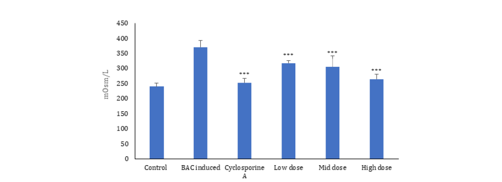
The low-dose group (318.50±8.69mOsm/L) showed a 14.3% reduction in tear osmolarity compared with the pathological control (P<0.001), whereas the mid-dose group (306.17±36.83mOsm/L) demonstrated a 17.6% reduction (P<0.001). The high-dose group exhibited the greatest improvement, with tear osmolarity decreasing to 265.50±15.72mOsm/L, representing a 28.6% reduction relative to the pathological control (P<0.001). Notably, the high-dose treatment restored tear osmolarity to a level approaching that of the normal control, with only a 10.5% higher osmolarity than the control group, indicating substantial preservation of tear film integrity. Overall, these findings demonstrate that the NS2130E03 effectively mitigated BAC-induced tear film hyperosmolarity in a dose-dependent manner, with the high dose producing efficacy comparable to the Cyclosporine A.
Effect of the NS2130E03 on serum pro-inflammatory cytokines and MUC5AC levels
Serum concentrations of the pro-inflammatory cytokines IL-1β, IL-6, and TNF-α, together with MUC5AC levels, were evaluated to assess systemic inflammatory responses associated with experimental dry eye disease (Table 2). The pathological control group exhibited a marked increase in IL-1β, IL-6, and TNF-α levels, accompanied by a substantial reduction in MUC5AC compared with the normal control group, confirming successful induction of ocular surface inflammation. Treatment with the Cyclosporine A significantly attenuated the inflammatory response, reducing IL-1β, IL-6, and TNF-α levels by 60.2%, 61.5%, and 61.0%, respectively, compared with the pathological control group (P<0.001). In parallel, MUC5AC levels increased by 95.4%, approaching those observed in the normal control group. Administration of the NS2130E03 produced a dose-dependent improvement in inflammatory biomarkers. The low-dose group (50mg/kg) significantly reduced IL-1β, IL-6, and TNF-α levels by 22.7%, 27.9%, and 26.7%, respectively (P<0.001), while increasing MUC5AC by 51.6% compared with the pathological control group.
Table 2:Effect of the NS2130E03 on Serum Pro-inflammatory Cytokines and MUC5AC Levels.
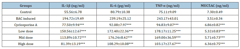
Values are presented as mean±SD (n=6). Serum concentrations of Interleukin-1β (IL-1β), Interleukin-6 (IL-6), tumor necrosis factor-α (TNF-α), and MUC5AC were determined at the end of the experimental period. Statistical comparisons were performed between the pathological control group (BAC-induced) and treatment groups (NS2130E03-low, mid and high dose, respectively). ***P<0.001. Higher IL-1β, IL-6, and TNF-α levels indicate an enhanced pro-inflammatory response, whereas reduced MUC5AC levels are indicative of impaired mucin-associated tear-film protection. NS2130E03 treatment reduced the BAC-induced elevations in pro-inflammatory cytokines and restored MUC5AC levels toward those observed in the control group. BAC; Benzalkonium Chloride.
The mid-dose group (150 mg/kg) produced greater reductions in IL-1β (41.5%) and TNF-α (30.5%), whereas IL-6 was reduced by 26.3% relative to the pathological control group (P<0.001). In addition, MUC5AC levels increased by 62.7%, indicating partial restoration of tear film mucin production. The high-dose group (500mg/kg) demonstrated the greatest therapeutic efficacy among all treatment groups. IL-1β, IL-6, and TNF-α concentrations were significantly reduced by 58.2%, 54.7%, and 57.6%, respectively, compared with the pathological control group (P<0.001). Concurrently, MUC5AC levels increased by 81.2%, indicating substantial recovery of ocular surface mucin secretion. The magnitude of improvement observed with the high dose closely approached that of the Cyclosporine A, demonstrating robust antiinflammatory activity and restoration of ocular surface homeostasis. Overall, these findings demonstrate that the NS2130E03 effectively suppressed dry eye-associated inflammatory cytokines while restoring MUC5AC expression in a dose-dependent manner.
Effect of NS2130E03 treatment on retinal histopathological alterations induced by BAC
Histopathological examination of retinal sections demonstrated that repeated topical administration of 0.2% BAC induced marked structural damage in the retina. Compared with the normal control group, the pathological control group exhibited pronounced thinning of the Outer Nuclear Layer (ONL), Inner Nuclear Layer (INL), and Photoreceptor Segment Layer (PSL), accompanied by inflammatory cell infiltration within the Retinal Ganglion Cell (RGC) layer and severe disruption of the normal retinal architecture (Figure 4). The photoreceptor nuclei appeared sparse and disorganized, indicating substantial retinal degeneration. Treatment with the Cyclosporine A and the NS2130E03 resulted in dose-dependent restoration of retinal morphology. The retinal layers exhibited progressively improved structural organization with better preservation of the ONL, INL, PSL, and RGC layers compared with the pathological control group.
Figure 4:Histopathological evaluation of retinal morphology following NS2130E03 treatment in the BAC-induced retinal injury model. Representative Hematoxylin and Eosin (H&E)-stained sections of the retina from (a) normal control, (b) BAC-induced pathological control, (c) cyclosporine A reference standard, (d) NS2130E03 low-dose, (e) NS2130E03 mid-dose, and (f) NS2130E03 high-dose groups. The normal control retina exhibited preserved retinal lamination and morphology, whereas the BAC-induced pathological control showed oedematous changes and inflammation accompanied by a reduction in the Inner Nuclear Layer (INL). The cyclosporine A reference group showed restoration of normal retinal morphology and preserved retinal laminar organization. NS2130E03-treated groups demonstrated comparatively preserved retinal architecture across the low-, mid-, and high-dose groups. RGC, retinal ganglion cell layer; INL, Inner Nuclear Layer; ONL, Outer Nuclear Layer (photoreceptor cell layer); PSL, Photoreceptor Segment Layer; RPE, Retinal Pigment Epithelium. Black scale bar = 50μm. BAC; Benzalkonium Chloride.
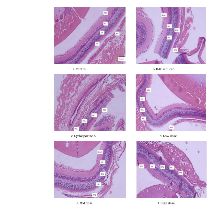
The photoreceptor nuclei appeared more densely packed and regularly aligned, while inflammatory changes were markedly reduced, particularly in the mid- and high-dose treatment groups. The high-dose group demonstrated retinal architecture that closely resembled that of the normal control, suggesting substantial protection against BAC-induced retinal injury. Quantitative morphometric analysis corroborated the histopathological observations (Table 3). The pathological control group showed severe reductions in retinal layer thickness compared with the normal control. Treatment with the Cyclosporine A significantly restored retinal thickness, increasing ONL, INL, and PSL thicknesses by 68.1%, 111.8%, and 174.3%, respectively, relative to the pathological control (p<0.001). Among the NS2130E03 treated groups, the low-dose treatment produced only modest improvement, with 7.0%, 19.7%, and a 16.8% reduction in ONL, INL, and PSL thickness, respectively, compared with the pathological control, with statistical significance observed only for INL (p<0.05).
Table 3:Effect of NS2130E03 on retinal layer thickness in the BAC-induced retinal injury model.

Retinal thickness of the Outer Nuclear Layer (ONL), Inner Nuclear Layer (INL), and Photoreceptor Segment Layer (PSL) was evaluated across the experimental groups. Data are presented as mean±SD. Statistical comparisons were performed between the BAC-induced pathological control group and the respective treatment groups. *P<0.05 and ***P<0.001 versus the BAC-induced pathological control group. NS2130E03 treatment increased the thickness of the retinal layers compared with the BAC-induced pathological control, with the high-dose group showing the greatest restoration toward control values. BAC; Benzalkonium Chloride.
In contrast, the mid-dose treatment significantly restored retinal morphology, increasing ONL, INL, and PSL thickness by 27.4%, 58.7%, and 45.1%, respectively (p<0.001). The greatest protective effect was observed in the high-dose group, which exhibited significant increases of 52.8%, 101.1%, and 153.3% in ONL, INL, and PSL thickness, respectively, compared with the pathological control (p<0.001). Notably, the retinal layer thicknesses in the high-dose group approached those observed in the normal control and Cyclosporine A groups, indicating nearcomplete restoration of retinal structural integrity following treatment. Overall, both qualitative histopathological assessment and quantitative morphometric measurements consistently demonstrated that the NS2130E03 effectively attenuated BACinduced retinal degeneration in a dose-dependent manner, with the high-dose treatment providing retinal protection comparable to that of the Cyclosporine A.
Discussion
Dry Eye Disease (DED) is a multifactorial ocular surface disorder in which disruption of tear-film homeostasis, tear-film instability, hyperosmolarity, epithelial damage, and inflammation interact in a self-perpetuating cycle. According to the TFOS DEWS II framework, tear-film instability and hyperosmolarity can promote epithelial and goblet-cell damage, which subsequently reduces surface wettability and accelerates tear-film breakup, further aggravating hyperosmolarity and inflammation [1-3]. The present study demonstrates that NS2130E03, a hydroalcoholic extract of purple carrot (Daucus carota subsp. sativus) standardized to ≥4.89% anthocyanins, attenuated several interconnected manifestations of BAC-induced experimental dry eye. The beneficial effects were evident across tear secretion, tear-film stability, tear osmolarity, inflammatory cytokines, MUC5AC, and ocular histopathology, with the 500mg/kg dose consistently producing the greatest response.
The experimental model was successfully established by repeated topical administration of 0.2% BAC. Compared with normal animals, the pathological control group demonstrated a pronounced reduction in Schirmer tear values and TBUT together with a substantial increase in tear osmolarity. These changes are consistent with the established characteristics of BAC-induced ocular surface injury. Lin et al. demonstrated that topical 0.2% BAC administration in mice produces decreased tear volume and tear break-up time, increased ocular surface inflammation, enhanced TNF-α expression, epithelial apoptosis, and a reduction in MUC5ACpositive conjunctival cells [16]. Similarly, the TFOS DEWS II reports recognize tear-film instability, increased osmolarity, epithelial injury, and inflammation as interconnected components of DED pathophysiology [1,2]. Thus, the marked changes observed in the present pathological control group indicate that the BAC protocol produced a robust experimental dry-eye phenotype.
A major finding of the present study was the ability of NS2130E03 to restore tear secretion. Following BAC induction, tear production was substantially reduced, whereas treatment with NS2130E03 significantly increased Schirmer values at all tested doses. By Day 21, the 50, 150, and 500mg/kg doses produced improvements of 52.8%, 54.7%, and 95.1%, respectively, relative to the pathological control. The high-dose group restored tear production to approximately 92% of the normal-control value and produced an effect comparable to that of cyclosporine A. These findings suggest that NS2130E03 can attenuate BAC-associated impairment of tear secretion and contribute to restoration of ocular surface hydration. Importantly, the progressive improvement with increasing dose provides evidence of a dose-responsive biological effect rather than an isolated change in tear production.
The improvement in tear secretion was accompanied by a marked recovery of tear-film stability. BAC reduced TBUT to approximately 2.3 seconds, whereas NS2130E03 increased TBUT to 5.33, 5.50, and 7.33 seconds at 50, 150, and 500mg/kg, respectively. The highest dose therefore restored TBUT to approximately 90% of the normal-control value and was comparable to cyclosporine A. Tear-film stability is a critical determinant of ocular surface homeostasis, and rapid tear-film breakup is a characteristic feature of DED [3]. The concurrent improvement in Schirmer values and TBUT is therefore important because it suggests that NS2130E03 did not merely increase tear volume but also improved the functional stability of the tear film. The reduction in tear osmolarity provides additional evidence for restoration of tear-film homeostasis. BAC-induced animals exhibited markedly elevated tear osmolarity, whereas NS2130E03 reduced osmolarity in a dosedependent manner.
The 500mg/kg dose reduced tear osmolarity by 28.6% compared with the pathological control, bringing the value close to that of normal animals. Hyperosmolarity is considered an important pathogenic driver of DED because it can directly induce cellular stress and promote inflammatory responses, thereby amplifying ocular surface damage [2]. Therefore, the simultaneous improvement in tear secretion, TBUT, and osmolarity suggests that NS2130E03 acts across several interconnected components of tearfilm dysfunction. The inflammatory findings further support this interpretation. BAC-induced dry eye was associated with substantial elevations of IL-1β, IL-6, and TNF-α. These cytokines are important mediators of ocular surface inflammation and are involved in the amplification of epithelial injury and disruption of ocular surface homeostasis. Treatment with NS2130E03 significantly reduced all three cytokines, with the greatest effect observed at 500mg/kg. At this dose, IL-1β, IL-6, and TNF-α were reduced by 58.2%, 54.7%, and 57.6%, respectively, relative to the pathological control.
These reductions approached those observed with cyclosporine A, which produced reductions of approximately 60-62%. The close similarity between the high-dose NS2130E03 and cyclosporine A responses suggests that suppression of inflammatory activity may contribute substantially to the ocular surface benefits observed with the extract. The anti-inflammatory response is particularly relevant because DED is not simply a disorder of inadequate tear production. Rather, inflammation and hyperosmolarity interact to maintain a vicious cycle of ocular surface injury [2]. Accordingly, an intervention capable of simultaneously improving tear-film parameters and reducing inflammatory mediators may provide broader protection than an intervention directed solely at tear replacement. The present results are consistent with this concept, as the improvements in Schirmer test and TBUT were accompanied by reductions in inflammatory cytokines rather than occurring independently of the inflammatory response.
The restoration of MUC5AC provides another potential link between the anti-inflammatory effects of NS2130E03 and the improvement in tear-film stability. Mucins contribute to the wettability and stability of the ocular surface, and BAC exposure has previously been shown to reduce MUC5AC-positive conjunctival cells in mice [16]. In the present study, BAC-induced dry eye was associated with a marked reduction in MUC5AC, whereas NS2130E03 increased MUC5AC levels by 51.6%, 62.7%, and 81.2% at 50, 150, and 500mg/kg, respectively. The restoration of MUC5AC was therefore broadly consistent with the improvement in TBUT and tear-film stability. However, because MUC5AC in the present study was quantified in serum rather than directly assessed in conjunctival goblet cells or ocular-surface tissue, these findings should be interpreted as evidence of a mucin-associated response rather than direct proof of restoration of conjunctival MUC5AC secretion. Future studies incorporating conjunctival MUC5AC immunostaining and goblet-cell density measurements would provide stronger evidence for this mechanism.
The biological activity of NS2130E03 may plausibly be related to its anthocyanin-rich composition. Purple carrot is recognized as a source of anthocyanins, particularly cyanidin derivatives, and anthocyanin-rich botanical preparations have demonstrated antioxidant and anti-inflammatory activities in experimental systems. Such properties are potentially relevant to DED because oxidative stress and inflammation are closely interconnected in ocular surface injury. However, oxidative-stress markers and specific intracellular inflammatory signaling pathways were not directly measured in the present study. Therefore, although modulation of oxidative and inflammatory pathways provides a plausible explanation for the observed activity of an anthocyaninrich extract, the current findings should not be interpreted as direct evidence of NF-κB, MAPK, NLRP3, or other specific pathway modulation. The present findings are nevertheless consistent with emerging evidence that anthocyanin-rich preparations may have beneficial effects on dry-eye-related outcomes. Fan et al. [10] reported in a randomized, double-blind, placebo-controlled clinical study that oral administration of an anthocyanin oligomer-rich grape skin extract improved dry-eye-related clinical outcomes.
Although grape skin and purple carrot differ in their botanical origin and anthocyanin composition, this clinical evidence provides relevant support for the concept that orally administered anthocyanin-rich preparations may influence ocular surface function. The current study extends this evidence by demonstrating a dose-dependent response to a chemically standardized anthocyanin-rich purple carrot extract in a controlled animal model and by examining multiple complementary endpoints. An additional and noteworthy observation was the preservation of ocular tissue architecture following NS2130E03 treatment. Histopathological examination of BAC-treated animals demonstrated disruption of retinal architecture, including thinning of the ONL, INL, and PSL and inflammatory changes. NS2130E03 attenuated these alterations in a dose-dependent manner. The 500mg/kg dose increased ONL, INL, and PSL thickness by 52.8%, 101.1%, and 153.3%, respectively, compared with the pathological control. The magnitude of recovery was particularly pronounced in the INL and PSL, and the overall retinal architecture in the high-dose group approached that observed in the normal and cyclosporine A groups.
The retinal findings suggest that the protective effects of NS2130E03 may extend beyond the measurable tear-film abnormalities. However, these results should be interpreted cautiously because the experimental model was primarily designed to investigate BAC-induced dry-eye pathology, and the study did not directly measure retinal oxidative stress, apoptosis, vascular alterations, or retinal-specific inflammatory signaling. Consequently, the present histopathological findings demonstrate structural preservation but do not establish a direct molecular mechanism of retinal protection. Additional investigations using retinal markers of oxidative stress, apoptosis, inflammation, and photoreceptor integrity would be necessary to determine whether the retinal effects represent a direct action of NS2130E03 or are secondary to attenuation of the broader ocular inflammatory environment. The dose-response pattern observed across the study is another important consideration. The 50mg/kg dose produced measurable but comparatively modest improvements, whereas the 150mg/kg dose produced intermediate effects. The 500mg/kg dose consistently demonstrated the strongest response across tear secretion, TBUT, tear osmolarity, cytokines, MUC5AC, and retinal morphology.
This concordance across independent endpoints strengthens the overall interpretation that NS2130E03 exerts a biologically meaningful protective effect in this model. Moreover, the similarity between the high-dose NS2130E03 group and cyclosporine A for several endpoints is noteworthy. Nevertheless, the present study was not designed as a formal non-inferiority or equivalence study; therefore, the results should be described as demonstrating a response approaching that of the reference treatment rather than establishing therapeutic equivalence. Several aspects of the study strengthen the relevance of these findings. The experimental design incorporated multiple complementary measures of DED, including tear secretion, tear-film stability, tear osmolarity, inflammatory cytokines, MUC5AC, and histopathological evaluation. This multidimensional assessment is consistent with the multifactorial nature of DED and reduces the likelihood that the observed activity is attributable to improvement in a single endpoint. Furthermore, the inclusion of cyclosporine A provided a pharmacological reference for interpreting the magnitude of the response.
The use of a standardized extract containing ≥4.89% anthocyanins also provide greater compositional definition than investigations using chemically uncharacterized botanical preparations. The study has several limitations that should be considered when interpreting the findings. First, the number of animals per group was relatively small (n=6), and the study was performed exclusively in male BALB/c mice. Sex-related biological differences are relevant to DED, and extrapolation to female animals or other strains should therefore be undertaken cautiously. Second, the study did not directly examine the molecular pathways underlying the anti-inflammatory or tissue-protective effects of NS2130E03. Finally, the use of serum cytokines and MUC5AC provides systemic rather than strictly ocular-surface-specific information. Future studies should therefore incorporate local ocular-surface cytokine measurements, conjunctival goblet-cell analysis, corneal epithelial integrity, oxidative-stress biomarkers, and mechanistic signaling studies.
Conclusion
Taken together, the findings indicate that NS2130E03 produces a broad protective effect against BAC-induced dry-eye-associated dysfunction. The extract improved tear secretion and tear-film stability, reduced tear hyperosmolarity, attenuated IL-1β, IL-6, and TNF-α elevations, increased MUC5AC-associated responses, and preserved ocular tissue morphology. These effects were generally dose dependent, with 500mg/kg producing the most pronounced response and approaching the effects of cyclosporine A across several endpoints. Given the known role of tear-film instability, hyperosmolarity, inflammation, and epithelial/gobletcell dysfunction in DED, the coordinated improvement of these parameters suggests that NS2130E03 may act through a multimodal mechanism rather than through a single pharmacological target. The anthocyanin-rich composition of the extract provides a plausible basis for these effects, although direct mechanistic studies are required to establish the pathways involved. Overall, the present findings provide preclinical evidence supporting further investigation of standardized anthocyanin-rich purple carrot extract as a potential nutritional or adjunctive approach for the management of dry-eye-associated ocular surface dysfunction.
Acknowledgement
The authors gratefully acknowledge Radiant Research Services Pvt. Ltd. for their contributions to this study..
Conflict of Interest
The authors declare that they have no conflicts of interest related to this work.
Author Contributions
Conceptualization and preclinical studies: Sameer Kamalakar
Akolkar.
Manuscript writing: Mohan Gowda C M
Extraction and Analytical: Madhukumar M.S.
Manuscript review: Anirudh Ranga
Competing Interests
Sameer Kamalakar Akolkar, Mohan Gowda C M, Madhukumar M S, Anirudh Ranga, are employed with Natural and Essential Oils Private Limited, Mysuru, Karnataka, India. This does not alter our adherence to Complimentary and Alternative Medicine policies on sharing data and materials.
Funding
The study was sponsored by Natural & Essential Oils Pvt. Ltd., Mysuru, India.
References
- Craig JP, Nichols KK, Akpek EK, Caffery B, Dua HS, et al. (2017) TFOS DEWS II definition and classification report. Ocul Surf 15(3): 276-283.
- Bron AJ, Paiva CSD, Chauhan SK, Bonini S, Gabison EE, et al. (2017) TFOS DEWS II pathophysiology report. Ocul Surf 15(3): 438-510.
- Willcox MDP, Argueso P, Georgiev GA, Holopainen JM, Laurie GW, et al. (2017) TFOS DEWS II tear film report. Ocul Surf 15(3): 366-403.
- Baudouin C, Labbe A, Liang H, Pauly A, Baudouin FB (2010) Preservatives in eyedrops: The good, the bad and the ugly. Prog Retin Eye Res 29(4): 312-334.
- Xiong C, Chen D, Liu J, Liu B, Li N, et al. (2008) A rabbit dry eye model induced by topical medication of a preservative benzalkonium chloride. Invest Ophthalmol Vis Sci 49(5): 1850-1856.
- Khoo HE, Azlan A, Tang ST, Lim SM (2017) Anthocyanidins and anthocyanins: Colored pigments as food, pharmaceutical ingredients, and the potential health benefits. Food Nutr Res 61(1): 1361779.
- Li D, Wang P, Luo Y, Zhao M, Chen F (2021) Health benefits of anthocyanins and molecular mechanisms: Update from recent studies. Crit Rev Food Sci Nutr 57(8): 1729-1741.
- Perez MB, Carvajal S, Beretta V, Bannoud F, Fangio MF, et al. (2023) Characterization of purple carrot germplasm for antioxidant capacity and root concentration of anthocyanins, phenolics, and carotenoids. Plants (Basel) 12(9): 1796.
- Sun T, Simon PW, Tanumihardjo SA (2009) Antioxidant phytochemicals and antioxidant capacity of biofortified carrots (Daucus carota) of various colors. J Agric Food Chem 57(10): 4142-4147.
- Fan M, Kim SA, Choi YJ, Tang Y, Yang HP, et al. (2023) Anthocyanin oligomer (grape skin extract) administration improves dry eye disease: a randomised, double-blind, placebo-controlled study. Clin Exp Ophthalmol 51(2): 122-130.
- Zaim M, Kara I, Muduroglu A (2021) Black carrot anthocyanins exhibit neuroprotective effects against MPP⁺-induced cell death and cytotoxicity via inhibition of oxidative stress-mediated apoptosis. Cytotechnology 73(6): 827-840.
- Nemet A, Belkin M, Rosner M (2007) Transplantation of newborn lacrimal gland cells in a rat model of reduced tear secretion. Isr Med Assoc J 9(2): 94-98.
- Kilic S, Kulualp K (2016) Efficacy of several therapeutic agents in a murine model of dry eye syndrome. Comp Med 66(2): 112-118.
- Marques DL, Alves M, Modulo CM, Silva LE, Reinach P (2015) Lacrimal osmolarity and ocular surface in experimental model of dry eye caused by toxicity. Rev Bras Oftalmol 74(2): 68-72.
- Pang J, Thomas N, Tsuchiya D, Parmely T, Yan D, et al. (2021) Step-by-step preparation of mouse eye sections for routine histology, immunofluorescence, and RNA in situ hybridization multiplexing. STAR Protoc 2(4): 100879.
- Lin Z, Liu X, Zhou T, Wang Y, Bai L, et al. (2011) A mouse dry eye model induced by topical administration of benzalkonium chloride. Mol Vis 17: 257-264.
© 2026 Sameer Kamalakar Akolkar. This is an open access article distributed under the terms of the Creative Commons Attribution License , which permits unrestricted use, distribution, and build upon your work non-commercially.
Editor In Chief







.jpg)






























Signup for Newsletter

Quick Links
 Editorial Board Registrations
 Submit your Article
 Best Paper of the Volume
 Reprints
 Refer a Friend
 Advertise With Us
Our Recent Edition
.jpg)


Top Editors




.jpg)

















.bmp)
.jpg)
.png)
.jpg)













.jpg)






.png)

.png)




.png)




Financial Support

Sponsors

Latest e-Books

Latest Video

 a Creative Commons Attribution 4.0 International License. Based on a work at www.crimsonpublishers.com.
Best viewed in
a Creative Commons Attribution 4.0 International License. Based on a work at www.crimsonpublishers.com.
Best viewed in 
 | Above IE 9.0 version