

- About Us
- Information
-

The Author ensures that the research has been conducted responsibly and ethically with adherence to all relevant regulations. read more..
- For Authors
- For Reviewer
- Manuscript Guidelines
- Membership
- Publication Ethics
-
- Journals
- Reprints
- e-Books
- Videos
- Policies
- Contact Us
 COVID-19
- Submissions



Full Text
Advances in Complementary & Alternative medicine
Retinoprotective Effects of a Standardized Anthocyanin-Rich Purple Carrot (Daucus Carota) Extract Against Blue Light-Induced Retinal Damage in Mice
Sameer Kamalakar Akolkar*, Mohan Gowda CM, Madhukumar MS and Anirudh Ranga
Natural & Essential Oils Private Limited, Mysuru, Karnataka, India
*Corresponding author:Sameer Kamalakar Akolkar, Natural & Essential Oils Private Limited, Mysuru, Karnataka, India
Submission: August 08, 2026;Published: August 24, 2026

ISSN: 2637-7802 Volume 9 Issue 3
Abstract
Excessive exposure to high-energy visible blue light has emerged as an important environmental risk factor for retinal damage by inducing oxidative stress, inflammation, angiogenesis, apoptosis, and photoreceptor degeneration. Anthocyanins possess potent antioxidant and anti-inflammatory properties and may provide nutritional protection against blue light-induced retinal injury. This study investigated the retinoprotective efficacy of NS2130E03, a standardized anthocyanin-rich hydroalcoholic extract of Daucus carota subsp. sativus (≥4.89% anthocyanins), against blue light-induced retinal damage in mice. Male BALB/c mice were randomly assigned to five groups (n=8/group): normal control, blue light-induced pathological control, and NS2130E03-treated groups receiving 50, 150, or 500mg/kg/day orally. Animals were pre-treated for 7 days, exposed to blue light (10,000 lux, 1h/day) for 2 weeks while continuing treatment, followed by an additional 2 weeks of treatment. Retinal antioxidant biomarkers (SOD, CAT, GPx, and TAC), inflammatory mediators (IL-6, TNF-α, and COX-2), angiogenic marker (VEGF), apoptotic marker (caspase-3), photoreceptor marker (rhodopsin), and retinal histopathology were evaluated.
Blue light exposure significantly impaired retinal antioxidant defence, increased inflammatory cytokines, COX-2, VEGF, and caspase-3, reduced rhodopsin expression, and induced marked retinal thinning and histopathological degeneration. NS2130E03 significantly ameliorated these pathological alterations in a dose-dependent manner. The 500mg/kg dose produced the greatest efficacy by restoring antioxidant enzyme activities, suppressing inflammatory and angiogenic mediators, reducing apoptosis, preserving rhodopsin expression, and maintaining retinal architecture, with retinal layer thickness approaching normal values. NS2130E03 effectively protected against blue light-induced retinal degeneration through coordinated attenuation of oxidative stress, inflammation, angiogenesis, and apoptosis while preserving photoreceptor integrity and retinal morphology. These findings suggest that standardized anthocyaninrich Daucus carota extract is a promising nutritional intervention for preventing retinal damage associated with chronic blue light exposure and warrants further mechanistic and clinical investigation.
Keywords:NS2130E03; Daucus carota; Anthocyanins; Blue light; Retinal degeneration; Oxidative stress; Inflammation; VEGF; Rhodopsin; Apoptosis
Abbreviations: AMD: Age-related Macular Degeneration; ANOVA: One-Way Analysis of Variance; BL: Blue Light; CAT: Catalase; COX-2: Cyclooxygenase-2; GPx: Glutathione Peroxidase; H/E: Hematoxylin and Eosin; IL-6: Interleukin-6; INL: Inner Nuclear Layer; LED: Light Emitting Diode; MAPK: Mitogen-Activated Protein Kinase; NF-κB: Nuclear Factor-kappa B; Nrf2: Nuclear factor erythroid 2-related factor 2; ONL: Outer Nuclear Layer; PSL: Photoreceptor Segment Layer; RPE: Retinal Pigment Epithelium; ROS: Reactive Oxygen Species; SOD: Superoxide Dismutase; TAC: Total Antioxidant Capacity; TNF-α: Tumor Necrosis Factor-α; VEGF: Vascular Endothelial Growth Factor
Introduction
The widespread use of smartphones, tablets, computers, and light-emitting diode (LED)- based lighting has substantially increased human exposure to high-energy visible blue light (400-500nm). Although blue light is essential for regulating circadian rhythm and normal visual function, prolonged or excessive exposure has emerged as an important environmental risk factor for retinal injury [1]. Owing to its short wavelength and high photon energy, blue light readily penetrates the ocular media and reaches the retina, where it induces photochemical damage to Retinal Pigment Epithelium (RPE) cells and photoreceptors through oxidative stress, mitochondrial dysfunction, and apoptosis [2]. Growing experimental and clinical evidence suggests that chronic blue light exposure contributes to retinal dysfunction and may accelerate the development of age-related retinal disorders, including Age-related Macular Degeneration (AMD), diabetic retinopathy, and other degenerative retinal diseases [2,3]. Oxidative stress is considered one of the primary mechanisms underlying blue light-induced retinal degeneration. The retina possesses one of the highest oxygen consumption rates in the body and is enriched with polyunsaturated fatty acids, making it particularly susceptible to oxidative injury [4,5].
Blue light stimulates excessive production of Reactive Oxygen Species (ROS), leading to lipid peroxidation, mitochondrial dysfunction, DNA damage, and depletion of endogenous antioxidant defence systems, including Superoxide Dismutase (SOD), Catalase (CAT), Glutathione Peroxidase (GPx), and Total Antioxidant Capacity (TAC) [2,6]. Persistent oxidative stress subsequently activates multiple intracellular signalling pathways that promote inflammation, apoptosis, and retinal degeneration [2,5,6]. Oxidative damage further initiates a robust inflammatory response characterised by increased production of pro-inflammatory cytokines such as Interleukin-6 (IL-6) and tumour necrosis factor-α (TNF-α), together with activation of Cyclooxygenase-2 (COX-2). These inflammatory mediators amplify retinal injury by disrupting the blood-retinal barrier, recruiting inflammatory cells, and promoting vascular dysfunction [7-9]. In parallel, increased expression of Vascular Endothelial Growth Factor (VEGF) contributes to pathological angiogenesis and vascular permeability, while activation of caspase-3 accelerates apoptosis of retinal neurons and photoreceptor cells [2,7,10].
Consequently, degeneration of photoreceptors is associated with reduced rhodopsin expression, impaired phototransduction, and progressive deterioration of visual function [11,12]. Natural products possessing antioxidant and anti-inflammatory properties have gained considerable attention as potential nutritional interventions for retinal protection. Among these, anthocyanins represent one of the most extensively investigated classes of dietary flavonoids because of their potent free radical scavenging activity, capacity to enhance endogenous antioxidant defence systems, suppress inflammatory signalling, preserve mitochondrial function, and inhibit apoptosis. Experimental studies have demonstrated that anthocyanin-rich plant extracts protect retinal cells against oxidative injury by activating the Nuclear Factor Erythroid 2-Related Factor 2 (Nrf2) pathway while suppressing Nuclear Factor-Kappa B (NF-κB) and Mitogen-Activated Protein Kinase (MAPK) signalling, thereby attenuating oxidative stress, inflammation, and retinal cell death [13-16]. Daucus carota is widely recognised as a rich source of bioactive phytochemicals, including anthocyanins, polyphenols, carotenoids, and phenolic acids, which exhibit strong antioxidant, anti-inflammatory, and cytoprotective activities.
While the nutritional benefits of carrot-derived phytochemicals have been extensively documented, their efficacy against blue lightinduced retinal injury has not been comprehensively investigated. Moreover, there remains limited experimental evidence demonstrating whether standardized anthocyanin-rich extracts can simultaneously modulate oxidative stress, inflammation, angiogenesis, apoptosis, photoreceptor preservation, and retinal structural integrity in an established in vivo model of blue lightinduced retinal damage. Therefore, the present study investigated the protective effects of NS2130E03, a proprietary hydroalcoholic extract prepared from Daucus carota subsp. sativus and standardized to contain ≥4.89% anthocyanins, in a mouse model of blue light-induced retinal injury. The study comprehensively evaluated retinal antioxidant defence (SOD, GPx, CAT, and TAC), inflammatory mediators (IL-6, TNF-α, and COX-2), angiogenic signalling (VEGF), apoptosis (caspase-3), photoreceptor integrity (rhodopsin), and histopathological alterations. We hypothesised that NS2130E03 would attenuate blue light-induced retinal degeneration through coordinated suppression of oxidative stress, inflammation, angiogenesis, and apoptosis while preserving retinal architecture and photoreceptor function.
Materials and Methods
Plant material
NS2130E03, a proprietary natural herbal supplement used in this study was prepared from purple carrot (Daucus carota subsp. Sativus) tubers. It’s a hydroalcoholic extract, developed and named by Natural and Essential oils Pvt Ltd, Mysore, India. NS2128E03 is phytochemically standardized to ≥4.89% anthocyanins w/w by using HPLC method [17].
Animals
Male BALB/c mice (10-12 weeks old; body weight 26-29g) were purchased from Spring Labs (Bengaluru, India). The animals were housed in standard polypropylene cages under controlled environmental conditions at a temperature of 23±3 °C, relative humidity of 30-70%, and a 12h light/12h dark cycle. Mice were provided with a standard laboratory rodent diet (National Institute of Nutrition, Hyderabad, India) and had free access to water throughout the study. All the experimental procedures were conducted with the approval of the Institutional Animal Ethics Committee (IAEC) of Radiant Research Services Pvt. Ltd., Bangalore, India (Approval no.: RR/IAEC/164-2025) and experiments were conducted in accordance with the guidelines of Committee for the Control and Supervision of Experiments on Animals (CCSEA Registration Number-1803/PO/RcBi/S/2015/CCSEA).
Experimental design
Following an acclimatization period of at least six days, forty mice were randomly allocated into five groups (n=8 per group). Group 1 served as the normal control, whereas Group 2 served as the Blue Light (BL)-induced pathological control. Groups 3-5 received NS2130E03 orally at doses of 50, 150, and 500mg/kg body weight/day, respectively, for seven days as a pre-treatment regimen. Following the pre-treatment period, BL-induced retinal damage was established by exposing the animals to BL irradiation (10,000 lux) once daily for 1 h over a period of two weeks, while continuing treatment with either vehicle or NS2130E03. NS2130E03 was administered 30min prior to each BL exposure. During the irradiation period, animals were maintained under dark-room conditions for the remainder of each 24-h cycle. After completion of the two-week BL exposure period, NS2130E03 administration was continued for an additional two weeks. At the end of the experimental period, the animals were anaesthetized with 2.8% isoflurane administered via an induction chamber, and terminal blood samples were collected. The animals were subsequently euthanized by carbon dioxide (CO₂) inhalation. Both eyes were carefully excised for histopathological examination and assessment of retinal antioxidant status. Retinal tissues were dissected, homogenized, and processed for the analysis of oxidative stress and inflammatory biomarkers (Table 1).
Table 1:Experimental design.

Biomarkers analysis
At the end of the experimental period, animals were euthanized, and blood samples were collected. Serum was separated by centrifugation at 6,000rpm for 10min and analyzed for the specified biochemical parameters using a fully automated clinical chemistry analyzer (MISPA Ace, Agappe Biomedicals Ltd., India). Serum samples were assessed using a BioTek ELISA microplate reader to quantify specific antioxidant markers, including Superoxide dismutase (SOD, Mouse Superoxide Dismutase ELISA Kit, cat. No.: E0290Mo, BT labs, China), Catalase (CAT, Mouse Catalase ELISA Kit, cat. No.: E0076Mo, BT labs, China), Glutathione peroxidase (GPx, Mouse Glutathione Peroxidase ELISA kit, cat. No.: E0394Mo, BT labs, China) and Total Antioxidant Capacity (TAC, Total Antioxidant Capacity colorimetric assay kit, cat. No.: SH0242, BT labs, China). Retinal tissue was homogenised according to the manufacturer’s instructions, and the resulting homogenates were analysed using commercially available ELISA kits to quantify inflammatory cytokines, including Tumor Necrosis Factor-α (TNF-α; (Mouse Tumor Necrosis Factor Αlpha ELISA Kit, cat. No: E0117Mo, BT labs, China); Interleukin-6 (IL-6; Mouse Interleukin-6 ELISA Kit, cat. No: E0049Mo, BT labs, China); the angiogenic mediator Vascular Endothelial Cell Growth Factor (VEGF; Mouse Vascular Endothelial Cell Growth Factor ELISA Kit, cat. No: E0114Mo, BT labs, China); the inflammatory enzyme Cyclooxygenase-2 (COX-2; Mouse Cyclooxygenase-2 ELISA Kit, cat. No: E0605Mo, BT labs, China); the apoptotic marker caspase-3 (Mouse Caspase 3 ELISA Kit, cat. No: E1513Mo, BT labs, China); and the photoreceptor specific protein Rhodopsin (Mouse RHO [Rhodopsin] ELISA Kit, Cat. No. ELK6782, ELK Biotechnology, China).
Histopathological examination of mouse eyes
Eyes were carefully enucleated from euthanized mice, and a small scleral window was created to facilitate fixative penetration and preserve ocular morphology. The tissues were fixed in Modified Davidson’s (Hartman’s) fixative (20:1 fixative-to-tissue ratio) for 20h at room temperature (20-25 °C) with gentle agitation. After fixation, samples were washed with Phosphate-Buffered Saline (PBS) and dehydrated through a graded ethanol series (30%, 50%, 70%, and 100%). Tissues were then processed in an automatic tissue processor, embedded in paraffin, and sectioned at 5μm using a rotary microtome. Sections were mounted on glass slides, dried overnight, deparaffinised in xylene, and rehydrated through graded ethanol. Hematoxylin and Eosin (H&E) staining was performed using an automated slide stainer by loading the slides and running a pre-set eye staining program that utilized the appropriate reagents and dyes. Finally, slides were mounted with a permanent mounting medium and examined under a light microscope for histopathological evaluation.
Statistical analysis
All data, including body weight and biomarker measurements, were statistically analyzed using GraphPad Prism software (version 5.01). Data are presented as the mean ± Standard Deviation (SD). Statistical differences between the treatment groups and the pathological control group were evaluated using one-way analysis of variance (ANOVA) followed by Dunnett’s multiple comparisons test. The results of the statistical analyses are summarized in the corresponding tables. A value of P<0.05 was considered statistically significant.
Results
Effect of NS2130E03 on retinal oxidative stress biomarkers
Blue light exposure markedly impaired the retinal antioxidant defence system, as evidenced by significant reductions in SOD, GPx, CAT, and TAC in the pathological control group compared with the normal control group (Table 2). SOD activity declined by 57.6%, GPx by 63.1%, CAT by 82.9%, and TAC by 87.9%, confirming severe oxidative stress following blue light exposure. Administration of the NS2130E03 significantly restored antioxidant enzyme activities in a dose-dependent manner. Compared with the pathological control group, SOD levels increased by 82.2%, 91.7%, and 117.3% in the low- (50mg/kg), mid- (150mg/kg), and high-dose (500mg/kg) groups, respectively. Similarly, GPx activity increased by 64.7%, 76.8%, and 126.3%, while CAT levels improved by 145.4%, 200.0%, and 417.0% in the respective treatment groups. TAC was also significantly restored, showing increases of 230.0%, 310.0%, and 510.0%, respectively. The high-dose group restored antioxidant capacity close to physiological levels, indicating potent protection against blue light-induced oxidative damage.
Table 2:Effect of NS2130E03 on retinal oxidative stress biomarkers in blue light-induced retinal damage. Levels of Superoxide Dismutase (SOD), Glutathione Peroxidase (GPX), Catalase (CAT), and Total Antioxidant Capacity (TAC) were determined in retinal tissue from the normal control, Blue Light (BL)-induced pathological control, and NS2130E03-treated groups (low, mid, and high dose). Blue light exposure significantly reduced retinal antioxidant enzyme activities and total antioxidant capacity compared with the normal control, indicating enhanced oxidative stress. Oral administration of NS2130E03 significantly restored SOD, GPX, CAT, and TAC levels in a dose-dependent manner, with the high-dose group exhibiting the greatest improvement toward normal values, demonstrating its antioxidant potential against blue light-induced retinal oxidative damage. Data are presented as mean±SD (n=8 animals per group). Statistical analysis was performed using one-way analysis of variance (ANOVA) followed by Dunnett’s multiple comparison test. Statistical significance was determined by comparison with the BL-induced pathological control group (***P < 0.001).

Effect of NS2130E03 on retinal inflammatory biomarkers
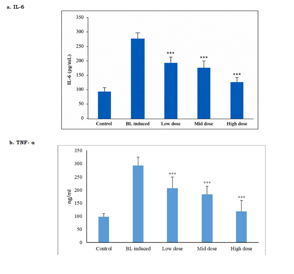
Blue light irradiation induced a pronounced inflammatory response characterized by marked elevations in IL-6 and TNF-α level. Compared with the normal control group, IL-6 increased by approximately 196.5%, while TNF-α increased by 199.5% in the pathological control group (Figure 1a and b). Treatment with the NS2130E03 significantly attenuated inflammatory cytokine production in a dose-dependent manner. Relative to the pathological control group, IL-6 concentrations were reduced by 30.6%, 37.1%, and 54.1% in the low-, mid-, and high-dose groups, respectively. Likewise, TNF-α levels decreased by 29.1%, 37.3%, and 59.4%, respectively. The high-dose group exhibited cytokine concentrations approaching those of the normal control group, demonstrating substantial suppression of blue light-induced retinal inflammation.
Figure 1:Effect of NS2130E03 on retinal inflammatory biomarkers in blue light-induced retinal damage. (a) Interleukin-6 (IL-6) and (b) Tumor necrosis factor-α (TNF-α) concentrations in retinal tissue of normal control, blue light (BL)-induced pathological control, and NS2130E03-treated groups (low, mid, and high dose). Exposure to blue light significantly increased retinal IL-6 and TNF-α levels compared with the normal control, indicating a pronounced inflammatory response. Oral administration of NS2130E03 attenuated the BL-induced elevation of both inflammatory cytokines in a dose-dependent manner, with the high-dose group showing the greatest reduction toward normal levels. Data are expressed as mean±SD (n=8 animals per group). Statistical analysis was performed using one-way analysis of variance (ANOVA) followed by Dunnett’s multiple comparison test. Statistical significance was determined by comparison with the BL-induced pathological control group (***P < 0.001).
Effect of NS2130E03 on COX-2 and VEGF levels
Blue light exposure significantly activated inflammatory and angiogenic pathways, as reflected by marked increases in COX- 2 and VEGF. Compared with normal controls, COX-2 increased by 141.4%, whereas VEGF increased by 205.9% in the pathological control group (Table 3). Treatment significantly suppressed both inflammatory mediators in a dose-dependent manner. Relative to the pathological control group, COX-2 levels decreased by 19.5%, 29.3%, and 49.4% in the low-, mid-, and high-dose groups, respectively. Similarly, VEGF concentrations declined by 30.6%, 46.1%, and 57.2%, respectively. The marked reduction in both biomarkers demonstrates that the NS2130E03 effectively interrupts the inflammatory angiogenic cascade associated with blue light-induced retinal injury.
Table 3:Effect of NS2130E03 on VEGF and COX-2 levels in blue light-induced retinal damage. Blue Light (BL) exposure significantly increased retinal VEGF and COX-2 levels compared with the normal control, indicating enhanced angiogenic signaling and inflammatory response. Oral administration of NS2130E03 significantly attenuated the BL-induced elevation of both VEGF and COX-2 in a dose-dependent manner, with the high-dose group demonstrating the greatest reduction toward normal levels, suggesting protection against blue light-induced retinal inflammation and pathological angiogenesis. Data are presented as mean±SD (n=8 animals per group). Statistical analysis was performed using one-way analysis of variance (ANOVA) followed by Dunnett’s multiple comparison test. Statistical significance was determined by comparison with the BL-induced pathological control group (***P < 0.001).

Effect of NS2130E03 on caspase-3-mediated apoptosis
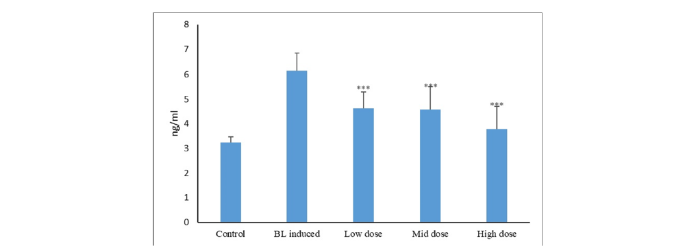
Blue light exposure significantly enhanced retinal apoptosis, as evidenced by a marked elevation in Caspase-3 expression. Caspase-3 levels increased by 89.5% in the pathological control group compared with the normal control group (Figure 2). Treatment with the NS2130E03 significantly attenuated apoptotic signalling. Relative to the pathological control group, Caspase-3 levels decreased by 25.0%, 25.6%, and 38.5% in the low-, mid-, and high-dose groups, respectively. The high-dose group exhibited Caspase-3 values approaching those of the normal control, suggesting substantial protection against blue light-induced retinal cell apoptosis.
Figure 2:Effect of NS2130E03 on retinal caspase-3 levels in blue light-induced retinal damage. Retinal caspase-3 levels in the normal control, Blue Light (BL)-induced pathological control, and NS2130E03-treated groups (low, mid, and high dose). Blue light exposure markedly increased caspase-3 levels compared with the normal control, indicating enhanced apoptotic activity in retinal tissue. Treatment with NS2130E03 significantly reduced BL-induced caspase-3 expression in a dose-dependent manner, with the high-dose group demonstrating the greatest inhibition of apoptosis. Data are presented as mean±SD (n=8 animals per group). Statistical analysis was performed using one-way analysis of variance (ANOVA) followed by Dunnett’s multiple comparison test. Statistical significance was determined by comparison with the BL-induced pathological control group (***P < 0.001).
Effect of NS2130E03 on rhodopsin levels
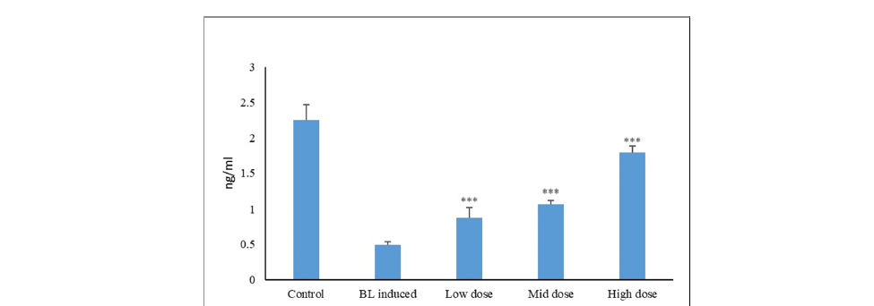
Blue light exposure caused severe photoreceptor dysfunction, with rhodopsin levels decreasing by 78.2% in the pathological control group compared with the normal control group (Figure 3). This reduction indicates significant impairment of phototransduction following oxidative retinal injury. Treatment with the NS2130E03 restored rhodopsin expression in a clear dose-dependent manner. Compared with the pathological control group, rhodopsin levels increased by 79.6%, 118.4%, and 265.3% in the low-, mid-, and high-dose groups, respectively. The high-dose treatment restored rhodopsin to approximately 80% of normal control values, demonstrating substantial preservation of photoreceptor integrity and visual signalling. Overall, the biochemical findings demonstrate that the NS2130E03 confers significant protection against blue light-induced retinal injury through coordinated enhancement of endogenous antioxidant defences, suppression of inflammatory cytokines and angiogenic mediators, inhibition of apoptosis, and preservation of photoreceptor function. The consistent dosedependent responses across all biomarkers indicate that the 500 mg/kg dose produced the greatest therapeutic efficacy.
Figure 3:Effect of NS2130E03 on retinal rhodopsin levels in blue light-induced retinal damage. Retinal rhodopsin levels in the normal control, Blue Light (BL)-induced pathological control, and NS2130E03-treated groups (low, mid, and high dose). Blue light exposure significantly reduced retinal rhodopsin levels compared with the normal control, indicating photoreceptor dysfunction and impairment of visual pigment integrity. Treatment with NS2130E03 significantly restored rhodopsin levels in a dose-dependent manner, with the high-dose group exhibiting the greatest recovery toward normal values, suggesting protection against blue light-induced photoreceptor damage. Data are presented as mean±SD (n=8 animals per group). Statistical analysis was performed using one-way analysis of variance (ANOVA) followed by Dunnett’s multiple comparison test. Statistical significance was determined by comparison with the BL-induced pathological control group (***P < 0.001).
Effect of NS2130E03 on eye histopathology
Quantitative morphometric analysis demonstrated that chronic blue light exposure induced marked retinal degeneration, characterised by significant thinning of the Outer Nuclear Layer (ONL), Inner Nuclear Layer (INL), and Photoreceptor Segment Layer (PSL) in the pathological control group compared with the normal control group (Table 4). The pathological control group exhibited a 33.8% reduction in ONL thickness, a 49.8% reduction in INL thickness, and a 66.0% reduction in PSL thickness, confirming severe retinal damage following blue light exposure. Treatment with the NS2130E03 produced a dose-dependent restoration of retinal thickness. Compared with the pathological control group, the low-dose group showed a 16.6% increase in INL thickness and a 22.5% increase in PSL thickness, whereas ONL thickness remained marginally reduced (7.5% lower than pathological control). The mid-dose group demonstrated substantial structural recovery, with 18.5%, 41.2%, and 55.2% increases in ONL, INL, and PSL thickness, respectively, compared with the pathological control group (P<0.001).
Table 4:Effect of NS2130E03 on retinal histomorphometric parameters in blue light-induced retinal damage. Histomorphometric analysis of the Outer Nuclear Layer (ONL), Inner Nuclear Layer (INL), and Photoreceptor Segment Layer (PSL) thickness in retinal sections from the normal control, blue light (BL)-induced pathological control, and NS2130E03-treated groups (low, mid, and high dose). Blue light exposure resulted in significant thinning of the ONL, INL, and PSL compared with the normal control, indicating retinal structural damage. NS2130E03 treatment preserved retinal architecture by significantly increasing the thickness of these retinal layers, with the mid- and high-dose groups demonstrating marked restoration toward normal values. Data are presented as mean±SD (n=8 animals per group). Statistical analysis was performed using one-way analysis of variance (ANOVA) followed by Dunnett’s multiple comparison test. Statistical significance was determined by comparison with the BL-induced pathological control group (*P < 0.05; ***P < 0.001).

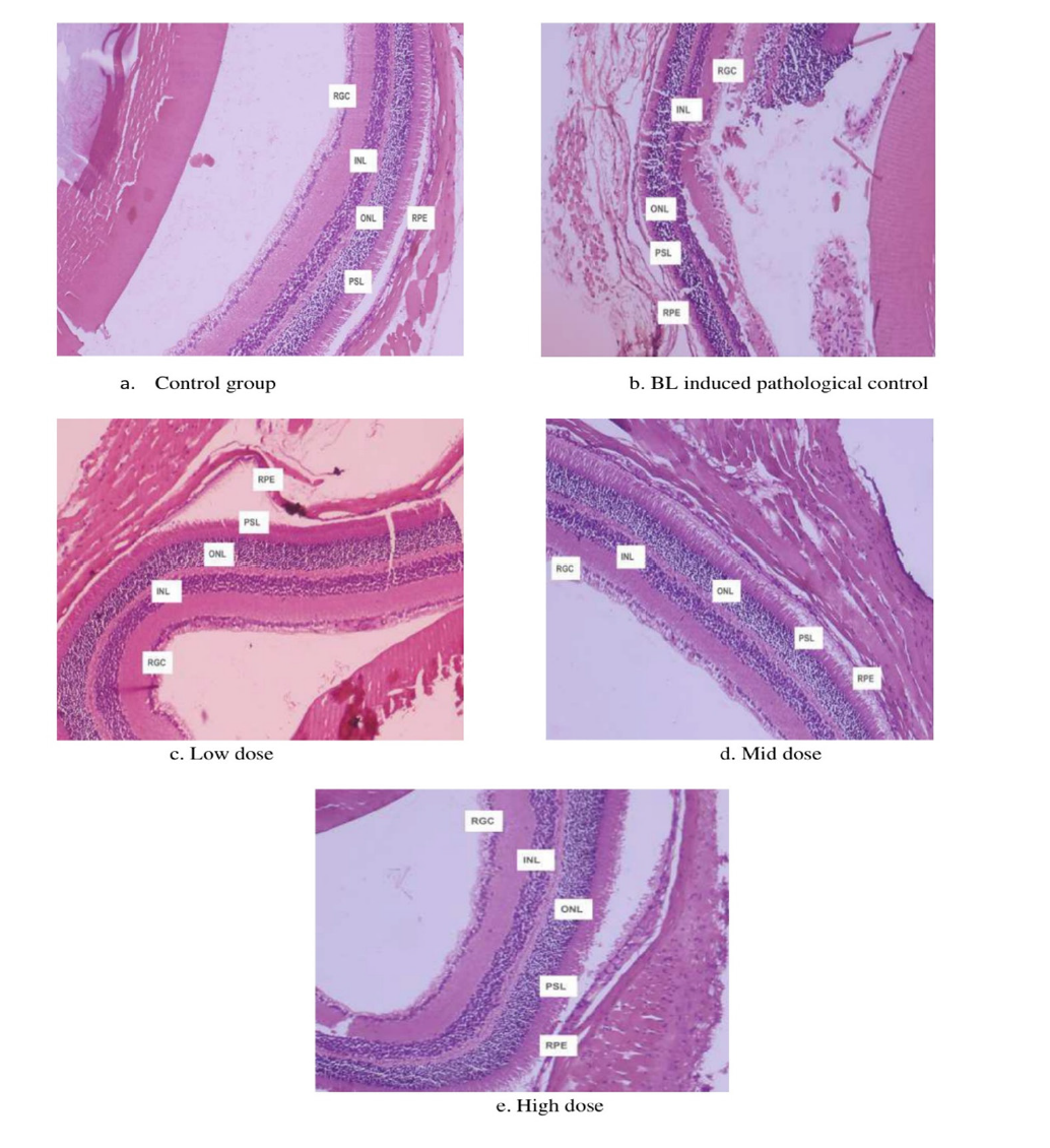
The high-dose group exhibited the greatest protective effect, restoring retinal thickness close to normal values, with 43.1%, 91.1%, and 162.1% increases in ONL, INL, and PSL thickness, respectively, relative to the pathological control group (P<0.001). At the highest dose, retinal thickness reached approximately 94.6% (ONL), 95.9% (INL), and 89.2% (PSL) of the normal control values, indicating near-complete preservation of retinal morphology. Histopathological examination corroborated the morphometric findings. Retinal sections from the normal control group displayed intact retinal architecture with well-organised retinal ganglion cell, INL, ONL, and PSL layers (Figure 4). In contrast, the pathological control group exhibited severe retinal degeneration characterised by pronounced thinning of the ONL, INL, and PSL, disruption of retinal lamination, inflammatory cell infiltration within the RGC layer, and marked disorganisation of the overall retinal structure. Administration of the NS2130E03 resulted in progressive histological improvement in a dose-dependent manner.
The low-dose group exhibited partial preservation of retinal architecture with moderate recovery of retinal layering. The mid-dose group demonstrated significant restoration of retinal organisation, characterised by increased thickness of the nuclear and photoreceptor layers, reduced inflammatory infiltration, and improved structural integrity. The high-dose group showed nearnormal retinal morphology, with well-preserved ONL, INL, PSL, and RGC layers, intact photoreceptor nuclei, minimal histopathological alterations, and restoration of normal retinal lamination. These qualitative observations are consistent with the quantitative morphometric analysis and indicate that the test substance effectively attenuated blue light-induced retinal degeneration by preserving retinal structure in a dose-dependent manner.
Figure 4:Histopathological evaluation of retinal tissue following blue light-induced retinal damage and NS2130E03 treatment (Hematoxylin and Eosin staining). Representative retinal photomicrographs from (a) normal control, (b) Blue Light (BL)-induced pathological control, (c) NS2130E03 low-dose, (d) NS2130E03 mid-dose, and (e) NS2130E03 high-dose groups. The normal control retina exhibited intact retinal architecture with well-organized Retinal Ganglion Cell (RGC), Inner Nuclear Layer (INL), Outer Nuclear Layer (ONL), Photoreceptor Segment Layer (PSL), and Retinal Pigment Epithelium (RPE). In contrast, the BL-induced pathological control showed marked retinal degeneration characterized by retinal edema, inflammatory cell infiltration, disruption of retinal architecture, and thinning of the INL. Treatment with NS2130E03 at low, mid, and high doses preserved retinal morphology, with restoration of retinal layer organization and reduced pathological alterations compared with the BL-induced pathological control, indicating a dose-dependent protective effect against blue light-induced retinal injury. Scale bar=50μm.
Discussion
The present study demonstrates that NS2130E03, a standardized anthocyanin-rich hydroalcoholic extract of Daucus carota subsp. sativus, confers significant protection against blue light-induced retinal injury in mice through coordinated modulation of oxidative stress, inflammation, angiogenesis, apoptosis, and retinal structural preservation. Blue light exposure markedly impaired endogenous antioxidant defence, increased inflammatory cytokines and angiogenic mediators, activated apoptotic pathways, reduced rhodopsin expression, and induced severe histopathological damage. Oral administration of NS2130E03 attenuated these pathological changes in a dose-dependent manner, with the highest dose (500mg/kg) producing near-complete restoration of retinal biochemical and morphological parameters. These findings support the hypothesis that anthocyanin-rich botanical interventions may represent an effective nutritional strategy for protecting the retina against phototoxic injury. Oxidative stress is widely recognised as the principal mechanism responsible for blue light-induced retinal degeneration. Because retinal tissue exhibits one of the highest oxygen consumption rates and contains abundant polyunsaturated fatty acids, excessive blue light irradiation readily generates reactive oxygen species (ROS), resulting in lipid peroxidation, mitochondrial dysfunction, DNA damage, and impairment of endogenous antioxidant systems.
Persistent oxidative stress subsequently initiates inflammatory and apoptotic signalling, ultimately leading to photoreceptor degeneration and retinal dysfunction [2,4-6]. Consistent with these reports, blue light exposure in the present study significantly reduced retinal SOD, CAT, GPx, and TAC, confirming severe oxidative injury. Treatment with NS2130E03 substantially restored all antioxidant biomarkers, indicating preservation of endogenous antioxidant defence. Anthocyanins possess potent free radical scavenging activity owing to their multiple hydroxyl groups and have been shown to activate the Nrf2/ARE pathway, thereby enhancing transcription of antioxidant enzymes including SOD, catalase, glutathione peroxidase, and heme oxygenase-1 (HO-1) [13,14]. Therefore, restoration of antioxidant capacity observed in the present study likely represents one of the primary mechanisms underlying retinal protection. Oxidative stress and inflammation are closely interconnected during retinal degeneration. ROS activate redox-sensitive transcription factors including nuclear factor-kappa B (NF-κB), resulting in increased production of proinflammatory cytokines such as IL-6, TNF-α, and COX-2. These mediators amplify retinal injury by disrupting the blood-retinal barrier, recruiting inflammatory cells, enhancing oxidative damage, and promoting vascular dysfunction [7-9].
In agreement with these observations, the pathological control animals exhibited marked elevations of IL-6, TNF-α, and COX-2, whereas NS2130E03 significantly suppressed these inflammatory mediators in a dose-dependent manner. Similar anti-inflammatory effects of anthocyanins have been reported in experimental retinal injury models where anthocyanin supplementation inhibited NF- κB activation and reduced inflammatory cytokine production [15,16]. The present findings therefore suggest that attenuation of retinal inflammation is a major contributor to the protective effects of NS2130E03. Another important finding of the present investigation was the marked reduction in VEGF expression following NS2130E03 treatment. Increased VEGF production represents a characteristic response to oxidative retinal injury and contributes to pathological angiogenesis, vascular leakage, and progression of retinal diseases including diabetic retinopathy and age-related macular degeneration [10]. Blue light-induced oxidative stress has previously been shown to increase VEGF expression in retinal pigment epithelial cells through ROS-dependent signalling pathways [2]. The significant suppression of VEGF observed in the present study indicates that NS2130E03 not only reduces oxidative stress but may also prevent secondary vascular complications associated with chronic retinal injury. Although angiogenesis was not directly assessed histologically, reduced VEGF levels strongly suggest inhibition of pathological angiogenic signalling.
Photoreceptor apoptosis represents the final common pathway of retinal degeneration following prolonged oxidative stress. Activation of caspase-3 results in irreversible photoreceptor cell death and progressive loss of visual function [11,12]. In the present study, blue light exposure significantly increased retinal caspase-3 concentrations, whereas NS2130E03 effectively suppressed apoptotic signalling. Concurrently, rhodopsin expression, which was markedly reduced following blue light exposure, was substantially restored after treatment. Rhodopsin is the principal visual pigment of rod photoreceptors, and preservation of its expression is considered an important indicator of photoreceptor integrity and functional retinal health. Restoration of rhodopsin together with reduced caspase-3 strongly suggests that NS2130E03 protects photoreceptors from oxidative apoptosis and preserves retinal function. Previous investigations have similarly demonstrated that anthocyanins protect retinal pigment epithelial cells and photoreceptors by maintaining mitochondrial integrity, reducing ROS generation, and inhibiting mitochondrial apoptotic pathways [14,16]. Histopathological findings further validated the biochemical observations. Blue light exposure produced characteristic retinal degeneration, including thinning of the ONL, INL, and photoreceptor segment layer, accompanied by disruption of retinal architecture and inflammatory infiltration.
Administration of NS2130E03 markedly preserved retinal morphology and restored retinal thickness in a dose-dependent manner, with the highest dose maintaining retinal architecture close to that of normal animals. Structural preservation of the outer nuclear layer is particularly important because this layer contains photoreceptor nuclei that are highly susceptible to oxidative injury. The concordance between biochemical biomarkers and histopathological outcomes substantially strengthens the evidence supporting the retinoprotective efficacy of NS2130E03. The observed protective effects are likely attributable to the diverse biological activities of anthocyanins naturally present in Daucus carota. Beyond direct antioxidant activity, anthocyanins regulate multiple signalling pathways, including Nrf2, NF- κB, MAPK, PI3K/Akt, and mitochondrial apoptotic cascades, thereby simultaneously reducing oxidative stress, inflammation, angiogenesis, and programmed cell death [13,14]. The anti-aging protein Sirtuin 1 (SIRT1) is critical for the regulation of oxidative stress, inflammation, retinal degeneration, neurodegeneration, angiogenesis, and programmed cell death. Anthocyanins have also been reported to activate SIRT1, suggesting a potential role in modulating multiple signalling pathways and protecting against mitochondrial apoptosis [18-20].
The potential involvement of SIRT1 in the protective effects of NS2130E03 remains to be established, as SIRT1 expression and activity were not assessed in the present study and will be investigated in future mechanistic studies. The present study possesses several strengths. It comprehensively evaluated multiple complementary endpoints encompassing antioxidant defence, inflammatory cytokines, angiogenic signalling, apoptosis, photoreceptor integrity, and histopathological alterations within a well-established blue light-induced retinal injury model. The consistent dose-dependent improvements observed across all biochemical and histological parameters increase confidence in the biological activity of the standardized extract. Furthermore, the use of a chemically standardized anthocyanin-rich preparation enhances reproducibility and facilitates future translational and clinical investigations. Nevertheless, several limitations should be acknowledged. The study did not directly quantify retinal ROS production, lipid peroxidation products, mitochondrial function, or activation of upstream molecular signalling pathways such as Nrf2, NF-κB, MAPK, or PI3K/Akt. Future investigations incorporating molecular analyses, retinal functional assessments, pharmacokinetic studies, and long-term safety evaluations will further clarify the mechanisms underlying the observed protective effects and support clinical translation.
Conclusion
The present findings demonstrate that NS2130E03 effectively protects against blue light-induced retinal degeneration by restoring endogenous antioxidant defences, suppressing inflammatory and angiogenic mediators, inhibiting apoptosis, preserving rhodopsin expression, and maintaining retinal structural integrity. The coordinated modulation of these interconnected pathological pathways suggests that standardized acylated anthocyanin-rich Daucus carota extract may represent a promising nutritional intervention for reducing retinal damage associated with chronic digital screen exposure, environmental blue light exposure, and early retinal degenerative disorders. These encouraging preclinical findings provide a strong scientific rationale for future mechanistic investigations and well-designed clinical trials evaluating the efficacy of NS2130E03 in human ocular health.
Acknowledgement
The authors gratefully acknowledge Radiant Research Services Pvt. Ltd. for their contributions to this study.
Conflict of Interest
The authors declare that they have no conflicts of interest related to this work.
Author Contributions
Conceptualization and preclinical studies: Sameer Kamalakar
Akolkar.
Manuscript writing: Mohan Gowda C M
Extraction and Analytical: Madhukumar M.S.
Manucript review: Anirudh Ranga
Competing Interests
Sameer Kamalakar Akolkar, Mohan Gowda C M, Madhukumar M S, Anirudh Ranga, are employed with Natural and Essential Oils Private Limited, Mysuru, Karnataka, India. This does not alter our adherence to Advances in Complementary and Alternative medicine policies on sharing data and materials.
Funding
The study was sponsored by Natural & Essential Oils Pvt. Ltd., Mysuru, India.
References
- Tosini G, Ferguson I, Tsubota K (2016) Effects of blue light on the circadian system and eye physiology. Mol Vis 22: 61-72.
- Shang YM, Wang GS, Sliney D, Yang CH, Lee LL (2014) White Light-Emitting Diodes (LEDs) at domestic lighting levels and retinal injury in a rat model. Environ Health Perspect 122(3): 269-276.
- Hunter JJ, Morgan JI, Merigan WH, Sliney DH, Sparrow JR, et al. (2012) The susceptibility of the retina to photochemical damage from visible light. Prog Retin Eye Res 31(1): 28-42.
- Beatty S, Koh H, Henson D, Boulton M (2000) The role of oxidative stress in the pathogenesis of age-related macular degeneration. Surv Ophthalmol 45(2): 115-134.
- Jarrett SG, Boulton ME (2012) Consequences of oxidative stress in age-related macular degeneration. Mol Aspects Med 33(4): 399-417.
- Roehlecke C, Schumann U, Ader M, Knels L, Funk RHW (2011) Influence of blue light on photoreceptors in a live retinal explant system. Mol Vis 17: 876-884.
- Kuse Y, Ogawa K, Tsuruma K, Shimazawa M, Hara H (2014) Damage of photoreceptor-derived cells in culture induced by light emitting diode-derived blue light. Sci Rep 4: 5223.
- Natoli R, Fernando N, Madigan M, Tan JAC, Valter K, et al. (2017) Microglia-derived IL-1β promotes chemokine expression by Müller cells and retinal pigment epithelium in retinal degeneration. Mol Neurodegener 12(1): 31.
- Tang J, Kern TS (2011) Inflammation in diabetic retinopathy. Prog Retin Eye Res 30(5): 343-358.
- Penn JS, Madan A, Caldwell RB, Bartoli M, Caldwell RW, et al. (2008) Vascular endothelial growth factor in eye disease. Prog Retin Eye Res 27(4): 331-371.
- Ardeljan D, Chan CC (2013) Aging is not a disease: Distinguishing age-related macular degeneration from aging. Prog Retin Eye Res 37: 68-89.
- Organisciak DT, Vaughan DK (2010) Retinal light damage: Mechanisms and protection. Prog Retin Eye Res 29(2): 113-134.
- Khoo HE, Azlan A, Tang ST, Lim SM (2017) Anthocyanidins and anthocyanins: Colored pigments as food, pharmaceutical ingredients, and the potential health benefits. Food Nutr Res 61(1): 1361779.
- Huang WY, Zhang HC, Liu WX, Li CY (2012) Survey of antioxidant capacity and phenolic composition of blueberry, blackberry, and strawberry in Nanjing. J Zhejiang Univ Sci B 13(2): 94-102.
- Ohgami K, Ilieva I, Shiratori K, Koyama Y, Jin XH, et al. (2005) Anti-inflammatory effects of aronia extract on rat endotoxin-induced uveitis. Invest Ophthalmol Vis Sci 46(1): 275-281.
- Osada H, Okamoto T, Kawashima H, Toda E, Miyake S, et al. (2017) Neuroprotective effect of bilberry extract in a murine model of photo-stressed retina. PLoS One 12(6): e0178627.
- Zaim M, Kara I, Muduroglu A (2021) Black carrot anthocyanins exhibit neuroprotective effects against MPP⁺-induced cell death and cytotoxicity via inhibition of oxidative stress-mediated apoptosis. Cytotechnology 73(6): 827-840.
- Martins IJ (2016) Anti-aging genes improve appetite regulation and reverse cell senescence and apoptosis in global populations. Adv Aging Res 5(1): 9-26.
- Martins IJ (2017) Single gene inactivation with implications to diabetes and multiple organ dysfunction syndrome. J Clin Epigenet 3(3): 1-8.
- Martins IJ (2017) Nutrition therapy regulates caffeine metabolism with relevance to NAFLD and induction of type 3 diabetes. J Diabetes Metab Disord 4(19): 1-9.
© 2026 Sameer Kamalakar Akolkar. This is an open access article distributed under the terms of the Creative Commons Attribution License , which permits unrestricted use, distribution, and build upon your work non-commercially.
Editor In Chief







.jpg)































Signup for Newsletter

Quick Links
 Editorial Board Registrations
 Submit your Article
 Best Paper of the Volume
 Reprints
 Refer a Friend
 Advertise With Us
Our Recent Edition
.jpg)



Top Editors




.jpg)

















.bmp)
.jpg)
.png)
.jpg)













.jpg)






.png)

.png)




.png)




Financial Support

Sponsors

Latest e-Books

Latest Video

 a Creative Commons Attribution 4.0 International License. Based on a work at www.crimsonpublishers.com.
Best viewed in
a Creative Commons Attribution 4.0 International License. Based on a work at www.crimsonpublishers.com.
Best viewed in 
 | Above IE 9.0 version