- Submissions

Full Text
Orthoplastic Surgery & Orthopedic Care International Journal
Ewing's Sarcoma (E.S) [Endothelial Myeloma]
Abdallah Attia*
Professor of Orthopedic surgery, Zagazig University, Egypt
*Corresponding author: Abdallah Attia, Orthopedic, Professor of Orthopedic surgery, Zagazig University, Egypt
Submission: October 4, 2017; Published: November 15, 2017

ISSN: 2578-0069Volume1 Issue1
Introduction
Ewing's sarcoma was first described by James Ewing in 1921. Ewing's sarcoma is the second most common primary malignancy of bone (after osteosarcoma) in patients younger than 30 years of age and the most common in patients younger than 10 years of age. Its maximum incidence is seen in 2nd decade of life. Ewing's sarcoma is derived from red bone marrow.
a) Age: 5-20 years; and rarely develops in adults older than 30 years.
b) Site: Diaphysis of long bones & pelvis. Both long and flat bones are affected because no bone is immune to tumor development. In the long bones, the tumor is almost always metaphyseal or diaphyseal. In reality, Ewing sarcoma more often originates in the metaphysis of a long bone, but frequently extends for a considerable distance into the diaphysis.
c) Sex: Male> Female: 3: 2.
Causes
a. A chromosome rearrangement between chromosomes #11 and #22. This rearrangement changes the position and function of genes, causing a fusion of genes referred to as a "fusion transcript."
b. Some physicians classify Ewing sarcoma as a primitive neuroectodermal tumor (PNET). This means the tumor may have started in fetal, or embryonic, tissue that has developed into nerve tissue.
Clinical Picture (Pain and Swelling of Weeks or Months Duration)
a) Pain: intermittent; recurs with T intensity & persistent; T by night.
b) Pyrexia: continuous or intermittent.
c) Attacks of T size; pain; fever.
d) Swelling: painful; warm; tender; ill defined; diaphyseal.
e) Erythema and warmth of the local area are sometimes seen.
f) Osteomyelitis is often the initial diagnosis based on intermittent fevers, leukocytosis, anemia and an increased ESR.
g) Weight loss.
h) Symptoms usually last a few weeks to a few months.
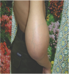
i) Eventually, most patients have a large palpable mass, which rapidly grows; with a tense and tender local swelling .There may be dilated skin veins (Figure 1).
Figure 1: Ewing’s sarcoma at lower third of the humerus showing dilated skin veins.

Laboratory studies
A. Tincreased white blood cell count
B. TESR
C. TC-reactive protein.
D. T Serum lactic dehydrogenase.
E. Leucocytosis.
Radiology
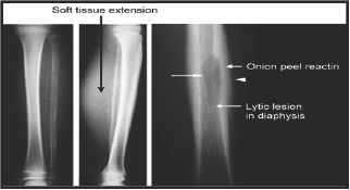
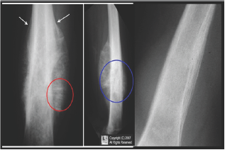
I. Plain X-ray (Figure 2-4)
Figure 2: Ewing's sarcoma.

Figure 3: Ewing's sarcoma of femur showing onion peel appearance

Figure 4: Ewing's sarcoma at metaphysis and diaphysis of humerus.

a) Poorly defined areas of central destruction in mid-shaft.
b) Onion-peel appearance: lamellar periosteal reaction and new bone formation.
a. It consists of densely packed uniform small blue round cells in sheets.
c) A long, permeative lytic lesion in the metadiaphysis and diaphysis of the bone with a prominent soft tissue mass extending from the bone.
d) The radiographic appearance of Ewing sarcoma may vary highly from a lytic one to a dominantly sclerotic one.
e) A periosteal reaction is usually present, and it often has an onion-skin or sunburst pattern, which indicates an aggressive process.
II. MRI of the entire bone to gives more accurate information about tumor size & extent of its relation to the surrounding structures.
III. CT scan of the lesion to define the extra-osseous extent of the tumor, especially in the spine, ribs and pelvis.
IV. CT scan of the chest because the lung is the most common site of metastases.
V. Isotope bone scan: The bone is the second most common site of metastases.
VI. PET scan for accurate localization of local and distant spread
Biopsy for histopathology
Biopsy is considered the gold standard test for diagnosis of Ewing's Sarcoma.
Pathology
i.Origin of ewing's sarcoma (Theories):
a.Endothelial cells of bone marrow (endothelioma).
b.Variant of reticulum cell sarcoma.
c.2ry tumor from neuroblastoma.
ii.N.E
a)The tumor is gray to white in color and poorly demarcated.
b)The consistency is soft and gray and sometimes semiliquid (like brain tissue) especially after breaking through the cortex.
c) Areas of hemorrhage and necrosis are common.
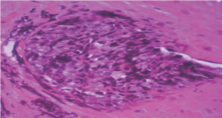
M.E (Figure 5)
Figure 5: M.E of Ewing's sarcoma showing pseudorossete structures.

a.It consists of densely packed uniform small blue round cells in sheets.
b. The cells have scant cytoplasm without distinct borders.
c. The cells are two to three times as big as lymphocytes and have a single oval or round nucleus without prominent nucleoli.
d. The tumor spreads through Haversian canals which cause the appearance of permeative margins on x-ray.
e. Glycogen is present within the cells causing a positive reaction to periodic acid-schiff (PAS) stain.
f. No matrix.
g. Pseudorossete structures (aggregation of tumor cells around blood vessels).
Spread
a) Blood born to other bonesa and lungs.
b) Lymphatic spread: to regional lymph nodes.
c) Fig.(3-53): M.E of Ewing's sarcoma showing pseudorossete structures.
Differential diagnosis
a. Osteomyelitis.
b. Neuroblastoma metastasis (less than 5 years and increase valinyl mandilic acid in urine)
c. Lymphoma of the bone(Reticulum cell sarcoma ):[more than 20 years] and Glycogen staining is positive.
d. Eosinophilic granuloma.
e. Lymphoma
f. Leukemia
Treatment
[Combination of Surgery+radiation+multi-drug chemotherapy]
A. The treatment of Ewing sarcoma must include neoadjuvant or adjuvant chemotherapy, or both, to treat distant metastases that may or may not be readily apparent at the initial staging.
B. The tumor is Radiosensitive.
C. Radiotherapy+chemotherapy.
D. Treatment often consists of neo-adjuvant chemotherapy generally followed by wide or radical excision, and may also include radiotherapy
E. Complete excision at the time of biopsy may be performed if malignancy is confirmed at that time. Radical chemotherapy may be as short as 6 treatments at 3 week cycles; however most patients will undergo chemotherapy for 6-12 months and radiation therapy for 5-8 weeks.
F. Definitive surgical margins are desirable (e.g., removal of fibula, limb salvage with extensive margins).
G. Amputation to get red from painful swelling.
Prognosis: Before the use of multiagent chemotherapy, longterm survival was less than 10%. Today, most centers report long-term survival rates of 60-75%. Poor prognostic signs include increased age, increased ESR and leukocytosis at presentation.
Summary of Ewing sarcoma
a. Ewing's sarcoma is a highly malignantbone tumor that affects mostly children.
b. It is a type of peripheral primitive neuroectodermal tumor(PNET).
c. It is a diaphyseal tumor most commonly found femur, tibia and humerus, as well as the pelvis.
d. Age: 5-20 years.
e. X-ray: Onion peels appearance.
f. N.E: Tumor tissue is SOFT; Grayish (Brain Like tissue).
g. An undifferentiated round cell sarcoma.
h. Treatment: Surgery+radiation+multi-drug chemotherapy.
i. Radiosensitive.
j. Spread to lung & other bones
© 2017 Abdallah Attia. This is an open access article distributed under the terms of the Creative Commons Attribution License , which permits unrestricted use, distribution, and build upon your work non-commercially.
 a Creative Commons Attribution 4.0 International License. Based on a work at www.crimsonpublishers.com.
Best viewed in


.jpg)


 Editorial Board Registrations
 Submit your Article
 Refer a Friend
 Advertise With Us
.jpg)


.jpg)


.bmp)
.jpg)
.png)
.jpg)


.jpg)


.png)

.png)


.png)


