- Submissions

Full Text
Techniques in Neurosurgery & Neurology
Is There a Protectiıve Effect of Prematurıty in Developmental Hıp Dysplasıa?
Özlem Bütün1, Birgül Vural1* and Orhan Balta2
1Child Health and Diseases, Gaziosmanpasa University, Türkiye
2Orthopedics and Traumatology Clinic, Gaziosmanpasa University, Türkiye
*Corresponding author:Birgül Vural, Child Health and Diseases, Gaziosmanpaşa University, Tokat, 60400, Türkiye
Submission: May 05, 2025;Published: June 20, 2025

ISSN 2637-7748
Volume5 Issue5
Abstract
This study was conducted to determine the prevalence of Developmental Dysplasia of the Hip (DDH), its risk factors, and the relationship between prematurity and DDH in infants aged 0-6 months who were brought to a university hospital orthopedic clinic for DDH screening. The hip ultrasonography records of 722 infants aged 0-6 months who presented for DDH screening were retrospectively reviewed. Among these infants, 341 (47.2%) were female, 381 (52.8%) were male; 64 (8.9%) had a positive family history; 95 (13.2%) were premature; 350 (48.5%) had been swaddled; 335 (46.4%) were delivered vaginally, and 384 (53.2%) were delivered by cesarean section. There were 21 infants (2.9%) from multiple pregnancies and 21 infants (2.9%) with a breech presentation. The findings, consistent with existing literature, showed that cesarean delivery, female sex, first birth, swaddling, and positive family history increased the risk of DDH, while breech presentation and birth weight did not show a significant association. Furthermore, similar to other limited studies, our data suggest that prematurity may have a protective effect on hip development. Hip ultrasonography results were classified according to the Graf method: right hip: 0.7% type Ia, 98.5% type Ib, 0.3% type IIa, 0.1% type IIb, 0.4% type III; left hip: 1.0% type Ia, 98.1% type Ib, 0.6% type IIa, 0.1% type IIb, 0.1% type D, 0.1% type III. Delayed ossification was detected in 2 infants, and subluxated hips were identified in 5 infants, all treated with a Pavlik harness. The prevalence of DDH in Tokat province was found to be 0.9%, lower than the reported national DDH rate in Turkey (1-1.5%). In conclusion, raising public awareness about DDH, ensuring timely and appropriate screening programs, and educating about the harms of traditional practices like swaddling are essential. This study emphasizes the importance of healthcare professionals having accurate knowledge of DDH and effectively informing families to minimize complications from late diagnosis or treatment.
Keywords:Developmental dysplasia of the hip; Prevalence; Prematurity; Ultrasonography; Risk factors
Introduction
Developmental Dysplasia of the Hip (DDH) is a common condition in childhood that has a good prognosis when treated at an early age [1]. It was first described in 1832 by Guillaume Dupuytren under the name “congenital hip dislocation” [2]. DDH was previously thought to be a congenital condition; however, it is now understood as a dynamic developmental condition that can either worsen or improve over time. Therefore, the term “Congenital Hip Dislocation (CHD),” which refers to the femoral head being dislocated from the acetabulum at birth, is no longer used [1,3]. Developmental dysplasia of the hip encompasses a broad spectrum ranging from mild dysplasia to complete hip dislocation, including unstable hips, subluxations, dislocations, and various malformations [4]. The exact etiology is uncertain, but there are several risk factors. According to the guidelines updated by the American College of Radiology in 2019, breech intrauterine position, a positive family history, and female sex are emphasized as the three main risk factors for DDH [5]. Other lower-risk factors associated with DDH include first birth, oligohydramnios, overly restrictive swaddling practices, and lower extremity anomalies [6]. Individuals with a positive family history have a 12-fold increased risk compared to those without a family history [7,8]. The incidence of developmental dysplasia of the hip in Turkey is reported to be approximately 5 to 10 per 1,000 live births (Ministry of Health, 2009).
Diagnosis of DDH is made through physical examination using the Ortolani and Barlow tests, Galeazzi sign, and asymmetry of skin folds [9]. Subsequently, hip ultrasonography, which is widely used in hip screening in many developed countries and was first implemented by Graf in the 1980s, is used to classify the hips according to the alpha and beta angles. According to the Graf method, hips with an alpha angle of 60˚ or above are evaluated as Type 1 (normal). If the child is younger than 3 months and the alpha angle is between 50˚ and 59˚, it is classified as Type 2a (physiologically immature); if the child is older than 3 months with the same angles, it is Type 2b (delayed ossification); regardless of age, an alpha angle between 43˚ and 50˚ is classified as Type 2c. Hips with an alpha angle below 43˚ are called Type 3 (subluxated), and hips where the angle cannot be measured are called Type 4 (dislocated) [10,11]. In our study, hip ultrasonography records were classified according to the Graf method. Early diagnosis and treatment of Developmental Dysplasia of the Hip (DDH) is extremely important. Early detection allows for minimal invasive intervention, helping achieve more effective outcomes. In contrast, late diagnosis of DDH may require surgical intervention and complex treatment [12]. Universal ultrasound screening programs implemented in many countries worldwide have significantly reduced the number and severity of surgical interventions associated with DDH [13]. In Turkey, with the implementation of the “Developmental Dysplasia of the Hip Screening Program Directive” in 2019, all newborns are screened for DDH (Ministry of Health, 2009). Our study covers the one-year period following the initiation of this screening program. Preterm infants are defined as babies born alive before completing the 37th week of gestation. The aim of this study was to determine the incidence of DDH in premature infants and to compare it with term infants. Additionally, the study aimed to determine the incidence of DDH specific to the Tokat province and to investigate the risk factors for DDH as defined in the literature.
Materials and Methods
Between January 1, 2020, and December 31, 2020, infants aged 0-6 months who presented to the Orthopedics and Traumatology Clinic of Gaziosmanpaşa University Hospital for developmental dysplasia of the hip screening were retrospectively reviewed through the hip ultrasonography record forms completed during examination and data obtained from the hospital automation system. Infants with incomplete, incorrect, or irregular medical records were excluded from the study. Infants were evaluated based on their gestational birth weeks, age at the time of ultrasonography (in weeks), alpha and beta angle values, and risk factors (positive family history, prematurity, swaddling, being the first child, female sex, breech presentation, and multiple pregnancy). Prematurity was defined as birth before the 38th gestational week. Only those who underwent ultrasonography using the Graf method were included in the study. The measured alpha and beta angles were classified according to the Graf method. Gestational weeks were grouped as 27-31, 32, 33, 34, 35, 36, 37, 38, 39, 40, 41, and 42; for each group, the number of infants, minimum alpha angle, maximum alpha angle, and mean alpha angle were calculated. For statistical analysis, descriptive statistics were used to provide general information about the study groups. Data related to quantitative variables were described using mean and standard deviation (x±sd), and data related to qualitative variables were described using numbers (n) and percentages (%). Differences between groups for quantitative variables were assessed using the Independent Sample T-Test or Kruskal-Wallis Variance Analysis. Differences between groups for qualitative variables were calculated using the Chi-Square Test. To examine relationships between variables, Pearson Correlation Analysis and Regression Analysis were applied. A p-value of less than 0.05 was considered statistically significant. Statistical calculations were performed using commercial statistical software (IBM SPSS Statistics 22, SPSS Inc., an IBM Co., Somers, NY).
Result and Discussion
Table 1:Distribution of qualitative variables (n=722) demographic characteristics and risk factors of infants undergoing hip ultrasonography.
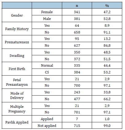
Our study includes the findings obtained from the retrospective screening of 1444 hips of 722 infants aged 0-6 months who applied to the Orthopedics and Traumatology Clinic for routine developmental hip dysplasia screening between January 2020 and December 2020. A total of 22 infants with incomplete records or who did not come for screening were excluded from the study. Of the 722 infants included in the study, 341 (47.2%) were female, 381 (52.8%) were male, 64 (8.9%) had a positive family history, 95 (13.2%) were premature, and 350 (48.5%) had been swaddled. Among the infants, 335 (46.4%) were born via vaginal delivery, and 384 (53.2%) by cesarean section. The number of infants from multiple pregnancies was 21 (2.9%), and the number of infants with breech presentation was also 21 (2.9%). Additionally, 7 infants (1.0%) had received Pavlik harness treatment (Table 1).
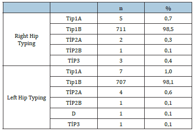
The classification of hip ultrasonography according to the Graf method and treatment options are presented in (Table 2). After ultrasonographic evaluation of the right and left hips, the types and frequencies of hips according to the Graf classification were as follows: right hip: 0.7% type Ia, 98.5% type Ib, 0.3% type IIa, 0.1% type IIb, 0.4% type III; left hip: 1.0% type Ia, 98.1% type Ib, 0.6% type IIa, 0.1% type IIb, 0.1% type D, and 0.1% type III (Table 3). Furthermore, delayed ossification was observed in 2 patients, and subluxation of the hip in 5 patients, who were treated with Pavlik harness. Studies in the literature report a success rate of Pavlik harness treatment between 84% and 91% [14].
Table 2:Classification of neonatal hip ultrasonography according to the graf method and treatment options.
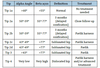
Table 3:Ultrasonographic hip typing according to graf classification (n=722).
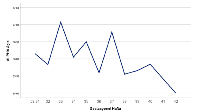
When the distribution by gender was evaluated with the Independent Samples T-Test, a statistically significant difference was found in the alpha angles of the right and left hips, whereas no statistically significant difference was found in the beta angles. The distribution of birth weight and gestational age according to right and left hip types was analyzed using the Kruskal-Wallis test, a non-parametric one-way variance analysis, and no significant difference was found. The relationship between gestational age and alpha angle was evaluated by independent samples T-Test, and a significant difference was detected. Infants with lower gestational age had higher alpha angles (Figure 1 & 2).
Figure 1:Alpha angle values according to pre-term and full-term status.
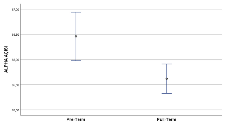
Figure 2:Changes in alpha angle according to gestational age.
Conclusion and Recommendations
As a result of our study, premature infants did not show a statistically higher frequency of Developmental Hip Dysplasia (DHD) compared to full-term infants. Additionally, infants born prematurely exhibited significantly higher alpha angles than those born at term. When examining alpha angles in more detail according to gestational age, hip development appears to follow a physiological curve peaking at the 33rd week of gestation. These findings suggest that prematurity is not a predisposing factor for DHD but may actually have a protective effect. This supports the study by Koob and colleagues in 2022, which also concluded that prematurity is not a risk factor for DHD [15]. The prevalence of developmental hip dysplasia in Tokat, based on the results of our study, was found to be 0.9%, which is below the reported rate in Turkey (1-1.5%). A major issue in screening programs in our country is the insufficient interest and attention of families towards these programs. Therefore, it is essential to raise public awareness about developmental hip dysplasia, ensure that screening programs are conducted timely and appropriately, and educate families about the harmful effects of traditional practices such as swaddling that increase risk factors.
References
- Pandey RA, Johari AN (2021) Screening of newborns and infants for developmental dysplasia of the hip: A systematic review. Indian Journal of Orthopaedics 55(6): 1388-1401.
- Dupuytren G (1964) Original or congenital displacement of the heads of thigh-bones. Clinical Orthopaedics and Related Research 33: 3-8.
- Zhang S, Doudoulakis KJ, Khurwal A, Sarraf KM (2020) Developmental dysplasia of the hip. British Journal of Hospital Medicine 81(7): 1-8.
- Furnes O, Lie S, Espehaug B, Vollset S, Engesaete LB, et al. (2001) Hip disease and the prognosis of total hip replacements: A review of 53,698 primary total hip replacements reported to the Norwegian arthroplasty register 1987-99. The Journal of Bone and Joint Surgery British Volume 83(4): 579-586.
- Nguyen JC, Dorfman SR, Rigsby CK, Iyer RS, Alazraki AL, et al. (2019) ACR appropriateness criteria® developmental dysplasia of the hip-child. Journal of the American College of Radiology 16(5S): S94-S103.
- Mulpuri K, Song KM, Goldberg MJ, Sevarino K (2015) Detection and nonoperative management of pediatric developmental dysplasia of the hip in infants up to six months of age. Journal of the American Academy of Orthopaedic Surgeons 23(3): 202-205.
- Stevenson DA, Minseau G, Kerber RA, Viskochil DH, Schaefer C, et al. (2009) Familial predisposition to developmental dysplasia of the hip. Journal of Pediatric Orthopaedics 29(5): 463-466.
- (2019) Republic of Turkey Ministry of Health. Developmental Dysplasia of the Hip (DDH) Screening Program.
- Arti H, Mehdinasab SA, Arti S (2013) Comparing results of clinical versus ultrasonographic examination in developmental dysplasia of hip. Journal of Research in Medical Sciences: The Official Journal of Isfahan University of Medical Sciences 18(12): 1051-1055.
- Graf R (1983) New possibilities for the diagnosis of congenital hip joint dislocation by ultrasonography. Journal of Pediatric Orthopedics 3(3): 354-359.
- Graf R (2006) Hip sonography. Diagnosis and management of infant hip dysplasia. Springer Berlin p. 114.
- Cha SM, Shin HD, Shin BK (2018) Long-term results of closed reduction for developmental dislocation of the hip in children of walking age under eighteen months old. International Orthopaedics 42(1): 175-182.
- Biedermann R, Eastwood DM (2018) Universal or selective ultrasound screening for developmental dysplasia of the hip? Discussion of the key issues. Journal of Pediatric Orthopaedics 12(4): 296-301.
- Wahlen R, Zambelli PY (2015) Treatment of the developmental dysplasia of the hip with an abduction brace in children up to 6 months old. Advances in Orthopedics 2015: 103580.
- Koob S, Garbe W, Bornemann R, Ploeger MM, Scheidt S, et al. (2022) Is prematurity a protective factor against developmental dysplasia of the hip? A retrospective analysis of 660 newborns. Ultraschall in der Medizin 43(2): 177-180.
© 2025 Birgül Vural. This is an open access article distributed under the terms of the Creative Commons Attribution License , which permits unrestricted use, distribution, and build upon your work non-commercially.
 a Creative Commons Attribution 4.0 International License. Based on a work at www.crimsonpublishers.com.
Best viewed in


.jpg)


 Editorial Board Registrations
 Submit your Article
 Refer a Friend
 Advertise With Us
.jpg)


.jpg)


.bmp)
.jpg)
.png)
.jpg)


.jpg)


.png)

.png)


.png)


