- Submissions

Full Text
Techniques in Neurosurgery & Neurology
Brain Parenchyma to Repair Spinal Cord Deficits
Abbas Alnaji*
Senior Consultant Neurosurgeon, Iraq
*Corresponding author: Abbas Alnaji, Senior Consultant Neurosurgeon, Iraq
Submission: February 14, 2020;Published: February 20, 2020

ISSN 2637-7748
Volume3 Issue2
Opinion
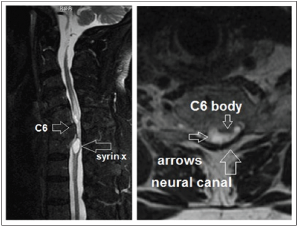
Decades of spinal cord repair efforts had been crowned by stem cells SC role as a very recent promising scientific prospect. Many and a lot of work all over the world used SC in very different ways and techniques to overcome the damage in spinal cord whether morbid or traumatic unfortunately until the date without yield. They used SC from placental/umbilical cord, bone marrow, perhaps blood components and recently the adipose tissue. I intended to use adipose tissue derived SC to treat a 70 years old man with a tetraplegia of 30 years due to a road traffic accident cervical 6 vertebral fracture. At the time of case discussion with the patient and his family my plan was to MRI imaging, to see the basic structural damage and to reveal any improvements after SC use, the MRI showed huge neural canal narrowing at the C6 due to fractured vertebral hypertrophy over these 30 years with adjacent inferior syrinx the case necessitated a laminectomy decompression with syrinx opening. The other factor in family discussion was the tissue biopsy for detection of any intracellular bacteria by molecular study as screen for unknown which was developed in a molecular biology lab. As I am increasing my knowledge in adipose tissue manipulation for being as SC, I realized that better if we can use brain tissue instead. Why brain tissue in a time it is highly differentiated and from where to take, do we want to build a place and spoil the other!! In some of my writings I mentioned the huge capability of brain to repair itself whatever the age and the damage is/ are just if we free its cells from the intracellular bacteria which I find in the biopsies I take in different diseases one of them is the cerebral palsy due to any cause standardly mentioned. This repair capacity comes from the bulk of brain parenchyma is as if being as a reserve to act as a spare part or as what SC do in other places of the body whatever the kind or source of these SC. Neurologists in general and neurosurgeons in particular know that some areas in brain can be incised and dipping deep in the white matter e.g. to remove a deep parietal big meningioma without apparent functional problem, the examples are much. Functionally we use a small portion of our brain, so the rest kept as reserve for this highly specialized and organized system. While spinal cord is with none of that completely. For that the generous brain looks like to act as a bank of cells to the spinal cord when needed for any cause. So, the foreign or non-CNS cells are not welcomed to do any task in the master system like in spinal cord highly sophisticated arrangement. Since no one is well knowledgeable of how stem cells CS come to work here or there, where we just armed with a single sentence that nondifferentiated turn into differentiated in place where it is needed to replace damaged native cells. So how to use highly differentiated or specialized cells like neurons and glial cells into spinal cord?! This can be analyzed by three hypotheses: First, CNS such a highly organized system the part of its sophistication lies in that its cells are also sophisticated where know what to do in such environment, the plain or blank cells from other where may not be qualified or even allowed. Secondly: as there is sufficient repair capacity of brain there should be some sort of cells can turn to what spinal cord needs so the glial cells the servant and framework of CNS take the task. Third: taking of a piece or a lump of cortex with white matter in a plaque and put it against the area of spinal cord that suffers defect as in our case the spinal cord is thread like in the area of fractured and hypertrophied vertebra with adjacent lower syrinx making the cord membrane like in all sides. This lump does not need to be turned into cells sap or suspension to keep the cellular scheme functionality in invading and opening the lines formed by morbid healing process as if it’s a patrol unit come to repair in the scene field. The same is true when the spinal cord is apparently normal in bulk, however there is no room for bulky plaque or lump some modifications may be done like cortex- white matter slicing put as much as possible to cover the sides of the level or the segment in question clinically. This may give rise to the or analyze why poor results up to date when other kind of repair or stem cells been used with different augmenting measures. Here talking as a neurosurgeon non-dominant parietal hemisphere as an initial career may taking safely. The size of lump is not in discussion where it follows the logic in every case. Animal studies are not necessary because the lines are straight forward and the scope of morbidity is negligible brain wise as for spinal cord I think it is wealthy to conduct where no loss more after 30 years of paraplegia and deformity in upper limbs due to weak lower cervical innervation. This is our case a male of 70 years old had RTA 30 years ago with hyperextension C6 fracture with quadriplegia, recent MRI study to show sever narrowing with syrinx (Figure 1).
Figure 1:

© 2020 Abbas Alnaji. This is an open access article distributed under the terms of the Creative Commons Attribution License , which permits unrestricted use, distribution, and build upon your work non-commercially.
 a Creative Commons Attribution 4.0 International License. Based on a work at www.crimsonpublishers.com.
Best viewed in


.jpg)


 Editorial Board Registrations
 Submit your Article
 Refer a Friend
 Advertise With Us
.jpg)


.jpg)


.bmp)
.jpg)
.png)
.jpg)


.jpg)


.png)

.png)


.png)


