- Submissions

Full Text
Research in Medical & Engineering Sciences
Dissection and Rheological Characterization of Cow Tail Discs as a Model for Human Spinal Discs
Katrina J Donovan1* and Skip Rochefort2
1Materials and Metallurgical Engineering, South Dakota Mines, South Dakota, USA
2School of Chemical, Biological, and Environmental Engineering, Oregon State University, Corvallis, USA
*Corresponding author:Katrina J Donovan, Materials and Metallurgical Engineering, South Dakota Mines, Rapid City, South Dakota, USA
Submission: February 12, 2024;Published: February 29, 2024

ISSN: 2576-8816Volume11 Issue1
Abstract
For this study, frozen tails of mature cows were obtained from a local slaughterhouse. The tails have 6-9 discs which are similar in size to human spinal discs. The discs were removed from the tail, thawed, dissected, and tested the same day. There was no added liquid, re-freezing, etc. Dynamic oscillatory compression tests were performed on the cow (bovine) spinal discs and compared to previously published human data.
Keywords:Spinal disc; Cow disc model; Dynamic modulus
Background
Low back (lumbar) pain is a medical condition that affects approximately 619 million people globally [1]. The spinal disc is composed of two main components: the annulus fibrosis (AF) and the nucleus pulposus (NP).
Herniation is the result of the weakening of the AF, the outer portion of the spinal disc [2]. This weakened region of the AF creates a failure point for the NP to extrude from the spinal disc cavity. The extruded NP compresses on the spinal nerve and induces severe back pain [3].
The traditional treatment for spinal disc herniation is spinal fusion surgery. Two or more bones are connected by bone or bonelike material in the space between the spinal bones, held together with metal screws or rods. The procedure eliminates movement to prevent pain, but the patient is left with severely limited spinal mobility [4]. Recent techniques have suggested removal of the herniated disc and replacement with mechanical or polymeric models, or simply removal of the extruded portion (discectomy) leaving a degenerated disc.
Several techniques have been investigated in our laboratories which involve removal of the extruded portion of the NP and replacement of the lost material with the injection of a polymer solution into the disc cavity, which would then form a hydrogel in-situ, “re-inflating” the disc. To formulate a hydrogel to simulate the mechanical properties of the original disc it is necessary to measure the mechanical properties of healthy human discs, which has proven to be a challenge. In 2009 Butterman [4] proposed that the discs in the tails of mature cows as a good model for human spinal discs [4]. The techniques to obtain and test bovine discs are presented in this paper.
Procedure
For this study, frozen tails of mature cows were obtained from a local slaughterhouse. The tails have 6-9 discs which are similar in size to human spinal discs. The discs were removed from the tail, thawed, dissected, and tested the same day. There was no added liquid, re-freezing, etc. Two procedures to prepare the bovine discs for mechanical compression testing were used. For mechanical analysis of the whole disc (NP and AF), the bovine disc was dissected from the tail (Figure 1A) and compression tested using an 8mm plate (Figure 1B). The NP of the bovine disc was dissected (punched) from whole disc (Figure 1C) for compression testing (Figure 1D).
Figure 1:A. Dissected bovine caudal disc with annulus fibrosis surrounding the nucleus pulposus.
B. The parallel plate analyzing a whole disc on the DHR.
C. Size comparison of punched and whole disc.
D. An 8mm parallel plate compressing a punched (10mm) disc.

Result and Discussion
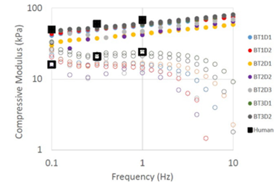
Dynamic oscillatory compression tests were performed on the bovine spinal discs to compare to the human data published by Freeman et al. [5]. The compressive moduli from a dynamic oscillatory axial compression frequency sweep at 1% strain reported (Figure 2). TA Instruments DHR3 in axial compression was utilized to perform this test. The bovine results in Figure 2 are from three separate bovine tails, the solid circles represent the elastic storage modulus (E’), and the open circles represent the viscous loss modulus (E”). The bovine discs compare favorably with the human values (black squares in Figure 2) reported in Freeman et al. [5]. The bovine specimens are of varying age, and discs were dissected from different portions of the tail. As mentioned previously, a typical cow tail will have 6-9 discs of comparable size to human spinal discs. Given that the discs were obtained from various cow tails at various locations on the tail, the reproducibility of the bovine data and favorable comparison to human data confirms the viability of bovine tails as a suitable compressive mechanical pseudo-model for human spinal discs. The bovine testing was extended to higher frequencies than previous studies, indicating the potential to predict the mechanical behavior of human spinal discs at higher frequencies.
Figure 2:C Compressive modulus (E) from the dynamic oscillatory axial compression frequency sweep at 1% strain is reported. TA Instruments DHR3 model in axial compression was utilized to perform the compression tests on the bovine specimen.

Summary
A technique is presented to obtain discs from cow tails that are comparable in size and structure to human spinal disc. The disc can be tested as dissected from a single frozen state without any artificial manipulation to hydrate, refreeze, etc. Typical cow tails contain 6-9 discs which meet the desired size and performance specifications. The compressive modulus as a function of deformation frequency for cow tail discs are reproducible over a range of specimens from different cow tails. The bovine results compare favorably to the human spinal disc data, indicating that cow tail discs provide a suitable mechanical model for healthy human spinal discs, which are often very difficult to obtain.
Acknowledgement
Special thanks to our dissection experts Emily Harding, Aileen Murphy, and Libby Williams, and to Dr. Glenn Butterman for pointing us to the cow tails.
References
- GBD 2021 Low Back Pain Collaborators (2023) Global, regional, and national burden of low back pain, 1990-2020, its attributable risk factors, and projections to 2050: A systematic analysis of the global burden of disease study 2021. Lancet Rheumatology 5(6): e316-e329.
- Rahman S, Das JM (2023) Anatomy, head and neck: Cervical spine. StatPearls.
- Mayo Foundation for Medical Education and Research (2022) Spinal fusion. Mayo Clinic.
- Buttermann GR, Beaubien BP, Saeger LC (2009) Mature runt cow lumbar intradiscal pressures and motion segment biomechanics. The Spine Journal 9(2): 105-114.
- Freeman AL, Buttermann GR, Beaubien BP, Rochefort WE (2013) Compressive properties of fibrous repair tissue compared to nucleus and annulus. Journal of Biomechanics 46(10): 1714-1721.
© 2024 Katrina J Donovan. This is an open access article distributed under the terms of the Creative Commons Attribution License , which permits unrestricted use, distribution, and build upon your work non-commercially.
 a Creative Commons Attribution 4.0 International License. Based on a work at www.crimsonpublishers.com.
Best viewed in


.jpg)


 Editorial Board Registrations
 Submit your Article
 Refer a Friend
 Advertise With Us
.jpg)


.jpg)


.bmp)
.jpg)
.png)
.jpg)


.jpg)


.png)

.png)


.png)


