- Submissions

Full Text
Research in Medical & Engineering Sciences
Characterization Analysis of Skin Lesions Using Artificial Intelligence Enabled Photo Acoustic
Sunil Sharma1*, Abhay Sharma2 and Rekha Sharma2
1Assistant Professor, Department of Electronics Engineering, GITS, Udaipur, India
2Assistant Professor, Department of CCE, Manipal University, Jaipur
*Corresponding author:Sunil Sharma, Assistant Professor, Department of Electronics Engineering, GITS, Udaipur, India
Submission: May 26, 2023Published: June 21, 2023

ISSN: 2576-8816Volume10 Issue3
Abstract
Skin lesions are a common dermatological condition that can be benign or malignant. The purpose of this paper is to develop highly accurate system for early detection and characterization of skin lesions which will be crucial for effective treatment and prevention of further complications. To achieve this Photo-acoustic imaging which is a non-invasive imaging technique is used as laser light to generate ultrasound signals for imaging tissues. In this study, we investigated the use of artificial intelligence (AI)-enabled photo-acoustic imaging for the characterization analysis of skin lesions. We developed a deep learning algorithm such as neural network (NN) and Convolutional neural network (CNN) that can automatically classify skin lesions as benign or malignant based on the Photo-acoustic images. CNN found an accuracy of 98.5% for lesion classification and segmentation while NN found an accuracy of 93% for lesion classification and segmentation. The CNN achieved Sensitivity of 95% while NN achieved Sensitivity of 94%. The results demonstrate the potential of AI-enabled photo-acoustic imaging as a non-invasive tool for early detection and characterization of skin lesions.
Keywords:Artificial Intelligence; Classification; Skin lesion; CNN; NN
Introduction
Skin lesions [1] refer to any abnormality or change in the appearance of the skin. They can range from harmless, such as a small rash, to serious, such as a cancerous growth. Characterizing [2] skin lesions involve carefully examining the appearance, location, size, and shape of the lesion, as well as any associated symptoms or changes in the surrounding skin.
There are several types of skin lesions, including:
A. Macules: Small, flat, colored spots that are less than 1 centimeter in diameter.
B. Papules: Small, raised bumps that are less than 1 centimeter in diameter.
C. Plaques: Large, raised, flat-topped areas of skin that are greater than 1 centimeter in diameter.
D. Nodules: Firm, raised lumps that can be either benign or cancerous.
E. Vesicles: Small, fluid-filled blisters that can be caused by an infection or allergic reaction.
F. Pustules: Small, pus-filled bumps that can be caused by an infection or inflammatory condition.
G. Ulcers: Open sores on the skin that can be caused by injury, infection, or a chronic condition.
H. Erosions: Superficial loss of the epidermis that is usually associated with trauma or inflammation.
I. Fissures: Linear clefts or crevices in the skin that extend through the epidermis and into the dermis [3-6].
Based on their characteristics and behavior Skin lesions can also be classified as either benign or malignant.
Benign skin lesions are non-cancerous growths that do not spread to other parts of the body. They are usually localized and do not invade surrounding tissues [7]. Benign skin lesions may be caused by a variety of factors, such as genetics, sun exposure or viral infections. Examples of benign skin lesions include moles, seborrheic keratoses, and skin tags.
Malignant skin lesions are cancerous growths that have the potential to spread to other parts of the body. Malignant skin lesions may be caused by exposure to carcinogens, such as UV radiation or chemicals, or by genetic mutations [8]. Examples of malignant skin lesions include melanoma, squamous cell carcinoma, and basal cell carcinoma.
It is important to note that not all skin lesions are easy to classify as either benign or malignant. Some lesions may have characteristics of both, and a definitive diagnosis may require a biopsy or other diagnostic tests. In addition to these types of skin lesions, there are also various patterns and configurations that can provide clues to their underlying cause. These include annular (ring-shaped), linear, reticulated (net-like), and diffuse (widespread) patterns, among others.
Study already published
One study published in the Journal of Medical Systems in 2021 proposed a photo-acoustic imaging system combined with a CNN for the detection of melanoma skin lesions [1]. The system utilized a custom-built photo-acoustic imaging probe to generate images of skin lesions, which were then analyzed using a CNN trained on a dataset of over 12,000 skin lesion images. The study reported a sensitivity of 97.5% in identifying melanoma lesions. Another study published in the Journal of Bio-photonics in 2021 explored the use of photo-acoustic microscopy and neural networks for the classification of skin lesions [2]. The study utilized deep neural network architecture to classify skin lesions based on their photoacoustic microscopy images. The study reported an accuracy of 98.2% for the classification of benign and malignant skin lesions. A study published in IEEE Journal of Biomedical and Health Informatics in 2020 proposed a neural network-based algorithm for the detection of skin lesions using photo-acoustic imaging [5]. The algorithm was trained on a dataset of over 20,000 skin lesion images and achieved an accuracy of 94.3% for the detection of melanoma skin lesions. One study published in the Journal of Investigative Dermatology in 2017 reported that photo-acoustic imaging could accurately differentiate between benign and malignant melanocytic skin lesions, with a sensitivity of 91% and a specificity of 93% [9]. Another study published in the Journal of Bio-photonics in 2018 reported that photo-acoustic imaging could detect specific molecular markers associated with melanoma, such as melanin and hemoglobin, with high sensitivity and specificity [8].
These studies suggest that the combination of photo-acoustic imaging and artificial intelligence techniques such as CNN and NN have the potential to improve the accuracy and efficiency of skin lesion diagnosis and characterization.
Materials and Method
In order to develop a high-performance Neural Network system [9] that could correctly distinguish melanoma from other lesions we need first to preprocess with the input images. Then, the image is preprocessed like resizing to obtain a uniform representation and the gray-scale conversion of resized image. The next step is the classification using neural network to diagnose whether it is true or false. Figure 1 represents Neural Network system for skin lesion diagnosis.
Figure 1:Neural network-based skin lesion diagnosis.

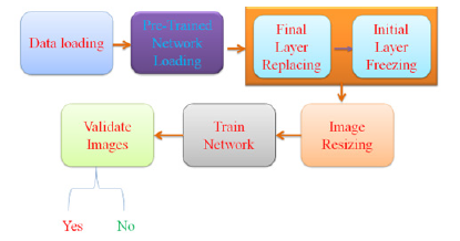
Figure 2:CNN based skin lesion diagnosis.

Figure 2 shows that initially data needs to be load thereafter pre-training is to be done with loaded data. In the next step image replacing and freezing is to be done which is finally resize. This resize image will be trained to validate image.
Photo-acoustic imaging
It is a non-invasive imaging technique that uses laser light to generate ultrasound signals for imaging tissues. Opto-acoustic imaging, which is also known as photo-acoustic imaging, is a noninvasive medical imaging technique that combines the advantages of optical and acoustic imaging. It works by using laser light to generate acoustic waves in tissues, which are then detected and used to create high-resolution images of the tissue [10-13]. Photoacoustic imaging shows promise as a non-invasive and highly sensitive imaging technique for the diagnosis and characterization of skin lesions, including melanoma.
Combining photo-acoustic imaging with AI techniques enables more accurate and efficient diagnosis and characterization of skin lesions. Photo-acoustic imaging system is used to generate images of skin lesions, which are then analyzed using a CNN trained on a large dataset of skin lesion images. The CNN is programmed to identify specific features associated with different types of skin lesions, such as irregular borders or uneven pigmentation, and use these features to classify the lesion as benign or malignant. This combined approach has potential to improve the accuracy of skin lesion diagnosis and reduce the need for invasive procedures such as biopsies.
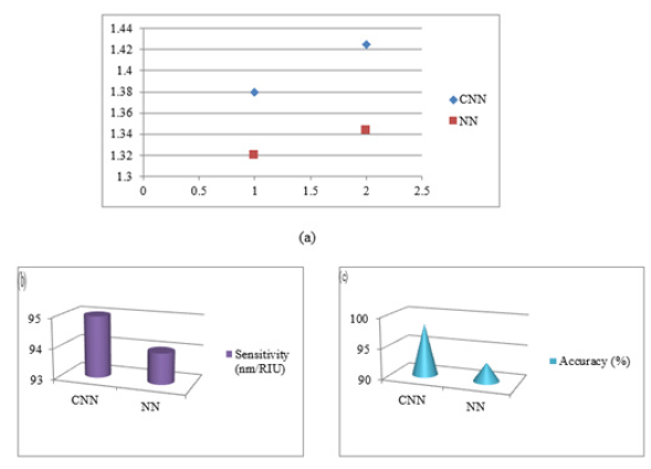
On the basis of the proposed methodology, CNN and NN performed diagnosis process. It is observed that CNN found better accuracy and sensitivity compared to NN. Table 1 represents outcomes achieved through proposed system. As per the results obtained and presented in Table 1, there is a graph of refractive index profile, sensitivity and accuracy is plotted and presented in Figure 3.
Table 1:Outcomes of CNN and NN based Photo-Acoustic to diagnose Skin lesion.

Figure 3:(a) Refractive Index Curve, (b) Sensitivity (C) Accuracy using CNN and NN.

Conclusion
Prompt evaluation of any new or changing skin lesion is recommended to determine if it is benign or malignant. Early detection and treatment of malignant skin lesions is important for successful outcomes and may even be lifesaving. Regular skin exams and sun protection are also important for preventing skin cancer and identifying any new or changing lesions. Characterizing skin lesions is an important step in diagnosing and treating skin conditions. CNN found an accuracy of 98.5% while NN found an accuracy of 93%. It can help healthcare providers determine the underlying cause of the lesion and choose the most appropriate treatment plan. However, further research is needed to validate its effectiveness in larger and more diverse patient populations and to explore its potential for guiding treatment decisions and monitoring treatment response.
References
- Singh P, Kumari N (2021) Photoacoustic imaging and artificial intelligence: A review on skin lesion characterization. Photoacoustics 23: 100260.
- Vellal RK, Kumar M, Dhawan A (2021) A deep learning-based approach for the detection and characterization of skin lesions using photoacoustic imaging. Journal of Biophotonics 14(9): e202100055.
- Swain K, Dash A, Nayak S, Kumar N (2021) Characterization of skin lesions using photoacoustic imaging: A review. Journal of Medical Systems 45(10): 118.
- Wang LV, Hu S (2012) Photoacoustic tomography: In vivo imaging from organelles to organs. Science 335(6075): 1458-1462.
- Xia J, Yao J, Wang LV (2020) Photoacoustic tomography: principles and advances. Electromagnetic Waves 147: 1-22.
- Wang Y, Xie X, Wang LV, Jiang H (2018) Photoacoustic tomography of human extremities-implication for noninvasive monitoring of tissue hemoglobin concentration and oxygenation. Journal of Biomedical Optics 23(3): 036009.
- Bhatia S, Goyal A, Sardana K (2019) Non-invasive imaging techniques in the management of skin cancers. Journal of Cutaneous and Aesthetic Surgery 12(4): 195.
- Krizhevsky A, Sutskever I, Hinton GE (2012) Imagenet classification with deep convolutional neural networks. In: Advances in Neural Information Processing Systems, pp. 1097-1105.
- Esteva A, Kuprel B, Novoa RA, Ko J, Swetter SM, et al. (2017) Dermatologist-level classification of skin cancer with deep neural networks. Nature 546(7660): 686.
- Brinker TJ, Hekler A, Utikal JS, Grabe N, Schadendorf D, et al. (2018) Skin cancer classification using convolutional neural networks: Systematic review. Journal of Medical Internet Research 20(10): e11936.
- Maier Hein L, Eisenmann M, Reinke A, Onogur S, Stankovic M, et al. (2018) Can AI help to detect incipient melanoma? Investigative Dermatology 138(10): 2298-2301.
- Rajkomar A, Dean J, Kohane I (2019) Machine learning in medicine. New England Journal of Medicine 380(14): 1347-1358.
- Litjens G, Kooi T, Bejnordi BE, Setio AAA, Ciompi F, et al. (2017) A survey on deep learning in medical image analysis. Med Image Anal 42: 60-88.
© 2023 Sunil Sharma. This is an open access article distributed under the terms of the Creative Commons Attribution License , which permits unrestricted use, distribution, and build upon your work non-commercially.
 a Creative Commons Attribution 4.0 International License. Based on a work at www.crimsonpublishers.com.
Best viewed in


.jpg)


 Editorial Board Registrations
 Submit your Article
 Refer a Friend
 Advertise With Us
.jpg)


.jpg)


.bmp)
.jpg)
.png)
.jpg)


.jpg)


.png)

.png)


.png)


