- Submissions

Full Text
Orthoplastic Surgery & Orthopedic Care International Journal
A rare manifestation of intraosseous synovial cyst of wrist scaphoid bone: Current concepts
Kastanis G1*, Magarakis G1, Kapsetakis P1, Spyrantis M3, Stavrakakis I1 and Pantouvaki A2
1Department of Orthopaedic, General Hospital of Heraklion-Venizeleio, Greece
2Department of Physiotherapy, General Hospital of Heraklion-Venizeleio, Greece
3Department of Orthopaedic and Traumatology, University Hospital of Heraklion, Greece
*Corresponding author: Grigorios Kastanis, Department of Orthopaedic, General Hospital of Heraklion-Venizeleio, Crete, Greece
Submission: May 25, 2021;Published: June 28, 2021
ISSN 2578-0069Volume2 Issue3
Abstract
Intraosseous wrist bone cyst lesion is a rare manifestation and presents an etiology of chronic wrist pain. We report a case presented at emergency department with a mild pain after a fall on outstretched hand with the initial x-rays views and computer tomography imaging diagnosed an intraosseous scaphoid ganglion cyst without fracture, while postoperative histopathological evidence shows a scaphoid synovial cyst. Finally, we analyzed the current concepts regarding the diagnosis and therapeutical management.
Keywords: Bone cyst; Wrist scaphoid cyst; Carpal bone; Wrist fractures
Introduction
Intraosseous cystic lesion of carpal bones is rare manifestation and in literature a small sample of cases has been reported [1]. With the term intraosseous cyst is described a closed cavity into bone which is surrounded by a membrane and stands out from the surrounding tissues [2]. The majority of them is localized in proximal carpal row bone and diagnosed incidentally on radiography for other causes [3]. The exact mechanism is unclear and many theories have been proposed with the most accepting one the minor trauma, leading to intraosseous lesion which initiated intramedullary metaplasia sequence by fibrous connective tissue proliferation [2]. Most of cases are asymptomatic for a long period of time but when patients typically present symptoms are generalized, resulting in delayed diagnosis and therapeutical handling [4].
In wrist scaphoid bone different lesions have been reported in literature which are characterized as intraosseous ganglion cysts or degenerative cysts secondary to osteoarthritis [5]. Intraosseous synovial cyst (IOSC) is very exceptional in carpal bones especially in scaphoid and until today only one case has been reported [6,7]. The aim of this study is to report a case of female with a painful wrist after a fall on outstretched hand examinated in our emergency department and on initial radiography and computed tomography imaging diagnosed with ganglion cyst while the postoperative histopathological evidence showed intraosseous synovial cyst and to analyze the current concept regarding the diagnosis and therapeutical management.
Case Report
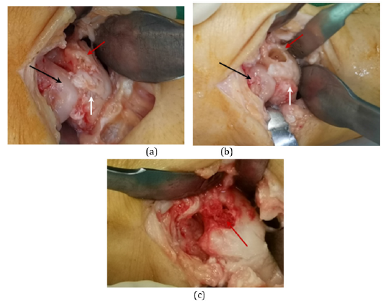
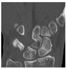
A 42 years-old female proceeded in emergency department, after a fall from a standing height, with a painful left wrist (dominant hand). During examination we diagnosed a mild dorsal swelling and some restriction of range motion of left wrist, as a result of pain. The patient did not present pain at radial styloid area or scaphoid tubercle during palpation. Initial x-rays (AP and oblique views) presented a well described intraosseous cyst with sclerosis extended from proximal pole until waist of left scaphoid without evidence of fracture (Figure 1a-b). A palmar half plaster of paris was applied for ten days and reexamination with computed tomography was suggested. CT scan delineated an intraosseous osteolytic lesion of the left scaphoid with surrounding anatomical element without sign of fracture at proximal pole near scapholunate joint and diagnosed as a ganglion cyst (Figure 2a-c). Because the patient presented painful wrist and restriction of range of motion of the wrist during daily activities, we decide to undergo to surgical excision of the lesion with the approval of the patient. Under regional anesthesia in supine position with arm tourniquet a dorsolateral approach performed to examinated the scaphoid. After trepanation of the lytic lesion, a yellow gelatinous liquid was found in the interior aspect of the bone which was given for histopathological examination. We curettage the cavity and cancellous bone graft from distal epiphysis of radius applied (Fig 3a-c). Post operatively a palmar functional brace was performed for 6 weeks (all day long) and for the other three weeks performed only during nights. From the first postoperative day, patient trained to follow a rehabilitation program. The treatment plan included passive and passive-assisted exercises of the fingers to control oedema, prevent joint stiffness and dysfunction initially and active movements to enhance range of motion after brace removal in a later stage.
Figure 1:Preoperative x-Rays (AP- oblique) which describe a intraosseous cyst on left scaphoid bone (red arrow).
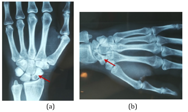
Figure 2:Preoperative ct/scan of left wrist (a-b) which diagnosed a ganglion cyst of left scaphoid bone localizated at proximal pole(red arrow).
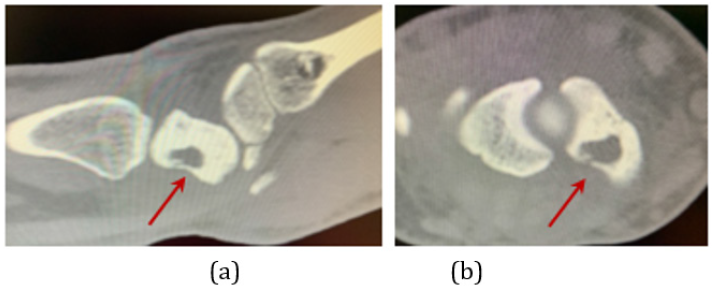
Figure 3:Intraoperative views of scaphoid. Sclerotic lesion (a) of the scaphoid (red arrow), cavity of scaphoid bone after curettage (b- red arrow) and after cancellous grafting of cavity (c) (hamate -black arrow, lunate- white arrow).
Histopathological evidence revealed an intraosseous synovial cyst (calcified fibrohyaline tissue with nonatypical fibroblastic cells). Bone consolidation of the scaphoid at x-ray examination appears at six months (Figure 4a-b). At final examination, one year postoperatively, the patient appeared with a good range of motion without pain and restriction of the wrist during daily living activities (Figure 5a-d), while ct/scan views did not appear recurrence but full consolidation of wrist scaphoid Figure (6a).
Figure 4:X-Rays views AP(a) and Profile(b) at six months postoperative.
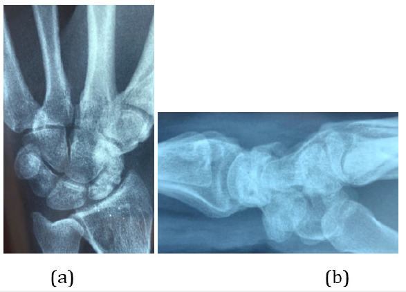
Figure 5:Range of motion of left wrist at one year (a,b,c,d).

Figure 6:CT/scan of left scaphoid at one year postoperative.
Discussion
Intraosseous carpal bone cysts lesions are very uncommon manifestations and for this reason a few studies have been reported in literature. The majority of them have a trend to be localized at proximal carpal row and especially at the lunate, scaphoid and triquetrum bones [8]. The most common type of these lesions is the intraosseous ganglion cyst. Hicks et al. [9] first described a lesion which developed slowly from an area of trabecular fading to define cavity walled off by a zone of radiopacity and characterized it as «synovial bone cyst» while Grabbe et al. [10] first introduced the term «intraosseous ganglion». Nowadays several names of the same lesion have been reported as: synovial bone cyst, ganglionic cystic defect of bone, subchondral bone cyst and juxta-articular bone cyst [11].
Intraosseous cyst lesion was diagnosed incidentally on plain radiography views because in majority of cases is asymptomatic. The lesion began to be symptomatic when the cyst lesion increased progressively into bone cavity and the patients report non-specific chronic wrist pain or created bone fracture from the increased volume of the cyst or after a minor injury [4,12]. Localization of intraosseous synovial cyst in wrist scaphoid bone (IOSC) is very rare, because the most common location is in epiphysometaphyseal zone of long bones principally in lower extremity [11]. For this reason the differential diagnosis included a variety of other pathology: ganglion cyst, aneurismal bone cyst, osteoid osteoma, osteoblastoma, chondroblastoma, osteosarcoma, benign cystic lesions, and osteomyelitis or preiser disease [13,14].
Two theories have been proposed for this lesion pathology. Magee et al. [15] proposed the traumatic theory that the cyst lesion comes from repetitive stress or micro trauma, causing metaplastic change of intramedullary mesenchymal stem cells into synovial type cells. This leads to traumatic bone necrosis, which following bone resorption and bone cavity is embody with metaplased cells [15]. In opposite Mnif et al. [16] proposed the penetration theory according to which cyst lesion is created by synovial inclusion from outside which penetrate the adjoining carpal bone cortise.17 In our case the patient refers prior injury, mild wrist pain and being manual worker, we believe that this cyst lesion is a result of traumatic etiology.
Scaphoid cyst lesions are frequently diagnosed on radiography views and according to Paparo et al. [3] is sufficient the diagnosed examination for controlled cyst progression and the percentage of post –treatment recurrence while Veseley et al. [19] suggest the poor sensitivity on radiographs when intramedullary cyst have small size. Dumas et al. [12] proposed that ct/scan is more sensitive examination offered two major advantages: first confirms the diagnosis of the cyst and eventual fracture and second is easier to preoperative surgical planning. Van den Dungen et al. [18] suggest magnetic resonance imaging to recognize identify soft tissue participation, with diagnosis further supported with nuclear bone scans. We believe that ct/scan offer more information about the characteristic pattern of the cyst (type, size, bone fracture), to differential diagnosis from other pathologies and follow the risk of postsurgical recurrence but we agree with Sbai et al. [11] that only anatomopathological examination of the curettage material is necessary to confirm the diagnosis. Initially in our case a diagnosed (x-rays and ct/scan) ganglion cyst is reported, while histopathological diagnosis approved a synovial cyst.
The treatment modality depended on the size of lesion, eventual fracture and symptoms. In initial stage with mild wrist symptoms Javdan et al. [19] suggested conservative treatment as anti-inflammatory drugs, immobilization, lifestyle modification and restriction of activity. Castellanos et al. [20] proposed conservative management of pathological fracture of scaphoid with belowelbow cast for eight weeks for bone union fracture and progressive disappearance of the cyst 12 months postinjury.
Indication for surgical management consists of persistent wrist pain, pathological fracture or cortical erosion and rapid progression,failure of conservative treatment (least six months) [4]. The main target of surgical therapy is to excise complete the cystic lesion and placed autologous cancellous bone graft with scope to restoration bone entity and avoid the risk of recurrence. The role of bone graft is to avoid fracture or wrist collapse while the type of graft is a matter of controversy in literature [4]. There are proponents that suggest vascularised bone graft (volar carpal artery) and other that proposed autogenous cancellous bone graft (from the radius or iliac crest) to improve bone union [2,6,7,11,15,21]. Other researchers have proposed other therapeutically management as: intracystic injection of methylprednisolone acetate, structural support with flexible intramedullary nailing, decompression with multiple drill holes, cannulated screws, or any combination of the above [22]. Bain et al. [5] introduce arthroscopically assisted minimally method of debridement and grafting under fluoroscopic guidance. Finally postsurgical immobilization is essential for 5 weeks to discharge the pain.
In our patient we apply cancellous bone graft from distal radius and postoperatively a functional wrist brace was applied for 6 weeks daily and for the next three weeks brace was applied only during night.
Conclusion
Intraosseous synovial cyst of scaphoid is a very rare lesion, diagnosed incidentally, usually after a light injury. Generally it has the same imaging characteristics with other lesions and especially with ganglion cyst. Computer tomography examination offers the initial diagnosis but histopathological evidence presents the accurate diagnosis. Indication for surgical treatment (curettage and bone grafting or osteosynthesis) is the presence of fracture or when the dimension of the cyst is such that it will create one.
Conflict of Interest
The authors declare that have no conflict of interest.
Ethical Approval
Our institution does not require ethical approval for reporting individual cases or case series.
Consent
Verbal informed consent was obtained from the patients for their anonymized information to be published in this article.
References
- Takahata S, Ogino T, Minami A (1992) Carpal bone cyst. In: Nakamura R, Linscheid RL, Miura T (Eds.,) Wrist Disorders: Current Concepts and Challenges. Springer, Tokyo, Japan, Pp: 257-260.
- Ikeda M, Oka Y (2000) Cystic lesion in carpal bone. Hand Surg 5(1): 25-32.
- Paparo F, Fabbro E, Piccazzo R, Revelli M, Ferrero G, et al. (2012) Multimodality imaging of intraosseous ganglia of the wrist and their differential diagnosis. Radiol Med 117(8): 1355-1373.
- Osagie L, Gallivan S, Wickham N, Urmarji S (2015) Intraosseous gaglion cysts of the carpus: current practice. Hand 10(4): 598-601.
- Bain GI, Turner PC, Ashwood N (2008) Arthroscopically assisted treatment of intraosseous ganglions of the lunate. Tech Hand Up Extrem Surg 12(4): 202-207.
- Sbai MA, Benzarti S, Sbei F, Maalla R (2016) A pathological fracture of the scaphoid revealing an intraosseous ganglion cyst. The Pan African Medical Journal 23(2): 185.
- Chouchene MO, Gallas A, Hashicha H, Mghirbi R, Sbai MA (2020) Intraosseous synovial cyst of carpal scaphoid: case report and review of literature. AJRS 3(3): 1-5.
- Eiken O, Jonsson K (1980) Carpal bone cysts: A clinical and radiographic study. Scand J Plast Reconstr Surg 14(3): 285-290.
- Hicks JD (1956) Synovial cysts in bone. Aust N Z J Surg 26(2): 138-143.
- Crabbe WA (1966) Intra-osseous ganglia of bone. Br J Surg 53(1): 15-17.
- Sbai MA, Benzarti S, Boussen M Msek H, Maalla R (2016) Intraosseous ganglion cyst of the lunate: a case report. Chin J Traumatol 19(3): 182-184.
- Dumas P, Georgiou C, Chignon SB, Balaguer T, Lebreton E, et al. (2012) Intra-osseous ganglion cyst of the carpal bones. A review of the literature underlining the importance of systematic computed tomography. Chir Main 32(1): 3-7.
- Bennett DC, Hauck RM (2002) Intra-osseous ganglion of the lunate. Ann Plast Surg 48(4): 439-442.
- Bulut M, Tosun HB, Simsek BC, Karakurt L (2012) Primary hydatid cyst of the scaphoid: case report. J Hand Surg Am 37(5): 1051-1053.
- Magee T, Rowedder A, Degnan G (1995) Intraosseous ganglia of the wrist. Radiology 195(2): 517–520.
- Mnif H, Koubaa M, Zrig M, Jawahdou R, Sahnoun N, et al. (2010) Ganglion cyst of the carpal navicular. A case report and review of the literature. Orthop Traumatol Surg Res 96(2): 190-193.
- Vesely M, Burge P (1999) Intraosseous ganglion of the trapezium in communication with the flexor carpi radialis tendon sheath. J Hand Surg Br 24(4): 486-488.
- Van den Dungen S, Marchesi S, Ezzedine R, Bindou D, Lorea P (2005) Relationship between dorsal ganglion cysts of the wrist and intraosseous ganglion cysts of the carpal bones. Acta Orthop Belg 71(5): 535-539.
- Javdan M, Zarezadeh A, Gaulke R, Eshaghi MA, Shemshaki H (2012) Unicameral bone cyst of the scaphoid: a report of two cases. J Orthop Surg (Hong Kong) 20(2): 239-242.
- Castellanos J, Bertràn C, Pérez R, Roca J (2001) Pathologic fracture of the scaphoid caused by intraosseous ganglion followed by regression after the healing of the fracture. J trauma 51(1): 141-143.
- Yakoubi M, Meziani N, Yahia Cherif M, Zemmouri A, Benbakouche R (2009) Pathological fracture of the carpal scaphoid. Chir Main 28(1): 37-41.
- Safran T, Hazan J, Al-Halabi B, Al-Naeem H, Cugno S (2019) Scaphoid cysts: Literature review of etiology, treatment and prognosis. Hand 14(6): 751-759.
© 2021 Kastanis G. This is an open access article distributed under the terms of the Creative Commons Attribution License , which permits unrestricted use, distribution, and build upon your work non-commercially.
 a Creative Commons Attribution 4.0 International License. Based on a work at www.crimsonpublishers.com.
Best viewed in


.jpg)


 Editorial Board Registrations
 Submit your Article
 Refer a Friend
 Advertise With Us
.jpg)


.jpg)


.bmp)
.jpg)
.png)
.jpg)


.jpg)


.png)

.png)


.png)


