- Submissions

Full Text
Open Access Research in Anatomy
Preliminary Report of Pachyosteosclerotic Bones in Seals
Irina Koretsky and Sulman J Rahmat*
Department of Anatomy, Howard University, USA
*Corresponding author: Sulman J Rahmat, Laboratory of Evolutionary Biology, Department of Anatomy, College of Medicine, Howard University, Washington DC, USA
Submission: August 08, 2017; Published: September 06, 2017

ISSN: 2577-1922 Volume1 Issue1
Introduction
Despite extensive knowledge about the distribution of pachyosteosclerosis (increased bone volume and density) among some modern groups of marine mammals, this aquatic adaptation is not well known in Phocidae (true seals). Pachyosteosclerotic bones reduce buoyancy and permit easier submergence for some marine mammals. Pachyostosis and osteosclerosis are two vastly different bone adaptations, which have co-occurred independently (termed pachyosteosclerosis). Pachyostosis describes the thickening of bone in cross-sectional area, whereas osteosclerosis is the replacement of cancellous bone with compact bone. Osteosclerosis, pachyostosis, and bone lightening consecutively occurred to various degrees as adaptations of marine mammals to different environmental niches and lifestyles.
Differing extents of pachyosteosclerosis has been demonstrated in Recent phocids (true seals) [1], otariids (sea lions) [2], odobenids (walruses) [3,4], cetaceans (whales and dolphins) [5], sirenians (manatees and dugongs) [6,7], seabirds [8], fish [9], reptiles [10] and other aquatic tetrapods [11]. There have been descriptions of pachyosteosclerosis among different modern groups of aquatic or semi-aquatic animals. However, this osteological condition has never been studied in fossil pinnipeds, specifically in Phocidae (true seals), and has not been compared with recent representatives.
Geological evidence suggests that marine mammals have evolutionarily undergone three distinct stages of bone modifications. Osteosclerosis first occurred in tetrapods secondarily adapted to life in water, seen in Sirenia and Cetacea in the Early to Middle Eocene period ~45-50 million years ago [12,13]. Subsequent to osteosclerosis (replacement of cancellous bone with compact bone) was pachyostosis (bone thickening), which appeared from the Middle Eocene. Thus, all lineages of aquatic tetrapods went through osteosclerosis and pachyostosis during the initial stages of aquatic adaptation [14]. The third stage of bone modification was an osteoporotic-like skeletal lightening [15], occurring only in advanced evolutionary stages as an adaptation for deep diving and fast swimming. Cetacea, the most abundant group of marine mammals, exhibits such light bones. During preliminary examination, the first two stages of bone modification (osteosclerosis and pachyostosis) are observed among both extinct and extant seals, extinct walruses and sirenians. Most likely, pachyosteosclerosis served as a hydrostatic adaptation (ballast), maintaining static equilibrium in water, during the transition from terrestrial to aquatic life.
Demonstration of variability in bone density is observed in two modern species of seals: Pusa hispida (Ringed seal) and Pagophilus groenlandicus (Harp seal). These two taxa are morphologically distinguishable as P. hispida has dense (increased compact bone), pachyosteosclerotic bones and Pag. groenlandicus has lighter (more cancellous bone), osteosclerotic bones (Figure 1). This difference may in part be explained by the diets of these two species as well as their diving habits. P. hispida feeds on cod, herring, smelt, whitefish, sculpin, perch, and other organisms primarily found in shallow Arctic waters, while Pag. groenlandicus routinely dives up to 100 meters to feed on capelin, cod, halibut, herring, redfish, and some crustaceans. Thus, P. hispida is able to reduce buoyancy and remain submerged underwater by having high bone density and Pag. groenlandicus can dive deeper and swim faster. Despite their sympatric populations in marine Arctic and northernmost Atlantic oceans, differing diving depths of these modern seals suggest dietary disparities due to availability of prey.
Figure 1: Pachyosteosclerosis in humeri of Recent and fossil seals: A: Recent Pagophilus groenlandicus (sagittal section); B: Recent Pusa hispida (sagittal section); and C: Middle Miocene (~11.2-12.3Ma), Middle Sarmatian Pachyphoca chapskii (NMNHU-P 64-708, National Museum of Natural History at the National Academy of Science of Ukraine, Kiev, Ukraine; R., distal end; caudal view)

Pachyosteosclerosis was observed in the bones of the first fossil record of the subfamily Cystophorinae [16], demonstrating that some extinct true seals did have pachyosteosclerotic bones. Morphological examination of these fossil postcranial bones, from the Middle Miocene, Middle Sarmatian (11.2-12.3 Ma) deposits of southern Ukraine, led to the description of a new genus (Pachyphoca), with two new species (Pachyphoca ukrainica and P. chapskii) that showed a mosaic of primitive characters. Anatomical traits were studied with corresponding morphological functionality. For example, the presence of a well-developed lesser trochanter of the femur in the smaller species (Pachyphoca ukrainica) suggests that this species was more adapted to terrestrial locomotion than its larger relative (P. chapskii). In addition, both new species are more primitive and better adapted for terrestrial locomotion than any living representatives of the subfamily Cystophorinae.
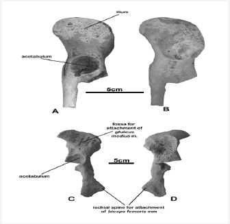
The larger species (P. chapskii) has innominate bones with a deep, conical acetabulum, and the margins of the acetabular fossa are raised high above the plane surface of the bone (Figure 2A,2B). In contrast, the smaller P. ukrainica (Figure 2C,2D) has: a pubis with a big, well-developed ridge for attachment of the obturator muscles (which cause outward rotation of the hip joint); a thick, wide and robust ischial spine for attachment of the biceps femoris muscle (an extensor of the hip joint); and a deep fossa on the medial aspect of the ilium for attachment of the gluteus medius muscle (also an extensor of the hip joint).
Figure 2: Innominate bone of Middle Miocene Pachyphoca chapskii (NMNHU-P 64-525, R.) in A: lateral and B: medial views and P. ukrainica (NMNHU-P 64-479, L.) in C: lateral and D: medial views.

Discussion
Koretsky [17] briefly detailed that some fossil postcranial elements of seals demonstrate thick and swollen (pachyosteosclerotic) bones that can be mistaken for those of sirenians such as Manatus maeoticus. If hyper saline closed basins developed when the ancient sea in Central Europe dried out, then pachyosteosclerotic seals and sirenians would have evolved in parallel, but separately, during the same time periods. Increased skeletal mass would allow taxa to remain submerged for longer periods of time and is likely a dietary adaptation for feeding in shallow waters. Pachyosteosclerotic bones would mean that these taxa swim at slow speeds and dive only shallow depths, suggesting that they ate slow-moving prey near the ocean floor.
Pachyosteosclerosis among fossil seals is a relatively new discovery and is hardly remarked at all in literature. Thus, future studies are needed to determine the cause and frequency of pachyosteosclerosis in marine mammals, especially in true seals. Upcoming morphological examinations will demonstrate whether pachyosteosclerosis is an adaptation of true seals that: may have helped them successfully adjust from terrestrial to fully aquatic life and to different salinity levels; or is an interspecific difference in bone mass resulting in varying dietary preferences, diving depths and/or ecological niches.
Pachyosteosclerois in fossil seals (~3.0-24.0Ma) from the Paratethys (Europe) and North America will be examined and compared to representatives of Recent seals who present this ostological condition. Future studies will examine diving depths, dietary specializations and ecological niches of taxa with and without pachyosteosclerosis to demonstrate the specific cause of this condition.
Acknowledgment
We sincerely thank Torres Advanced Enterprise Solutions LLC (TAES) for grant support to examine pachyosteosclerosis in seals.
References
- Le Boeuf BJ, Morris PA, Blackwell, SB, Crocker DE, Costa DP (1996) Diving behavior of juvenile northern elephant seals. Canadian Journal of Zoology 74: 1632-1644.
- Boyd IL, Croxall JP (1996) Dive duration in pinnipeds and seabirds. Canadian Journal of Zoology 74(9): 1696-1705.
- Barnes LG (1993) Fossil pinnipeds, including walruses, from near Santa Rita, Baja California Sur, México. Memoria del IV Congreso Nacional de Paleontología, Sociedad Mexicana de Paleontología AC, México City, México, 19: 20-22.
- Barnes LG (1994) A Pliocene pinniped assemblage, including strange new walruses, from near Santa Rita, Baja California Sur, México. Journal of Vertebrate Paleontology 14(3): 16A.
- de Buffrénil V, de Ricolès A, Ray CE, Domning DP (1990) Bone histology of the ribs of the Archeocetes (Mammalia: Cetacea). Journal of Vertebrate Paleontology 10(4): 455-466.
- Domning D, de Buffrénil V (1991) Hydrostasis in the Sirenia: quantitative data and functional interpretations. Marine Mammal Science 7(4): 331- 368.
- Amson E, de Muizan C, Domning D, Argot C (2015) Bone histology as a clue for resolving the puzzle of a dugong rib in the Pisco Formation, Peru. Journal of Vertebrate Paleontology 35(3): e922981.
- Boyd IL , Croxall JP (1992) Diving behaviour of lactating Antarctic fur seals. Canadian Journal of Zoology 70(5): 919-928.
- Bedosti N (1999) Pachystosis in Aphanius crassicaudus (Agassiz) (Teleostei, Cyprinodontidae) From the Upper Miocene of Monte Castellaro, Studi e Richerche sui Giacimenti Terziari di Bolga, VIII. Miscellanea Paleontologica Verona, Italy: 151-155.
- Houssaye A, de Buffrenil V, Rage JC, Bardet N (2008) An analysis of vertebral “Pachyostosis” in Carentonosaurus mineaui (Mosasauroidea, Squamata) from the Cenomanian (Early late Cretaceous) of France, with comments on its phylogenetic and functional significance. Journal of Vertebrate Paleontology 28(3): 685-691.
- Wall WP (1983) The correlation between high limb-bone density and aquatic habits in Recent mammals. Journal of Paleontology 57(2): 197- 207.
- de Buffrénil V, Astibia H, Suberbiola XP, Berreteaga A, Bardet N (2008) Variation in bone histology of middle Eocene sirenians from western Europe. Geodiversitas 30(2): 425-432.
- de Buffrénil V, Canoville A, Anastasio R, Domning D (2010) Evolution of sirenian pachyosteosclerosis, a model-case for the study of bone structure in aquatic tetrapods. Journal of Mammalian Evolution, 17(2): 101-120.
- Ricqles A (1989) Les mécanismes hétérochroniques dans le retour des tétrapodes au milieu aquatique. Geobios, mémoire spécial 22(2): 337- 348.
- Ricqles A, Buffrénil V (2001) Bone histology, heterochronies and the return of tetrapods to life in water: where are we? In: Mazin JM & Buffrénil V de (Eds.), Secondary Adaptation to Life in Water. Verlag Dr F Pfeil, Munchen, pp. 289-306.
- Koretsky IA, Rahmat SJ (2013) First Record of Fossil Cystophorinae (Carnivora, Phocidae): Middle Miocene Seals from the Northern Paratethys. Rivista Italiana di Paleontologiae Stratigrafia 119(3): 325- 350.
- Koretsky IA (2001) Morphology and Systematics of Miocene Phocinae (Mammalia: Carnivora) from Paratethys and the North Atlantic Region. Geologica Hungarica Budapest 54: 109.
© 2017 Irina Koretsky, et al. This is an open access article distributed under the terms of the Creative Commons Attribution License , which permits unrestricted use, distribution, and build upon your work non-commercially.
 a Creative Commons Attribution 4.0 International License. Based on a work at www.crimsonpublishers.com.
Best viewed in


.jpg)


 Editorial Board Registrations
 Submit your Article
 Refer a Friend
 Advertise With Us
.jpg)


.jpg)


.bmp)
.jpg)
.png)
.jpg)


.jpg)


.png)

.png)


.png)


