- Submissions

Full Text
Novel Research in Sciences
Crohn’s Disease and Appendicitis: Case Report
Marina Gabrielle Epstein1*, Gabriel Maccapani1, Franco Milan Sapuppo2, Luis Roberto Manzione Nadal1 and Marilia Fernandes1
1Department of General, Gastric and Minimally Invasive Surgery, Brazil
2Department of General Surgery Resident, Brazil
*Corresponding author: Marina Gabrielle Epstein, General Gastric and Minimally Invasive Surgery, Brazil
Submission: March 31, 2022;Published: April 21, 2022
.jpg)
Volume11 Issue1April, 2022
Introduction
Crohn’s Disease (CD) is a relapsing systemic inflammatory disease, mainly affecting the gastrointestinal tract with extraintestinal manifestations and associated immune disorders [1]. Appendiceal CD is a rare disease but has been well summarized in the various reports The incidence of appendicitis with granulomatous reaction varies from 0.1% to 2.0%. The prognosis of such patients seems to be good, and additional treatment is rarely needed [2,3].
Objective
We present a 30-year-old female with isolated appendiceal Crohn’s disease presenting as acute appendicitis.
Case Report
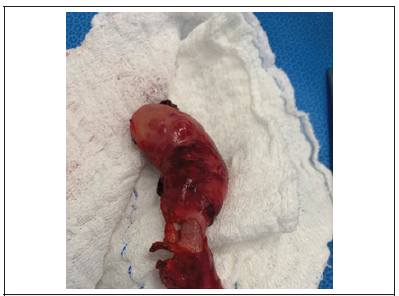
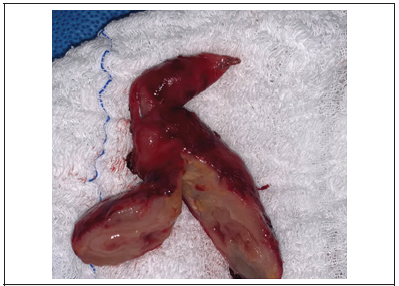
A 30-year-old female patient who was previously healthy presented with a history of right iliac fossa pain for 2 days duration without fever. There were no other associated urinary or gastrointestinal symptoms. On examination, she was in good general condition with pain in the right iliac fossa and positive sudden decompression. Laboratory tests were slightly altered and the ultrasound of the abdomen showed a small amount of free fluid in the cavity, a thickened cecal appendix with a diameter of approximately 2cm. In view of the findings, laparoscopic appendectomy was indicated. During the procedure highlighted. an excessively swollen, edematous, and reddish appendix with swelling extending to the base of the caecum (Figure 1). The swelling was more prominent at the tip of the appendix. An appendectomy was performed and the hypothesis of an appendix tumor was made. Freezing drove away malignancy. Cut section of the appendix showing an edematous wall with significant mural thickening (Figure 2). The anatomopathological examination ruled out tuberculosis and infectious causes and revealed prominent lymphoid hyperplasia and numerous non-caseous epithelioid granulomas in the wall of the appendix-aggregates, muscular hypertrophic changes, and fibrous reaction of the appendix wall. The patient evolved satisfactorily and was discharged the following day. A colonoscopy was performed showing no particular changes. She is under ambulatory follow-up, asymptomatic.
Figure 1: Irregular, distended and inflamed appendix.

Figure 2:Cut section of the appendix.

Discussion
Granulomatous inflammation of the appendix is rare, with a reported frequency of <2% in appendectomy specimens [3]. Idiopathic granulomatous appendicitis is a very rare disease. The most common symptom of appendiceal CD is right lower quadrant pain, very similar to that observed in acute appendicitis, which can be acute or chronic [4,5]. Histopathologic features are characterized by transmural chronic inflammation with marked fibrous thickening of the wall, lymphoid aggregates, small non-caseous granulomas, ulcerative mucosal change, crypt abscesses, muscular hypertrophy, and neural hyperplasia. The treatment of choice for appendiceal CD is appendectomy. Appendiceal CD shows lower recurrence rate compared with CD arising in other parts of the intestine [1-3]. Differential diagnosis should include intestinal tuberculosis, foreign body reaction, and diverticulitis of the appendix, actinomycosis, yersinia infection and even carcinoma [4,5].
Conclusion
The disease limited to the appendix is usually benign and has indolent course than that developed elsewhere. Appendectomy is often sufficient for patients. who are diagnosed with radiographic and histologic evidence that show the disease is restricted to the appendix alone, however studies suggest ambulatory follow up for at least 5 years.
References
- Loureiro ACCF, Barbosa LER (2019) Appendectomy and crohn’s disease. J Coloproctol 39(4): 373-380.
- Shaoul R, Rimar Y, Toubi A, Mogilner J, Polak R, et al. (2005) Crohn's disease and recurrent appendicitis: a case report. World J Gastroenterol 11(43): 6891-6893.
- Han H, Kim H, Rehman A, Jang SM, Paik SS (2014) Appendiceal crohn's disease clinically presenting as acute appendicitis. World J Clin Cases 2(12): 888-892.
- Yang SS, Gibson P, McCaughey RS, Arcari FA, Bernstein J (1979) Primary crohn’s disease of the appendix: report of 14 cases and review of the literature. Ann Surg 189(3): 334-339
- Otsuka R, Shinoto K, Okazaki Y, Sato K, Hirano A, et al. (2019) Crohn’s disease presenting as granulomatous appendicitis. Case Rep Gastroenterol 13: 398-402.
© 2022 Marina Gabrielle Epstein. This is an open access article distributed under the terms of the Creative Commons Attribution License , which permits unrestricted use, distribution, and build upon your work non-commercially.
 a Creative Commons Attribution 4.0 International License. Based on a work at www.crimsonpublishers.com.
Best viewed in


.jpg)


 Editorial Board Registrations
 Submit your Article
 Refer a Friend
 Advertise With Us
.jpg)


.jpg)


.bmp)
.jpg)
.png)
.jpg)


.jpg)


.png)

.png)


.png)


