- Submissions

Full Text
Novel Approaches in Cancer Study
Unstipulated and Inexact-Indeterminate Dendritic Cell Tumour
Anubha Bajaj*
Department of Pathology, Panjab University, India
*Corresponding author:Anubha Bajaj, Department of Pathology, Panjab University, India
Submission: June 20, 2024;Published: July 05, 2024

ISSN:2637-773XVolume8 Issue1
Opinion
Indeterminate dendritic cell tumour emerges as an extremely exceptional tumefaction composed of proliferation of dendritic cells or cells of histiocytic lineage. Additionally designated as indeterminate cell histiocytosis, indeterminate cell tumour or indeterminate dendritic cell tumour, true cutaneous dendritic cell tumour may depict solitary or multifocal lesions. Generally, tumefaction is confined to diverse cutaneous surfaces wherein deep seated visceral or regional lymph node involvement is exceptional. Tumour forming indeterminate cells simulate Langerhans cells vis-à-vis morphological and antigenic features. However, indeterminate cells appear devoid of Birbeck granules and lack immune reactivity to langerin (CD207). Median age of disease emergence is 45 years although no age of disease occurrence is exempt. An almost equivalent gender predilection is encountered. Indeterminate dendritic cell tumour predominantly(~88%) implicates diverse cutaneous surfaces. Infrequently, regional lymph nodes (9%) or spleen (2.3%) may be involved [1,2]. Of obscure a etiology, neoplasm expounds varied concurrence between indeterminate cells and Langerhans cells as ~indeterminate cells manifest as Langerhans cells devoid of Birbeck granules ~indeterminate cells represent as immature Langerhans cells. A subset of neoplasms express dendritic cell marker ZBTB46, thereby indicating the emergence of neoplasms directly from bone marrow progenitors, in contrast to embryonic precursors which undergo localized cutaneous regeneration [1,2].
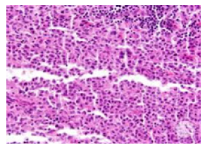
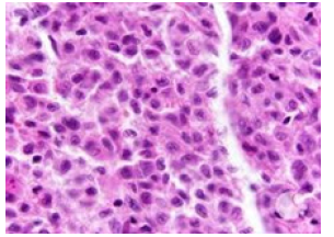
Indeterminate dendritic cell tumour may concur with various haematological neoplasms such as B cell lymphoma, T cell lymphoma, acute myeloid leukaemia or chronic myelomonocytic leukaemia. Indeterminate dendritic cell tumour may demonstrate clonal anomalies of concordant lymphomas or leukaemia [2,3]. A subset of indeterminate dendritic cell tumours demonstrate ETV3::NCOA2 genetic fusion. Exceptionally, BRAF V600E or TP53 gene may be encountered [2,3]. Commonly, a singular, cutaneous papule or nodule is observed. Alternatively, few to innumerable, multiple lesions may ensue. Besides, plaques and tumours may concur with leonine facies [2,3].Tumefaction is confined to cutaneous region wherein lesions within diverse organs or regional lymph nodes are exceptional. Indeterminate dendritic cell tumour demonstrates a variable clinical countenance as spontaneous retrogression of the lesion, stable disease, rapidly progressive lesions or tumour reoccurrence [3,4]. Upon microscopy, neoplasm is configured of pale staining or eosinophilic histiocytic cells impregnated with reniform nuclei. Occasionally, neoplasm is composed of spindle shaped cells (Figures 1 & 2). Tumour cells appear to infiltrate subjacent dermis. Neoplastic cells appear commingled with a population of reactive lymphocytes and multinucleated cells. Eosinophils appear minimal to absent. Ultrastructural examination depicts histiocytic cells with nuclear infolding and peripheral dissemination of villi. Tumour cells are permeated with lamellated dense bodies and appear devoid of Birbeck granules [3,4] (Table 1).
Figure 1:Indeterminate dendritic cell neoplasm demonstrating aggregates of pale histiocytic cells with reniform nuclei admixed with reactive lymphocytes and multinucleated cells. Tumor cells appear to infiltrate subjacent dermis (Source: Haematology image bank).

Figure 2:Indeterminate dendritic cell tumor delineating aggregates of pale histiocytic cells with reniform nuclei commingled with reactive lymphocytes and multinucleated cells, Tumor cells appear to infiltrate subjacent dermis (Source: ASH image bank).

Table 1:WHO classification of haematolymphoid Tumors-Stroma derived neoplasms of lymphoid tissues 5th edition [4].

Indeterminate dendritic cell tumour appears immune reactive to S100 protein, CD1a and CD68. Besides, tumour cells appear immune reactive to CD4, CD45, CD56 and lysozyme [5,6]. Tumour cells appear immune non-reactive to langerin (CD207), histiocytic markers as CD163, B cell markers, T cell markers and follicular dendritic cell markers as CD21, CD23, CD35, CD30 and Myeloperoxidase (MPO) [5,6]. Flow cytometric analyses of peripheral blood samples obtained from true cutaneous indeterminate dendritic cell tumour appear to lack specific anomalies. However, the disease may concur with chronic myelomonocytic leukaemia [7,8]. Indeterminate dendritic cell tumour requires segregation from neoplasms as Langerhans cell histiocytosis, juvenile xanthogranuloma, multicentric reticulohistioyctosis, interdigitating dendritic cell sarcoma, histiocytic sarcoma, chronic myelomonocytic leukaemia, reactive indeterminate cell infiltrates, tick bite or scabies [7,8]. Localized lesions of indeterminate dendritic cell tumour may be appropriately ascertained by histological assessment of excised cutaneous tissue. Deep seated lesions necessitate precise workup of associated systemic features [7,8]. Computerized Tomography (CT) and bone marrow biopsy may be optimally employed for disease discernment. Neoplasm expounds elevated levels of serum lactate dehydrogenase, Erythrocyte Sedimentation Rate (ESR) and C-Reactive Protein (CRP). Upon imaging, true cutaneous indeterminate dendritic cell tumour is characteristically devoid of features of involvement of deep seated organs or various viscera [8,9]. Isolated, singular neoplasms may be subjected to surgical extermination of the neoplasm. Alternatively, tumefaction may be treated with Psoralen and Ultraviolet A (PUVA) therapy. Adjuvant chemotherapy may be employed for alleviating aggressive disease or lesions associated with concurrent myeloid or lymphoid neoplasms [8,9]. Characteristically, cutaneous lesions demonstrate indolent biological behavior. Lesions with involvement of deepseated viscera or bone marrow may delineate inferior prognostic outcomes [8,9].
References
- Chhangte MZ, Paul D, Lamba R, Biswajit D (2021) Disseminated indeterminate dendritic cell tumor: A rare presentation. Indian Dermatol Online J 12(3): 462-464.
- Li Y, Zhang C, Xiong J (2023) Recurrent chemotherapy treated indeterminate dendritic cell tumor: Case report and literature review. Clin Cosmet Investig Dermatol 16: 2985-2993.
- Kong FW, Read J, Pitney L, Fiona T, Samantha D, et al. (2021) S100‐negative indeterminate cell histiocytosis: In an eight‐month‐old boy. Australas J Dermatol 62(1): e124-e12
- Alaggio R, Amador C, Anagnostopoulos I, Ayoma DA, Iguaracyra BOA, et al. (2022) The 5th edition of the world health organization cassification of haematolymphoid tumours: Lymphoid neoplasms. Leukemia 36(7): 1720-1748.
- Thurner L, Bewarder M, Rosar F, Patrick O, Raoul BM, et al. (2020) Indeterminate dendritic cell tumor with persistent complete metabolic response to BRAF/MEK inhibition. Hemasphere 5(1): e511.
- Xia DM, Zhou QT, Wang L (2023) Typical leonine facies in a patient with indeterminate dendritic cell tumor. Int J Dermatol 62(1): 128-129.
- Moon J, Yang JH, Lee J, Jong SP, Kwang HC (2018) A case of indeterminate dendritic cell tumor: A rare neoplasm with langerhans cell lineage. Ann Dermatol 30(6): 744-746.
- Joo JW, Chung T, Cho YA, Sang KK (2018) Recurrent indeterminate dendritic cell tumor of the skin. J Pathol Transl Med 52(4): 243-247.
- Davick JJ, Kim J, Wick MR, Alejandro AG (2018) Indeterminate dendritic cell tumor: A report of two new cases lacking the ETV3-NCOA2 translocation and a literature review. Am J Dermatopathol 40(10): 736-748.
© 2024. Anubha Bajaj. This is an open access article distributed under the terms of the Creative Commons Attribution License , which permits unrestricted use, distribution, and build upon your work non-commercially.
 a Creative Commons Attribution 4.0 International License. Based on a work at www.crimsonpublishers.com.
Best viewed in


.jpg)


 Editorial Board Registrations
 Submit your Article
 Refer a Friend
 Advertise With Us
.jpg)


.jpg)


.bmp)
.jpg)
.png)
.jpg)


.jpg)


.png)

.png)


.png)


