- Submissions

Full Text
Experiments in Rhinology & Otolaryngology
Otitis Media with Effusion: A Silent Killer of Middle Ear -At Least In a Few Patients
George MV1*, Aravind Sen V2
1 Professor in ENT, Jubilee Mission Medical College, India
2 Assistant Professor in ENT, Jubilee Mission Medical College, India
*Corresponding author: George MV, Professor of ENT, Head and Neck Surgery, Jubilee Mission Medical College, Thrissur, India
Submission: September 23, 2018;Published: September 28, 2018

ISSN 2637-7780 Volume2 Issue5
Abstract
Otitis Media with Effusion (OME) is a very common disease among catarrhal children of younger age group who are immunologically in the developing stage. At school they are exposed to similar kids of the same age group among whom someone may be having upper respiratory tract infection. It may be thus shared among them in crowded class rooms. Disease remain asymptomatic due to absence of ear symptoms other than mild hearing loss. Thus, it may remain unnoticed. Only intelligent parents or teachers notice it. Those self-resolving, due to high prevalence of the disease, a lot of them go in for sequelae like atelectasis, adhesive otitis media, ossicular erosion, posterosuperior retraction pocket and finally cholesteatoma. Hence in ears which are not picked up early it may be a silent killer of middle ear.
Keywords: Anterior; Ethmoidal; Artery; Endoscopic; Surgery
Opinion
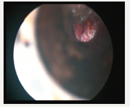
Clinical practice guidelines of OME-update 2016 defines OME as fluid in the middle ear without signs or symptoms of acute ear infection. The condition is so common enough to be called as occupational hazard of early childhood. For a lay man it is ear fluid and AOM is ear infection [1]. Chronic OME is, OME for more than three months from the date of onset, or from the date of diagnosis. OME may occur during an upper respiratory tract infection (URTI), spontaneously because of poor Eustachian Tube function or as an inflammatory response after AOM (Figure1). When children of age 5 to 6 years are screened for OME, about one in eight are found to have fluid in the middle ear. Most of the textbooks describe it as a self-resolving condition spontaneously within three months’ time. About 30-40% will get repeated OME and 5-10% lasts for more than one year [1].
Figure 1:

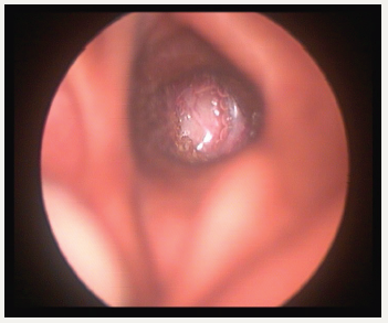
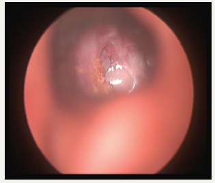
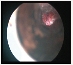
Persistent fluid in the middle ear causes not only reduced hearing, can go in for poor scholastic performances, speech-language development also. In long standing effusion, structural damage of the tympanic membrane (T.M) can go in for atelectasis, adhesion, ossicular damages, retraction pocket, and finally cholesteatoma. Stuart Mawson had described many of these complications after a long term follow up 129 glue ear cases [2]. Auto inflation and treatment URTI is the only treatment required. But the child should not sniff in. He should learn how to blow the nose correctly by occluding ne side first and then the other side, not two sides together (Figure2). Otovent is a rounded plastic balloon with a plastic nose piece. When the balloon is inflated auto inflation take place [3]. Even though the condition is considered self-resolving, since URTI is very frequent in childhood, by the time one incidence of OME is coming down, another URTI may start and the middle ear goes back to the previous stage. OME being a silent condition, many of the children present only in the late stages of the spectrum of the disease. By that time, irreversible damages might have taken place. Experimental studies in Chinchillas have demonstrated that the relationship between the time of evolution of effusion and structural changes of mucoperiosteum are related [4]. In this context, we should regularly follow up patients who are having OME and likely to have OME because of very frequent URTI (Figure3). If there is a structural damage tympanostomy must be done. Further episodes of URTI must be treated promptly (Figure4). The update of guidelines points out that under the age of 4 years adenoidectomy must be combined only in children having nasal obstruction or chronic adenoiditis. Above 4years adenoidectomy can be combined. It is learned that earlier, there was an epidemic of tympanostomy in U.K. [5].
Figure 2:Glue ear with atelectasis.

Figure 3:Adhesive otitis media.

Figure 4:Ossicular necrosis.

Conclusion
In countries like India, where the number of students in a class are more and URT Infection being very common, these children are very likely to be targets of OME. It being a silent condition, if not picked up by parents or teachers, it is likely to go in for more serious damages and finally the middle ear gets silently killed. Regular check up surveillance and treatment of the condition at each point of time is necessary to avoid these problems.
References
- Rosenfeld RM, Shin JJ, Schwartz SR, Coggins R, Gagnon L, et al. (2016) Clinical practice guideline: otitis media with effusion executive summary (update). Otolaryngol Head Neck Surg 154(2): 201-214.
- Mawson, Stuart R, John Brennand (1969) Long-Term follow up of chronic exudative otitis media (glue ears) long-term follow up of 129. Glue Ears (1969): 460-464.
- Blanshard JD, Maw AR, Bawden R (1993) Conservative treatment of otitis media with effusion by autoinflation of the middle ear. Clin Otolaryngol Allied Sci 18(3): 188-192.
- Hueb MM, Marcos VG (2009) Experimental evidence for early intervention in mucoid otitis media. Acta oto-laryngologica 129(4).
- Black N (1995) Surgery for glue ear: the English epidemic wanes. Journal of Epidemiology & Community Health 49(3): 234-237.
© 2018 George MV. This is an open access article distributed under the terms of the Creative Commons Attribution License , which permits unrestricted use, distribution, and build upon your work non-commercially.
 a Creative Commons Attribution 4.0 International License. Based on a work at www.crimsonpublishers.com.
Best viewed in


.jpg)


 Editorial Board Registrations
 Submit your Article
 Refer a Friend
 Advertise With Us
.jpg)


.jpg)


.bmp)
.jpg)
.png)
.jpg)


.jpg)


.png)

.png)


.png)


