- Submissions

Full Text
Clinical Research in Animal Science
Surgical Correction of Cleft Palate and Leporine Lip in Canine: A Case Report
Pereira JCC1*, Oliveira VC2, Siqueira AN2, Brandão EM3 and Melo SA4
1Medicine veterinary academic, Brazil
2Veterinary Doctor, Brazil
3Post Graduation Program in Animal Science, Brazil
4Department of Pathology, Brazil
*Corresponding author:Pereira JCC, Medicine veterinary academic, São Luís, Maranhão, Brazil
Submission: November 18, 2022;Published: January 23, 2023

ISSN: 2770-6729Volume 2 - Issue 4
Abstract
A two-month-old canine male patient presents with unilateral cleft palate and lip. The clinical inspection showed normal parameters. In the radiological study of oral cavity, failure in the left labial muscles and a presumptive discontinuity of the incisor bone, showing caudal displacement of the lateral incisors in relation to the contralateral one was seen. The patient was treated with a palatorrhaphy technique performed along the hard palate, joining the two edges and with cheiloplasty of the lips. Tissue from the mucosa was separated and deciduous 501, 502, 503, 601, 602 and 603 were extracted closing the cavity. The nasal and oral cavity were separated by creating a flap, which was sutured to the nasal mucosa using modified Z-plasty technique to close the defect. The animal was discharged without an esophageal tube. After 7 days of treatment, due to the non-use of the Elizabethan collar, the animal returned with suture dehiscence located where the cleft lip correction procedure was performed. The same suture technique was used to repair stitch dehiscence. The patient was monitored for 20 days and had a satisfactory postoperative period, without observing recurrence of the condition and adverse effects to the treatment. The patient was fed a diet of pasty consistency until his complete recovery. Thus, it is a fact that early diagnosis, food management, correct surgical technique and adequate postoperative care are essential for a patient’s survival and appropriate juvenile development.
Keywords:Canine; Oral cavity; Nasal cavity; Palatoschisis; Cheiloschisis; Surgery
Abbreviations:CP: Cleft Palate; CL: Cleft Lip; CLP: Cleft lip and cleft palate; NSAIDs: Non-steroidal antiinflammatory drugs
Introduction
Cleft Palate (palatoschisis) and Cleft Lip (cheiloschisis) are congenital malformations that can affect some facial regions due to failure of tissue fusion [1]. The etiology of these abnormalities is multifactorial. Cleft Palate (CP) occurs when the tissues separating the oral and nasal cavity do not grow together properly, creating communication between the nasal and oral cavity through an orifice in the palate [2]. Otherwise, the Cleft Lip (CL) comprises a cleft in the lip region that occurs in most cases in the upper part of the lip [3]. It disrupts the important circumoral orbicularis oris musculature [4]. The lack of continuity of this muscle allows the developing parts of the maxilla to grow in an uncoordinated manner so that the cleft in the alveolus is accentuated [5]. Many techniques have been described for repair of the palate and lip, and therefore, the combination of palatorrhaphy and cheilorrhaphy are the most indicated treatment for these pathologies. In the present report, we describe the successful management of a canine that was diagnosed with cheiloschisis and unilateral primary palatoschisis, as well as the description of the surgical techniques used to effectively treat it.
Case Presentation
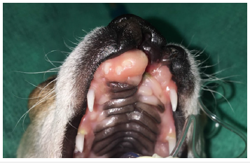
A two-month-old canine male, Brazilian Terrier breed, patient with unilateral cleft palate and Cleft Lip (CLP) was attended to at the University Veterinary Hospital of the State University of Maranhão. The clinical inspection showed normal parameters. The animal was attentive and reactive to the environment. Otherwise, anatomical abnormalities in the oral and nasal region were seen (Figure 1). According to the information provided by the owner, the animal presented signs of choking after feeding and drinking water. One x-ray view (craniodorsal) was obtained from the oral and nasal cavity of the patient, which relieved evident failure in left labial muscles and presumptive discontinuity of the incisor bone, showing caudal displacement of the lateral incisors in comparison to contralateral ones. Based on the clinal findings and anatomical abnormalities shown by the x-ray performed suggested a diagnosis of dysfunction of unilateral cheiloschisis and primary palatoschisis.
Figure 1:Animal before surgery. Anatomical abnormalities observed in the oral and nasal region (unilateral cleft palate and cleft lip).

Treatment
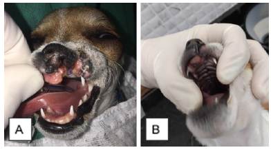
The patient was treated with cleft palate reduction using the palatorrhaphy technique performed along the hard palate, joining the two edges. Simple interrupted sutures and Polyglactin 910 4-0 suture line were performed along the hard palate, joining the two edges. Concomitantly, the cleft lip was corrected using the cheiloplasty technique. Tissue from the mucosa was separated and deciduous 501, 502, 503, 601, 602 and 603 were extracted closing the cavity. The nasal and oral cavity were separated by creating a flap, which was sutured to the nasal mucosa using modified Z-plasty technique. The labial defect was closed, making the distance between the ventral nostril and the free ventral edge of the lip the same on both sides. The animal was discharged without an esophageal tube. After 7 days from the surgical procedure, due to the non-use of the Elizabethan collar, the animal returned with suture dehiscence located where the cleft lip correction procedure was performed (Figure 2). The same suture technique was used to repair stitch dehiscence. The patient was monitored for 20 days and had a satisfactory postoperative period, without observing recurrence of the condition and adverse effects to the treatment (Figure 2).
Figure 2:A) Patient after the first surgical produce with suture dehiscence located at the site of cleft lip correction, B) Patient after the final surgical procedure, availed after 20 days showing satisfactory postoperative.

Discussion
Cheiloschisis is a defect that affects structures of the primary palate such as dental alveoli, incisor bone and upper lip [4]. In the above case report, the patient presented with these characteristics and only one side was affected. These congenital malformations affect several species; however, their etiology is still unknown [4].
Diagnosis was based on anamnesis, physical examination (oral and nasal cavity inspection), combined with x-ray to provide a view of the affected bone and its bone commitment. Computed tomography is considered the gold standard for the diagnosis of this alteration, as it is possible to reconstruct three-dimensional images of the bone defect and analyze the local dentition [5]. However, due to the low cost, greater accessibility to extraoral radiography was performed, as well as intraoral radiography for evaluation of the dental arch. With the imaging exams, it was possible to analyze the extent of the lesion, thus contributing to the surgical planning and consequently the correction of the defect [5].
A surgical procedure was performed to correct the fissure; however, there was dehiscence of the stitches in the postoperative period, thus it was possible to restore the flaw again. Repair sometimes requires multiple surgeries to correct the defect, as it is a complex technique [4]. However, the dehiscence occurred as a result of the absence of adequate postoperative management. There was a prescription of analgesics, Non-Steroidal Anti-Inflammatory Drugs (NSAIDs), Elizabethan collar, it was advised to restrict play during the recovery period, remove hard toys, keep the food soft during the first weeks; however, it was not necessary to prescribe therapeutic antibiotics and hospitalization to the patient, thus diverging from [5].
References
- Shibukawa B, Rissi G, Higarashi I, Oliveira R (2019) Factors associated with the presence of cleft lip and/ or cleft palate in Brazilian newborns. Rev Bras Saude Mater Infant 19(4): 947-956.
- Sahoo NK, Desai AP, Roy ID, Kulkarni V (2016) Oro-Nasal Communication. J Craniofac Surg 27(6): e529-533.
- Vyas T, Gupta P, Kumar S, Gupta R, Gupta T, et al. (2020) Cleft of lip and palate: A review. J Family Med Prim Care 9(6): 2621-2625.
- Peralta S, Nemec A, Fiani N, Verstraete FJ (2015) Staged double-layer closure of palatal defects in 6 dogs. Veterinary Surgery 44(4): 423-431.
- Fiani N, Verstraete FJ, Arzi B (2016) Reconstruction of congenital nose, cleft primary palate, and lip disorders. Veterinary Clinics: Small Animal Practice 46(4): 663-675.
© 2023 Pereira JCC. This is an open access article distributed under the terms of the Creative Commons Attribution License , which permits unrestricted use, distribution, and build upon your work non-commercially.
 a Creative Commons Attribution 4.0 International License. Based on a work at www.crimsonpublishers.com.
Best viewed in


.jpg)


 Editorial Board Registrations
 Submit your Article
 Refer a Friend
 Advertise With Us
.jpg)


.jpg)


.bmp)
.jpg)
.png)
.jpg)


.jpg)


.png)

.png)


.png)


