- Submissions

Full Text
Experimental Techniques in Urology & Nephrology
Cutaneous Metastasis of Transitional Cell Carcinoma of Urinary Bladder-An Unusual Case
Vazir Singh Rathee*, Sartaj Wali Khan, Aditya Kumar, Shukla PK, Trivedi S and Dwivedi US
Department of Urology, BHU, India
*Corresponding author: Vazir Singh Rathee, Department of Urology, IMS, BHU, Varanasi, UP, India
Submission: September 10, 2017; Published: January 11, 2018

ISSN: 2578-0395 Volume1 Issue2
Introduction
The reported incidence of cutaneous spread from primary urologic malignancies is 1.3%. Urinary bladder malignancies altogether account for 0.84% of cutaneous metastases [1]. In this case report, we present a patient with transitional cell carcinoma bladder who developed a solitary subcutaneous nodular metastasis in the right gluteal region of two months duration [2].
Case Report
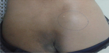
A 60M presented with total recurrent painless gross haematuria with passage of clots for one year. No history of LUTS. GPE-normal. LE-tender s/c mass of size 4x3 cm, firm, freely mobile, on the (R) gluteal region, local tempt normal (Figure 1) [3].
Figure 1: Photograph showing the tender s/c mass of size 4x3cm, firm, freely mobile,on the right gluteal Region.

Results
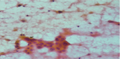
Figure 2: FNAC of the s/c mass-micro photograph showing the infiltration of the skin by the metastatic TCC.

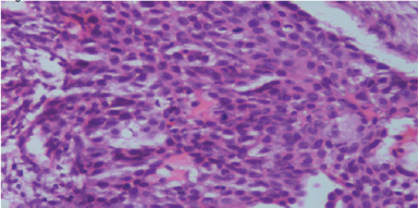
Cystoscopy done a papillary growth of approximately of size 4x3cm in the right posterolateral wall and trigone close to bladder neck. No changes suggestive of carcinoma in situ on cystoscopy. TURBT was done. Biopsy-low grade papillary urothelial carcinoma (lamina invasive) [4]. FNAC (fine needle aspiration cytology) of the subcutaneous nodule was strongly suggestive of metastatic deposits of urothelial carcinoma. C.T urography showed metastatic bony lesions in the spine. The patient was referred to radiotherapy department for further treatment (Figure 2).
Conclusion
This is a rare case of early presentation of cutaneous metastasis of transitional cell carcinoma particularly of low grade type. In many cases, as in our patient, treatment is mainly supportive and prognosis is poor (Figure 3) [5]. Therefore, it is important for the medical community to have access to each case, through case reports, so as to advance our understanding of this particular disease.
Figure 3: Cold biopsy from bladder growth-microphotograph showing low grade papillary urothelial carcinoma (lamina invasive).

References
- Mueller TJ, Wu H, Greenberg RE, Hudes G, Topham N, et al. (2004) Cutaneous metastases from genitourinary malignancies. Urology 63(6): 1021-1026.
- Fujita K, Sakamoto Y, Fujime M, Kitagawa R (1994) Two cases of inflammatory skin metastasis from transitional cell carcinoma of the urinary bladder. Urol Int 53(2): 114-1
- Messing EM, Catalona W (1998) Urothelial tumors of the urinary tract. Bladder cancer. In: Walsh PC, Retik AB, Vaughan ED, et al. (Eds.), Campbell's Urology. (7th edn), W.B. Saunders Co, Philadelphia, USA, pp. 2329-2383.
- Swick BL, Gordon JR (2010) Superficially invasive transitional cell carcinoma of the bladder associated with distant cutaneous metastases. J Cutan Pathol 37(12): 1245-1250.
- Block CA, Dahmoush L, Konety BR (2006) Cutaneous metastases from transitional cell carcinoma of the bladder. Urology 67(4): 846.
© 2018 Vazir Singh Rathee, et al. This is an open access article distributed under the terms of the Creative Commons Attribution License , which permits unrestricted use, distribution, and build upon your work non-commercially.
 a Creative Commons Attribution 4.0 International License. Based on a work at www.crimsonpublishers.com.
Best viewed in


.jpg)


 Editorial Board Registrations
 Submit your Article
 Refer a Friend
 Advertise With Us
.jpg)


.jpg)


.bmp)
.jpg)
.png)
.jpg)


.png)

.png)


.png)


