- Submissions

Full Text
Techniques in Neurosurgery & Neurology
Retrospective Review of the Presentation of Erb’s Palsy as Seen in a Tertiary Hospital in Southeastern Niger
Anyachukwu Canice Chukwudi1,2, Adadinfiari Ateke Peter1, Ajare Enyereibe Chuks3, Ede Stephen Sunday1,4*, Onyeso Ogochukwu KK1 and Ezema Charles Ikechukwu1
1Department of Medical Rehabilitation, Nigeria
2Molecular Pathology Institute, Nigeria
3Department of Radiation Medicine, Nigeria
4Department of Gerontology, UK
*Corresponding author: Ede Stephen Sunday, Department of Medical Rehabilitation, Nigeria
Submission: February 1, 2022;Published: February 23, 2022

ISSN 2637-7748
Volume4 Issue5
Abstract
Background: The presentations of Erb’s palsy due to birth-related peripheral nerve injury of the brachial plexus could be based on the type and extent of child’s injury sustained.
Purpose: This study was aimed at verifying the presentation of Erb’s palsy between 2001−2009.
Methodology: This was a retrospective study design, which determined the presentation of Erb’s palsy as reported on the folders of the patients between 2001−2009 in the Physiotherapy Department of University of Nigeria Teaching Hospital, Ituku-Ozalla, Enugu Nigeria.
Findings: This study showed that among the presentations of Erb’s palsy, inability of the subjects to move their upper limb had the highest occurrence (n=22; 24.2%), followed by their inability to raise the limb (n=19; 20.9%) and inability to raise the arm above the shoulder (n=9; 9.9%). The study reveals that higher birth weight (>4.0kg) may have led to the increased risk factors, resulting to increase in assisted delivery (n=46; 50.7%). In addition, the study reveals that Erb’s palsy injury is more on the right upper limb than left limb, with females (n=52; 57.1%) on a higher incidence than males.
Conclusion: This study showed that there is various clinical presentation of Erb’s palsy and patients may present differently or similarly depending on the level of injury at the Brachial plexus. In addition, the causes and precipitating risk factors in Erb’s palsy includes assisted delivery because of increased birth weight, forced delivery, prolonged delivery, breech delivery and some unknown factors.
Introduction
Erb’s Palsy is a type of paralysis that is commonly thought to be caused by damage to the upper brachial plexus at the time of birth [1]. Erb’s palsy may also be referred to as Erb- Duchenned paralysis, Klumpke paralysis, or brachial palsy and while each of these falls under the umbrella of Erb’s palsy, they each represent a specific level of impairment. Erb’s palsy is most likely to occur during difficult deliveries in which the attending physician may decide to use forceps to in order to move the baby’s head through the birth canal [2]. The first medical description of obstetric brachial plexus palsy was by William Smellie in his 1768 treatise on midwifery who reported a case of transient bilateral arm paralysis in a newborn after difficult labor [3]. The description of inability of children to move their arm’s following birth has been recorded since the days of Hippocrates [4]. Although the name Erb’s palsy is named after Wilhelm Heinrich Erb (1840-1921) a German neurologist who described a case in 1874, but Duchene described an earlier case in 1872. However, Erb was also a pioneer in a description of the electrophysiological nature of tetany, characterization of the physiological response to stimulation of the superior root of the brachial plexus and describing the deep tendon reflex [5].
Erb’s palsy is a paralysis of the muscles of the shoulder and arm supplied by (C5 and C6 spinal nerves) [5]. This is because of injury to the superior parts of the brachial plexus [6]. It is also defined as a peripheral nerve disorder [7,8]. When there is injury at level of Erb’s point (C5,C6), it causes paralysis of the deltoid, supraspinatus, infraspinatus, biceps and brachialis muscle. The arms hang by the side with shoulder in internal rotation, elbow in extension and the forearm pronated with the palm facing backwards; often called “police tip” position the hand and finger function is preserved [9,10]. In industrialized countries, the incidence of Erb’s palsy ranges from 0.38–3.0 per 1,000 live births making it a very common injury in neonates and clinically common presentation [11]. Studies also in France and Saudi Arabia have suggested an incidence of 1.09-1.19 cases of Brachial plexus palsy per 1,000 births [12]. This work is thus carried out to add to literature; knowledge on the pattern of presentation of Erb’s palsy in the eastern Nigeria, as well as find out the possible causes and precipitating risk factors.
Material and Methods
Design
This is a retrospective review of patients’ case note to determine the presentation of Erb’s palsy as reported at the physiotherapy department of University of Nigeria Teaching Hospital, Ituku- Ozalla, Enugu, Nigeria between 2001-2009. Ethical clearance for this study was obtained from the Ethical and research committee of University of Nigeria teaching hospital.
Participants
The study utilized a non-probability sampling technique in the physiotherapy department of university of Nigeria Teaching Hospital, Ituku-Ozalla, Enugu. Ninety-one (91) Erb’s palsy cases within the age range of 0 – 4 years, managed within 2001-2009 were identified for this review. The Erb’s palsy cases that were older and cases with other major neurodevelopmental disorder were excluded from this study.
Procedure
Approval was sort from the ethical review committee of University of Nigeria Teaching Hospital, Ituku− Ozalla, Enugu before the commencement of the study with a written proposal to inform the permission of the head of record department and the head of department of physiotherapy for use of folders of patients
Subsequently, a preformed proforma was prepared to collect data on demography (age,sex), weight at birth, mode of delivery, causes/precipitating risk factors, presentations of baby, and upper limb affected by the injury.
Data analysis
Descriptive statistics of frequencies and percentages were used to summarize the data in a simple pattern with the use of tables. Data were analyzed with SPSS software version 20.0 for data analysis (SPSS Inc., Chicago, IL).
Result
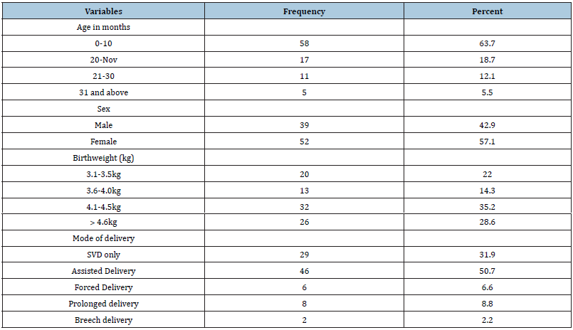
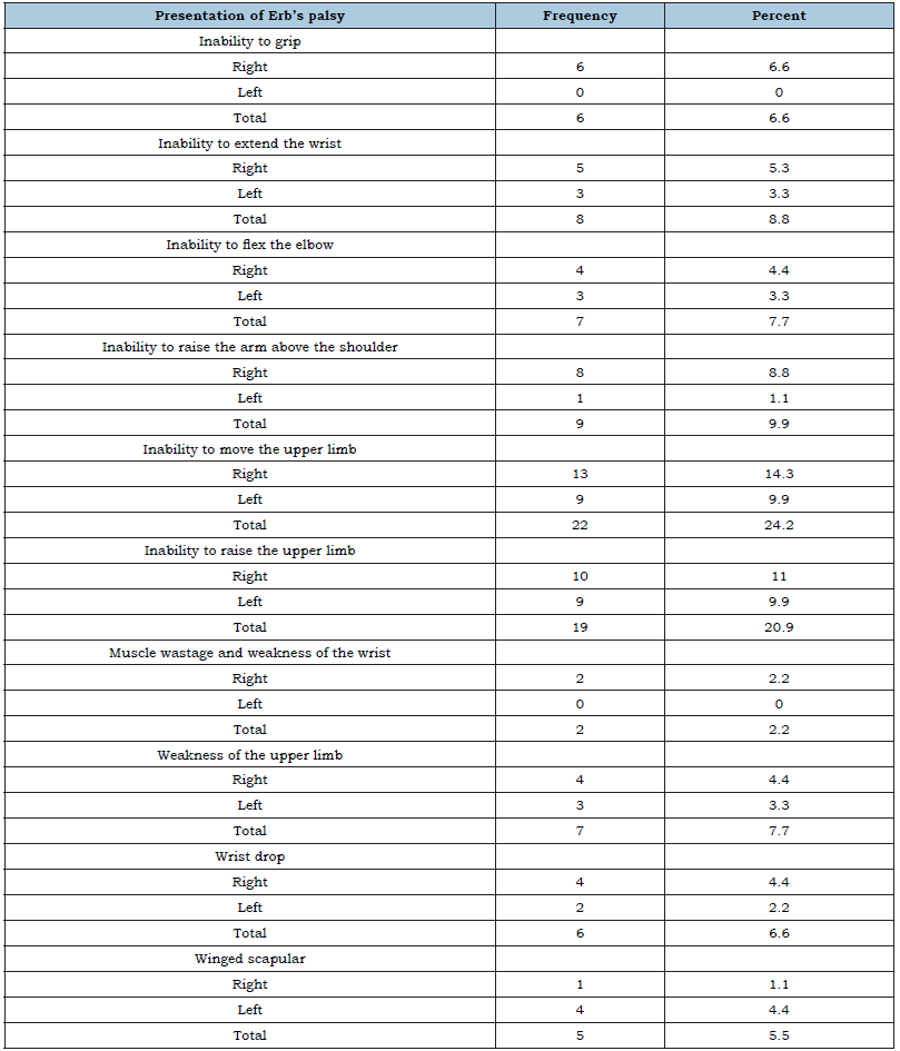
From Table 1, majority of the cases where within 0-10 months (43.1%), had birth weight ˃greater than 4.6kg (28.6%) and majority were delivered through assisted mode of delivery (50.7%). Table 1 also showed that out of the 91 subjects that participated in the study, incidence of Erb’s palsy is more in females with percentage frequency of 57.1%. Table 2 shows that inability of the subjects to move their upper limbs had the highest frequency of 14.3% on the right and 9.9% on the left. This is followed by inability to raise the upper limb with frequency of 11.0% on the right and 9.9% on the left, while muscle wastage and weakness of the wrist had the least frequency of 2.2% percent on the right and 0% on the left. This same Table 2 also showed that cases presented more at the right upper limb 57(62.6%). Table 3 presented the relationship between Subjects’ Birthweight and their mode of delivery. It reveals that more babies with birth weight within 4.1-4.5kg, while assisted delivery occurred more in babies with birth weights greater 4.6kg.
Table 1:General characteristics of the Erb’s palsy cases (n=91).

Table 2:Evaluation of Erb’s palsy presentation and the affected limbs.

Table 3:Relationship between Subjects’ birthweight and their mode of delivery.

Discussion
The prevalence of Erb”s palsy in Africa has been shown to be on the increase despite improved medical care [13]. It has also been reported to be one of the most common birth injury hospitals in Nigeria [14]. A number of predisposing factors have been linked to increased incidence of Erb’s palsy including gestational diabetes, fetal macrosomia, instrument-assisted delivery, prolonged labor and/or breech delivery [15], male gender [16], primigravida [17], babies’ weight at birth, and place of birth [18], macrosomia and shoulder dystocia [5], etc. This study was designed to examine the pattern of clinical presentations of Erb’s palsy presentation of Erb’s palsy and it is associated with their levels of injury on the Brachial plexus, the causes and precipitating risk factors. Understanding of the pattern of presentations and their associated factors will help in drawing out awareness and preventive campaign. The main finding from this study showed that most cases of Erb’s palsy reported to physiotherapy very early from onset within the tenth months after birth. This is a positive indication because is that early and effective treatment for Erb’s palsy makes a real difference to it prognosis [5,18]. Secondly, this study result showed that an increase in birth weight depicts increase chances of Erb’s palsy. This is because at weight between 4.1-4.5kg there is an increase in the number of cases needing assisted delivery [1], which also showed a strong correlation to birth injury. This finding corresponds with findings reported by Gudmundsson et al. [19] who carried out a study to verify Correlation of birth injury with maternal height and birthweight and confirmed a strong correlation. This result is also in accordance with Weizsaecher et al. [20] who found that larger babies are likely to be stocked in the birth canal. Grassi [21] reported that Brachial plexus injury occur with average vertex of 3.8kg-5.0kg and average breech of 1.8-3.7kg. The most striking factor as a cause in Erb’s palsy was the large size of babies at birth; a big baby causes greater trouble in delivery, particularly in bringing down the arms if caught above the head [5,22]. Unlike findings from previous studies [14,16,23], the incidence of Erb’s palsy as found from this present study is more in females with percentage frequency of 57.1%. Although Yarfi et al. [24] and Getson [25] also found higher incidence in females, this difference in results could be linked to the difference in methodology utilized for these studies or the difference may be due to change of trend in sex as more females may have been born than males recently [26].
More so, the result of this study showed that more cases presented at the right upper limb, which is coherent with Ugbonna [16] who in their retrospective work with 116 Erb’s palsy patients noted a higher frequency on the right upper limb. Getson [25] also reveals that right sided injury is more with 51 percent frequency in brachial plexus injury while left was 45% and 4% in bilateral cases. In addition, increase presentation of Erb’s palsy on the right upper limb may imply that the right shoulder is usually posterior during delivery. Gherman et al. [27] in their study of dystocia as a cause of brachial plexus injury noticed that children who did not have shoulder dystocia but sustained brachial plexus injury often presents the injured shoulder posterior during delivery. Generally, inability of the subjects to move their upper limbs had the highest frequency followed by inability to raise the upper limb with frequency, while Muscle wastage and weakness of the wrist had the least frequency. Other literature in line with these finding states that injury to C5, C6 nerve root of the brachial plexus leaves patients with inability to lift arm above the shoulder, weakness, of the affected arm and elbow contracture [28,29]. Literature on the description of Erb’s palsy as given by Joey et al. [4] stated that the inability of children to move their arm following birth with Erb’s palsy has been recorded since the days of Hippocrates. In addition, studies from literature on the identification of presentation in Erb’s palsy identified the most obvious feature as lack of mobility of the arm [1,17]. The easy identification of child’s inability to move the affected arm could be linked to this frequent report. The precipitating factors of Erb’s palsy identified in this study include Birth-eight and their mode of delivery. Assisted delivery occurred more in babies with birth weights greater 4.6kg. This supports previous studies, which found that assisted delivery might be a major cause/ precipitating risk in Erb’s palsy [20,22]. Especially, prolonged second phase of labor is a risk factor in Erb’s palsy [20] and mothers who have had their labor induced or speeded up by drugs leads to difficult deliveries and Erb’s palsy [30].
Conclusion
This study shows the various clinical presentation of Erb’s palsy. Patients present differently or similarly depending on the level of injury on Brachial plexus on the affected upper limb. In addition, the causes and precipitating risk factors in Erb’s palsy include assisted delivery because of increased birth weight, forced delivery, prolonged delivery, breech delivery and some unknown factors. The study recommends for further studies on presentation, classification and any related disorders of Erb’s palsy among nuliparous and multiparous women.
Ethics
This study was approved by the University of Nigeria Research Ethics Committee.
Authors’ contributions
CC Anyachukwu: Protocol development, Manuscript writing, Data collection, Data analysis; AP Adadinfiari: Project development, Data collection, SS Ede: Data collection, Manuscript writing/editing, Ajare EC, Onyeso OKK, and Ezema CI: Protocol development, Manuscript editing. All authors read and approved the final manuscript.
References
- Chater M, Camfield P, Camfield C (2004) Erb's palsy who is to blame and what will happen? Paediatr Child Health 9(8): 556-560.
- Chauhan S, Blackwell S, Ananth C (2014) Neonatal brachial plexus palsy: incidence, prevalence, and temporal trends. Semin Perinatol 38: 210-218.
- Gilbert A, Giorgio P (2005) Obstetrical palsy: the french contribution. Seminars in Plastic Surgery 19(1): 5-16.
- Joey A, Joel A, Junjoe A (2008) Chiropractic care of a patient with Erb's Palsy with a review of the literature. Clinical Chiropractic 11(2): 70-75.
- Watt AJ, Niederbichler AD, Yang LJ (2007) Historical perspective on Erb's palsy 119(7): 2161-2166.
- Basit H, Ali CDM, Madhani NB (2020) Erb Palsy. In: Stat Pearls. Treasure Island (FL), Stat Pearls Publishing.
- Moore KL, Dalley AF (2006) Clinically oriented anatomy 5th edn, Philadelphia Lippincott Williams and Wilkins, USA.
- Nawabi DH, Jayakumar P, Carlstedt T (2006) Brachial plexus and peripheral nerve injury. Ann R Coll Surg Engl 88(3): 328.
- Singh J (2005) Textbook of electrotherapy. (1st edn), Jaypee brothers. New Delhi, India.
- Hemandy N, Noble C (2006) Newborn with abnormal arm posture. Am Fan Physician 73(11): 2015-2016.
- Pollack R, Buchman A, Yaffe H, Divon M (2000) Obstetrical brachial palsy: pathogenesis, risk factors, and prevention. Clin Obstet Gynecol 43(2): 236-246.
- Kay SEJ (1998) Obstetrical brachial palsy. Br J Plast Surg 51(1): 43-50.
- Omole J, Bankole K, Abiodun A, Omole K, Afolabi M (2016) Prevalence and pattern of birth brachial plexus injuries seen in Nigeria. Journal of Pediatric Neurology 14(2): 51-56.
- Ogwumike OO, Adeniyi AF, Badaru U, Onimisi JO (2014) Profile of children with new-born brachial plexus palsy managed in a tertiary hospital in Ibadan, Nigeria. Niger J Physiol Sci 29(1): 1-5.
- Louden E, Marcotte M, Mehlman C, Lippert W, Huang B, et al. (2018) Risk factors for brachial plexus birth injury. Children 5(4): 46.
- Ugbonna HA, Omojunikanbi A (2010) Journal of Clinical Medicine and Research 2(5): 74-78.
- Ojumah N, Ramdhan RC, Wilson C, Loukas M, Oskouian RJ, et al. (2017) Neurological neonatal birth injuries: a literature review. Cureus 9(12): e1938.
- Dumpa V, Kamity R (2020) Birth Trauma. Treasure Island, Stat-Pearls Publishing.
- Gudmundsson S, Henningsson A, Lindqvist P (2005) Correlation of birth injury with maternal height and birthweight. BJOG 112(6): 764-767.
- Weizsaecker K, Deaver JE, Cohen WR (2007) Labour characteristics and neonatal Erb's palsy. BJOG 114(8): 1003-1009.
- Grassi AE, Giuliano M (2000) The neonate with macrosomia. Clin Obstet Gynecol 43(2): 340-348.
- Wolman B (1948) ERB'S palsy. Arch Dis Child. 1948 23: 129-131.
- Sharma D, Pandita A, Shastri S, Sharma PK (2016) Duchenne Erb's palsy in newborn: result of birth trauma. Med J DY Patil Univ 9: 274-276.
- Yarfi C, Elekusi C, Banson AN, Angmorterh SK, Kortei NK, et al. (2019) Prevalence and predisposing factors of brachial plexus birth palsy in a regional hospital in Ghana: a five-year retrospective study. Pan Afr Med J 32: 211.
- Eng GD, Binder H, Getson P, Dannell R (1996) Obstetrical Brachial plexus palsy (OBPP) outcome with conservative management. Muscle nerve.19(7): 884-991.
- Austad SN (2015) The human prenatal sex ratio: a major surprise. Proc Natl Acad Sci USA 112(16): 4839-4840.
- Gherman RB, Ouzouman JG, Miller DA (1998) Spontaneous vaginal delivery a risk factor for Erb’s palsy. Am J Obstet Gynecol 178(3): 423-427.
- Saliba S, Saliba EN, Pugh KF, Chhabra A, Diduch D (2009) Rehabilitation considerations of a brachial plexus injury with complete avulsion of c5 and c6 nerve roots in a college football player: a case study. Sports Health 1(5): 370-375.
- Seo TG, Kimdu H, Kim IS, Son ES (2016) Does C5 or C6 radiculopathy affect the signal intensity of the brachial plexus on magnetic resonance neurography? Ann Rehabil Med 40(2): 362-367.
- Sturdee D, Otah k, Keane D (2001) Yearbook of Obstetrics and Gynecology. Royal college of Obstetrics and Gynecology press, London. 2001.
© 2022 Ede Stephen Sunday. This is an open access article distributed under the terms of the Creative Commons Attribution License , which permits unrestricted use, distribution, and build upon your work non-commercially.
 a Creative Commons Attribution 4.0 International License. Based on a work at www.crimsonpublishers.com.
Best viewed in


.jpg)


 Editorial Board Registrations
 Submit your Article
 Refer a Friend
 Advertise With Us
.jpg)


.jpg)


.bmp)
.jpg)
.png)
.jpg)


.jpg)


.png)

.png)


.png)


