- Submissions

Full Text
Techniques in Neurosurgery & Neurology
Recognition and Cognitive Risk Assessment of Brain Dysfunction in Patients with Migraine by Resting-state Functional Magnetic Resonance Imaging
Ziye Jia, Jialu Gao, Jing Li, Xin Wang, Xinxiu Shi and Ying Xing*
Department of Neurology, China
*Corresponding author: Ying xing, Department of Neurology, China
Submission: July 22, 2021;Published: August 09, 2021

ISSN 2637-7748
Volume4 Issue3
Abstract
Abstract: Using Resting-State Functional Magnetic Resonance Imaging (rest-fMRI) to post-process the data of the resting-state fMRI image of migraine patients and healthy controls without external conditions, analyze the spontaneous activity of nerve cells in the brain area without external stimulus, and analyze the changes of its brain functional network signals, so as to further analyze the relationship between the functional changes of multiple brain regions and clinical manifestations of migraine. At the same time, the Montreal Cognitive Assessment (MoCA) scale was used to screen the cognitive function of the above participants, which was associated with the functional changes of brain areas obtained by fMRI, so as to evaluate whether migraine will affect cognitive function.
Method: Twenty-nine patients with migraine in our hospital from September 2020 to March 2021 were included. Patients with migraine were the case group (group A), then according to whether there was Patent Foramen Ovale (PFO), they were divided into migraine group with patent foramen ovale (group A1,20) and migraine group without patent foramen ovale (group A2,9). and selected 14 healthy people whose age, and education level match the case group, that is the control group (group B). Montreal cognitive assessment scale was used to test the cognitive function domains such as visual space, executive function, attention, memory, abstraction and orientation, using statistical software SPSS (Statistic a Product and Service Solutions) version 23.0, using Kruskal-Wallis H test, the P value is less than 0.05 as statistically different. Resting state fMRI pre-processing and post-processing auxiliary software MATLABR2010a, REST for subsequent processing of the data.
Result: Alff values of bilateral cerebellar hemisphere, bilateral frontotemporal occipital parietal lobe, thalamus and basal ganglia in group A were significantly higher than those in group B (P< 0.05).Alff values of right temporal lobe in group A1 were significantly higher than those in group A2 (P<0.05). Alff values of bilateral cerebellar hemisphere, bilateral frontotemporal parietal lobe, left thalamus and basal ganglia in group A1 were significantly lower than those in group A2 (P< 0.05). The statistical results of MOCA scale scores in group A were lower than those in group B, and the difference was statistically significant (P<0.05), manifested in visual space and executive function, attention, memory, language ability, abstraction, and orientation. There was no significant difference in naming, language, abstraction, and delayed recall between A1 group and A2 group Montreal Cognitive Assessment Scale score statistics (P>0.05). The difference between visual space and execution, attention, and orientation was statistically significant (P<0.05).
Conclusion: Migraine patients have abnormal connections in multiple brain areas in the resting state, resulting in abnormal brain function information integration, which is consistent with a variety of clinical manifestations of migraine patients. Migraine patients cause cognitive dysfunction, which can be manifested as damage to visual space and executive function, attention, memory, language ability, abstraction, and orientation; compared with migraine patients without patent foramen ovale, there is oval Migraine patients with patent foramen have more prominent cognitive impairments in visual space and execution, attention, and orientation.
Keywords: Migraine; Cognition; Patent foramen ovale; Resting state functional magnetic resonance imaging
Introduction
Migraine is characterized by paroxysmal pulsatile pain accompanied by other characteristics [1] and its repeated attacks can also cause the decline of cognitive function [1,2]. At present, the clinical diagnosis of migraine mainly depends on family history, typical clinical characteristics and the use of relevant auxiliary examination means to exclude other diseases for comprehensive judgment. In order to better understand the brain function changes and attack mechanism of migraine, according to our strict inclusion criteria and exclusion criteria, 20 migraine patients with patent foramen ovale, 9 migraine patients without patent foramen ovale and 14 healthy people matched with age and education were selected as the control group. By comparing the Alff values obtained by post-processing of resting state fMRI imaging changes, the relationship between the functional changes of multiple brain regions and clinical manifestations of migraine was confirmed. At the same time, combined with MOCA score scale, compared with patients without patent foramen ovale, patients with patent foramen ovale have more prominent cognitive impairment in visual space and execution, attention and orientation. Migraine is a recurrent headache disease with heredity. The World Health Organization (WHO) has classified it as one of the functional disability diseases [3,4]. Functional Magnetic Resonance (fMRI) measures brain functional activity by detecting the changes of local cerebral blood flow caused by nerve cell activity, which has high temporal and spatial resolution, and is of great significance to the study of brain function and connectivity [5]. In order to explore the pathogenesis of migraine, we compared the resting state functional MRI Alff values of different groups to evaluate the changes of brain functional areas in migraine patients, and proved that migraine can cause cognitive dysfunction.
Data and Methods
Inclusion and exclusion criteria
Inclusion criteria of migraine patients
a) Meet the International Classification of headache diseases
third diagnostic criteria of migraine
b) Ages are between 12 and 70
c) There was no headache during the scan
d) There was no MRI contraindication
e) The consent was obtained from the patients themselves
and their families
Exclusion criteria of migraine patients
a) Other types of headaches, except headache caused by
other reasons, such as encephalitis, meningitis, vascular
disease, high intracranial pressure, etc.
b) The somatization symptoms caused by depression and
anxiety were excluded
c) Migraine attack at the time of or within 24 hours after the
scan
d) There was a history of alcohol and tobacco abuse, and
there was a history of preventive drug use within one year
e) There are magnetic resonance scan contraindications or
claustrophobia
f) Female subjects were pregnant or in menstrual period
Inclusion criteria of control group
a) Healthy people matched with age and education level of
migraine group
b) No history of migraine or other pain, no family history of
headache
c) Ages are between 12 and 70
d) The consent was obtained from the patients themselves
and their families
Exclusion criteria of control group
The exclusion criteria were the same as the above case group.
Resting state data acquisition
The participants were asked to wear earplugs, close their eyes, breathe quietly, avoid head movement, keep awake and relaxed, and try not to make systematic thinking, so as to obtain the activity data of brain regions in the resting state.
Statistical Analysis
In the operating environment of MATLAB R2010a, rest software was used to analyze the Alff. The data of MOCA scale were analyzed by SPSS 23.0 and Kruskal Wallis h test. The P value is less than 0.05 as statistically different.
Results
Baseline data statistics of enrolled personnel
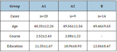
The age, course of disease and education level of 43 enrolled persons were collected (Table 1).
Table 1: p>0.05.

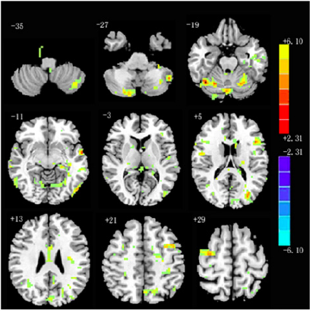
Alff values of bilateral cerebellar hemisphere, bilateral frontotemporal occipital parietal lobe, thalamus and basal ganglia in group A were significantly higher than those in group B (P < 0.05) (Figure 1). Alff values of right temporal lobe in group A1 were significantly higher than those in group A2 (P < 0.05). Alff values of bilateral cerebellar hemisphere, bilateral frontotemporal parietal lobe, left thalamus and basal ganglia in group A1 were significantly lower than those in group A2 (P < 0.05) (Figure 2).
Figure 1:

Figure 2:

Comparison of MOCA scale among groups
Normality test of variables
Kolmogorov Smirnov (KS) was used to test the normality of visuospatial and executive, naming, attention, language, abstraction, delayed recall, orientation and MOCA scores of 43 participants. The results showed that the P value of the total scores of them were less than 0.05 showing a non-normal distribution (Table 2).
Table 2: Normality test results of each variable(n=43).

Comparison of MOCA in each group
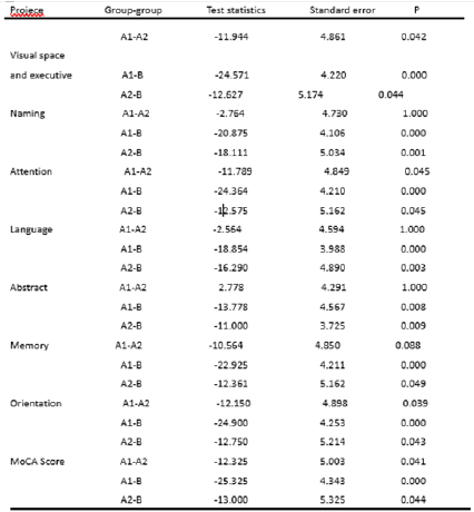
Kruskal Wallis H test was used to compare the differences of visuospatial and total scores of execution, naming, attention, language, abstraction, delayed recall, orientation and MOCA among the three groups. The statistical results of MOCA scale scores in group A were lower than those in group B, and the difference was statistically significant (P<0.05), manifested in visual space and executive function, attention, memory, language ability, abstraction, and orientation; there was no significant difference in naming, language, abstraction, and delayed recall between A1 group and A2 group Montreal Cognitive Assessment Scale score statistics (P>0.05); the difference between visual space and execution, attention, and orientation was statistically significant (P< 0.05 ) (Figure 3).
Figure 2:

Discussion
At present, the pathogenesis of migraine is not clear. At present, the mainstream theories of migraine pathogenesis mainly include trigeminal neurovascular theory and cortical spreading inhibition theory [6]. It is very important to explore the relationship between the changes of migraine brain functional areas and clinical manifestations, so as to achieve effective treatment results and improve the quality of life of migraine patients [7]. In this clinical study, the resting state fMRI technology was used to compare the brain functional activity areas of group A and group B. It was found that there were significant differences in Alff signal values of bilateral cerebellum, bilateral frontotemporal occipital parietal lobe, thalamus and basal ganglia in the case group (group A). The brain dysfunction areas in this study are similar to those in previous studies [8-10], and most of them are related to brain function areas about pain processing, such as frontal lobe, parietal lobe, temporal lobe, cerebellum, basal ganglia, etc. Studies have confirmed that there are lateral and medial pain systems in the brain, and the lateral pain system is responsible for pain recognition; The medial pain related system is responsible for processing the emotional and physical responses to pain. These two pain related systems can be combined into a pain related network, namely “pain matrix”. The pain matrix mainly includes frontal lobe, primary and secondary sensorimotor cortex, Anterior Cingulate Cortex (ACC), thalamus, Insular Cortex (IC), basal ganglia, cerebellum, amygdala, hippocampus, parietal lobe and temporal lobe [8,11]. Repeated migraine attacks can also lead to the reduction of cognitive function [4] and the damage of related brain functional areas, resulting in a vicious circle. This study found that compared with group B, migraine in group a can lead to cognitive dysfunction, specifically in visual space and executive, attention, orientation. In conclusion, according to whether there was patent forman ovale, this study divided migraine patients into different groups, and revealed the abnormal changes of brain functional areas in migraine patients more intuitively by resting state fMRI. At the same time, through the comparison of MOCA score, it revealed that migraine can lead to cognitive dysfunction in patients, manifested in visual space and executive, attention and orientation. Therefore, this study is helpful to explore the pathogenesis of migraine and guide the treatment according to the clinical manifestations.
Limitations
The number of cases included in this study is relatively small, and we can exclude the influence of research bias on the conclusion. We need to expand the sample size in the future. The subjects were not followed up regularly. The changes of clinical characteristics and functional magnetic resonance cannot be observed dynamically.
References
- Macgregor EA (2010) Prevention and treatment of menstrual migraine. Drugs 70(14): 1799-1818.
- Vos T, Barber RM, Bell B (2015) Global, regional, and national incidence, prevalence, and years lived with disability for 301 acute and chronic diseases and injuries in 188 countries, 1990-2013: A systematic analysis for the global burden of disease study 2013. Lancet 386(9995): 743-800.
- Jensen R, Stovner LJ (2008) Epidemiology and comorbidity of headache. Lancet Neurol 7(4): 354-361.
- Shun Wei Li, Yan Sheng Li, Ruo Zhuo Liuet (2011) Chinese guidelines for the diagnosis and treatment of migraine. Chinese J Pain Medicine 17(02): 65-86.
- Xiao JY, Xing YH (2005) Research Progress on pathogenesis of migraine. Foreign Medical Neurology Neurosurgery 32(3): 280-283.
- Yuan YG, Jun W, Hui HP (2016) Research progress of resting state fMRI in migraine. Contemporary Chinese Medicine 23(15): 13-15.
- Head and face pain group of Pain Society of Chinese Medical Association. Chinese guidelines for migraine prevention and treatment. Chinese Journal of Pain Medicine 22(10): 721-727.
- Schmidt W (2008) Subtle grey matter changes between migraine patients and healthy controls. Cephalalgia 28(1): 1-4.
- Kim JH, Suh SI , Seol HY (2010) Regional grey matter changes in patients with migraine: A voxel-based morphometry study. Cephalalgia 28(6): 598-604.
- Russo A, Tessitore A, Esposito F (2012) Pain processing in patients with migraine: An event-related fMRI study during trigeminal nociceptive stimulation. J Neurol 259(9): 1903-1912.
- Peyron R, Laurent B, García L (200) Functional imaging of brain responses to pain. A review and meta-analysis (2000). Neurophysiol Clin 30(5): 263-288.
© 2021 Ying Xing. This is an open access article distributed under the terms of the Creative Commons Attribution License , which permits unrestricted use, distribution, and build upon your work non-commercially.
 a Creative Commons Attribution 4.0 International License. Based on a work at www.crimsonpublishers.com.
Best viewed in


.jpg)


 Editorial Board Registrations
 Submit your Article
 Refer a Friend
 Advertise With Us
.jpg)


.jpg)


.bmp)
.jpg)
.png)
.jpg)


.jpg)


.png)

.png)


.png)


