- Submissions

Full Text
Surgical Medicine Open Access Journal
Our Experience in Removing Foreign Bodies from the Esophagus in Children
Mehmet Sarıkaya* and Fatma Ozcan Sıkı
Selcuk University Faculty of Medicine, Turkey
*Corresponding author:Mehmet Sarıkaya, Selcuk University Faculty of Medicine, Turkey
Submission: August 17 2023;Published: September 13, 2023

ISSN 2578-0379 Volume5 Issue4
Introduction
Ingestion of Foreign Bodies (FB) is a prevalent problem among children of all ages worldwide. That’s why 80% of foreign body ingestion cases are children [1]. Babies and toddlers want to put just about anything in their mouths and eat it. The majority of Esophageal Foreign Bodies (EFB) in children occur in children between the ages of six months and six years, which, if not treated in time, can be life-threatening [2,3]. EFB can be asymptomatic or present with symptoms such as difficulty swallowing, feeling stuck, cyanosis, and drooling [4]. Ingested FB tends to become lodged in areas of physiological constriction, such as the upper esophageal sphincter, the level of the aortic arch, and the lower esophageal sphincter [4].
Materials and Methods
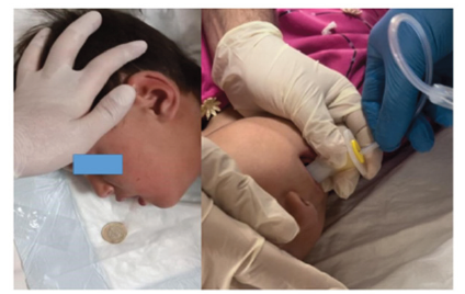
The files of 98 patients who were followed up and treated by us due to FB ingestion between July 2019 and December 2022 were reviewed retrospectively. Forty-two patients with EFB whose data could be accessed were included in the study. The clinical and demographic characteristics of the patients, the characteristics of the FB, and the imagings (x-ray, computed tomography, or esophagography) were recorded. While blunt foreign bodies such as coins in the esophagus were removed with the Foley Catheter Technique (FCT), Figure 1 rigid esophagoscopy was performed under general anesthesia in cases that failed with FCT and sharp foreign bodies.
Figure 1:Removal of FB with FCT.
Result
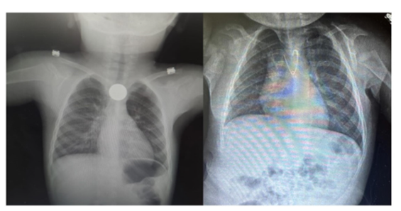
A total of 42 EFB patients followed in our clinic between July 2019 and December 2022 were included in the study. An overwhelming majority were males, i.e. 27 (64.2%), while 15 (35.8%) were females, with male to female ratio of 1.8:1. Twenty-eight (66.6%) patients were younger than 6 years and 14 (33.4%) were older than 6 years. FCT was performed in 20 (47.7%) patients with EFB and esophagoscopy was performed in 22 (52.3%) patients. The clinical and demographic characteristics of the patients are listed in Table 1. The most common complaint in patients was drooling in 30 (71.4%) patients, followed by difficulty in swallowing in 22 (52.3%) patients. Fifteen (35.7%) patients had sore throat and 12 (28.5%) had unexplained crying. The characteristics and locations of FBs are listed in Table 2. It was observed that chest and neck X-rays were taken in all cases included in the study Figure 2. The foreign body was opaque in 36 (85.7%) patients and did not require additional imaging. Computed tomography was performed in 6 (14.3%) cases that did not show opacity. In one of the cases with tomography, the diagnosis could not be clarified and esophagography was performed.
Table 1:Demographic and clinical characteristics of the patients.
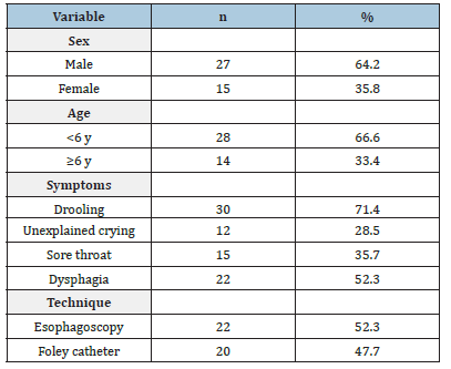
Table 2:Characteristics of foreign bodies.
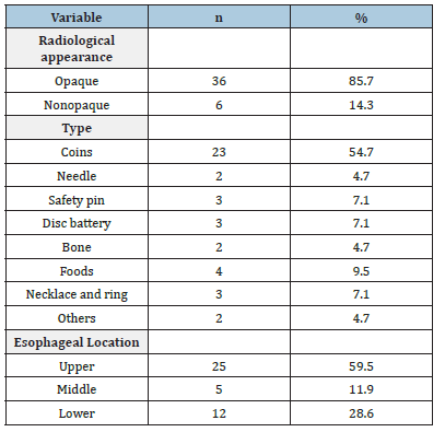
Figure 2:Chest X-ray anteroposterior view showing the radio-opaque FB with serrated margin in upper esophagus.
The most common localization of foreign bodies was in the upper esophagus in 25 (59.5%) patients, followed by the lower esophagus in 12 (28.5%) and the middle esophagus in 5 (11.9%) patients. It was observed that the overwhelming majority of patients [23 (54.7%)] swallowed coins. Needle in 2 (4.7%), safety pin in 3 (7.1%), disc battery in 3 (7.1%), bone-in 2 (4.7%), food in 4 (9.5%), necklace or ring in 3 (7.1%) patients removed from the esophagus. It was observed that in 24 patients with blunt FB in the upper or middle esophagus, FCT was attempted to be removed by FCT, successful in 20 patients, and esophagoscopy was performed in 4 unsuccessful patients. No major complications or mortality were observed in any of the patients. The hospital stay was 3±0.7 hours in patients who underwent FCT, while it was 14±2.2 hours in patients who underwent esophagoscopy.
Discussion
EFBs can be asymptomatic or present with symptoms such as drooling from the mouth, difficulty swallowing, a feeling of being stuck in the throat, coughing and dispnea. Long-standing FBs may cause recurrent pneumonia and related developmental delays [5]. In our series, the most common reason for patients to present was drooling. It was remarkable that our patients did not have cough and dispnae. We did not have a case of long-term EFB.
Physical examination and radiological imaging are usually sufficient for the diagnosis of the disease. The gold standard diagnostic method is esophagoscopy, but in some cases it may be difficult to diagnose even with esophagoscopy [6]. It was observed that x-ray was used as the first diagnostic tool in all of our cases. X-ray alone was diagnostic in 85.7% of our patients. Tomography was performed for diagnosis in 6 cases that did not appear opaque on x-ray, and esophagography was performed in one of them.
The most commonly ingested FBs are coins by children [7]. In our series, the most common EFB was coins, consistent with the literature (%54.7). Various techniques have been described for the removal of EFBs, the most known of which are; flexible endoscopy, rigid endoscopy, FCT, push with bougie, Magill forceps, penny pincher technique [8-11]. The use of the FCT was found to be safe and effective, especially in blunt FB such as coins [11]. In a series of 43 patients who underwent FCT, the authors reported that they were able to remove EFB without complications with a success rate of 81% [11]. In our series, the FCT was tried first in 24 patients with blunt FBs, such as money, in the middle and upper part of the esophagus, and the FB was successfully removed in 20 patients (83.3%). FB was removed by performing rigid esophagoscopy in 4 unsuccessful cases. The FCT is difficult to apply, especially in children older than 1 year, due to teeth eruption and biting the catheter. In order to overcome this difficulty, we provided our cases with appropriate conditions and applied sedation with various agents, and sent the catheter through the airway so that it would not be bitten. We observed that with this technique, the catheter advances more easily and it can be intervened more quickly because it prevents patients from biting. Although FCT is easy to apply and has a high success rate, it should be applied by creating intubation and emergency intervention conditions, since it carries the risk of esophageal damage and more importantly, tracheal aspiration. Esophageal damage or aspiration did not occur in patients to whom we applied the FCT.
Alcaline disc batteries are the leading FBs that can cause serious morbidity and mortality if they remain in the esophagus [12]. Apart from pressure necrosis caused directly by the battery, necrosis and perforation may develop in the esophagus with the dissolution of heavy metals such as mercury, silver and lithium in the battery [13]. In studies performed on rats, mucosal necrosis was observed within one hour after the batteries were removed, and ulceration was observed after two hours, and perforation was observed after eight hours [14]. In our series, only 3 cases had disc battery in the esophagus, one of them was brought to the hospital 2 hours after swallowing and the battery was in the upper part of the esophagus. FTT was applied to this case. In the other two cases, the battery esophagus was successfully removed by rigid esophagoscopy, since the esophagus was at the lower end. In our cases, no necrosis or perforation was observed in the esophagus. We think that this is due to early intervention in the cases.
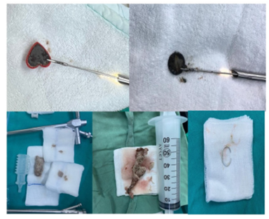
Sharp objects that are likely to be swallowed by children are needles, safety pins, screws, bones, staples and pieces of glass. If these sharp objects remain in the esophagus, the risk of perforation is very high and must be removed immediately [15] In our series, chicken bones were found in 2 cases, needles in 2 cases and safety pins in 3 cases. FBs were removed with rigid esophagoscopy without complications Figure 3. In our country, there is a practice traditionally applied by parents, especially in infants, to attaching an evil eye bead or amulet to the child’s dress with safety pins. For this reason, safety pin ingestion cases are more common in infants. It is a known fact that the cases of needle swallowing in our country are mostly young girls who wear headscarves . In our series, both patients who swallowed needles were young female patients wearing a headscarf.
Figure 3:Examples of FBs we have extracted by esophagoscopy.
FBs are often lodged in the esophagus at three levels; thoracic outlet (upper)(70%), Carina Level (middle) (15%), lower Esophageal Sphincter (lower) (15%) [16]. In our patients, FBs were found most frequently in the upper esophagus (59.5%), secondly in the lower esophagus (28.6%) and less frequently in the middle esophagus (11.9%). Our findings were consistent with the literature. It can be waited up to 24 hours for EFBs, or the objects can be pushed into the stomach with the help of a catheter [16]. If FBs remaining in the esophagus for more than 24 hours, or larger than 2cm, or sharp tip, or the disc batteries, or the patient has respiratory distress, or there are signs of esophageal perforation, should be removed [5]. Since foreign bodies remaining in the esophagus for more than one week carry the risk of serious erosion and perforation, surgical team and equipment should be prepared before endoscopy [5]. All of our cases were treated within 24 hours, we did not have any delayed cases.
Conclusion
EFBs are frequently encountered in children younger than 6 years of age and can cause mortality. Therefore, it is important to inform parents about taking precautions against FB ingestion by children. Foley catheter technique has been found to be very successful and safe in removing FBs such as coins, and sending the catheter over the airway will increase the chance of success. Foreign bodies in the esophagus are frequently encountered in children younger than 6 years of age and can cause mortality. Therefore, it is important to inform parents about taking precautions against foreign body ingestion by children. Foley catheter technique has been found to be very successful and safe in removing blunt foreign bodies such as money, and sending the catheter over the airway will increase the chance of success. Despite this, rigid esophagoscopy is still popular in all EFB cases.
References
- Waltzman ML, Baskin M, Wypij D, Mooney D, Jones D, et al. (2005) A randomized clinical trial of the management of esophageal coins in children. Pediatrics 116(3): 614-619.
- Uyemura MC (2005) Foreign body ingestion in children. American Family Physician 72(2): 287-291.
- Singh A, Bajpai M, Panda SS, Chand K, Jana M, et al. (2014) Oesophageal foreign body in children: 15 years’ experience in a tertiary care paediatric centre. African Journal of Paediatric Surgery 11(3): 238-241.
- Sekmenli T, Çifci I (2022) Çocuklarda Özofagus Yabancı Cisimlerine Genel Yaklaşım. Pediatr Pract Res 10(1): 38-43.
- Eisen GM, Baron TH, Dominitz JA, Faigel DO, Goldstein JL, et al. (2002) Guideline for the management of ingested foreign bodies. Gastrointest Endosc 55(7): 802-806.
- Athanassiadi K, Gerazounis M, Metaxas E, Kalantzi N (2022) Management of esophageal foreign bodies: a retrospective review of 400 cases. Eur J Cardiothorac Surg 21(4): 653-656.
- Waltzman ML (2006) Management of esophageal coins. Curr Opin Pediatr 18(5): 571-574.
- Gmeiner D, Rahden BH, Meco C, Hutter J, Oberascher G, et al. (2007) Flexible versus rigid endoscopy for treatment of foreign body impaction in the esophagus. Surgical Endoscopy 21(11): 2026-2029.
- Janik JE, Janik JS (2003) Magill forceps extraction of upper esophageal coins. Journal of Pediatric Surgery 38(2): 227-229.
- Gauderer MW, Decou JM, Abrams RS, Thomason MA (2000) The 'penny pincher': A new technique for fast and safe removal of esophageal coins. Journal of Pediatric Surgery 35(2): 276-278.
- Lim D, Kim JK, Kim YJ, Cho YJ, Cho JW, et al. (2021) Factors affecting successful esophageal foreign body removal using a Foley catheter in pediatric patients. Clinical and Experimental Emergency Medicine 8(1): 30-36.
- Votteler TP, Nash JC, Rutledge JC (1983) The hazard of ingested alkaline disk batteries in children. JAMA 249(18): 2504-2506.
- Maves MD, Carithers JS, Birck HG (1984) Esophageal burns secondary to disc battery ingestion. Ann Otol Rhinol Laryngol 93(4 Pt 1): 364-369.
- Krey H (1952) On the treatment of corrosive lesions in the oesophagus; an experimental study. Acta Otolaryngol Suppl 102: 1-49.
- Boo SJ, Kim HU (2018) Esophageal foreign body: Treatment and complications. Korean J Gastroenterol 72(1): 1-5.
- Chen MK, Beierle EA (2001) Gastrointestinal foreign bodies. Pediatr Ann 30(12): 736-742.
© 2023 Mehmet Sarıkaya. This is an open access article distributed under the terms of the Creative Commons Attribution License , which permits unrestricted use, distribution, and build upon your work non-commercially.
 a Creative Commons Attribution 4.0 International License. Based on a work at www.crimsonpublishers.com.
Best viewed in


.jpg)


 Editorial Board Registrations
 Submit your Article
 Refer a Friend
 Advertise With Us
.jpg)


.jpg)


.bmp)
.jpg)
.png)
.jpg)


.jpg)


.png)

.png)


.png)


