- Submissions

Full Text
Surgical Medicine Open Access Journal
A Rare Case of All 4 Permanent Canines Impacted-Radiographic Finding: Case Report
Nielsen S Pereira*
Private Practice in Oral and Maxillofacial Radiology, Brazil
*Corresponding author:Nielsan S Pereira, Private Practice in Oral and Maxillofacial Radiology, Brazil
Submission: November 21, 2022Published: May 01, 2023

ISSN 2578-0379 Volume5 Issue2
Abstract
Impacted maxillary or mandibular canines is frequently encountered in clinical practice, but encountered the 4 canines impacted is very rare. The importance of all canines in a healthy, esthetic dentition and occlusion is very large.
Keywords:Maxillary canines; Mandibular canines; Impacted
Introduction
The canines play an important role in facial appearance, dental esthetics, arch development, and occlusion and its impaction of the maxillary canines is twice common as the mandible [1]. The maxillary canine is second only to the mandibular third molar in its frequency of impaction, with a rate that varies from 0.2 to 3.0% [2-5]. The impaction of the mandible canine is much lower than the maxillary [2]. Impacted teeth may not only interfere with esthetic aspect but as a source of an oral pathology, such as dentigerous cyst [6], so the early detection and treatment of this condition is fundamental. 2D X-ray examination, such as Panoramic has been used to locate impacted tooth, but with the appearance of the CBCT-Cone Beam Computed Tomography, this 3D examination has become the golden standard for the location and spatial position of the impacted tooth [3,4,7,8]. The treatment of the impacted canines varies from orthodontic extrusion, aligners, fixed appliance, surgical removal [2,5,9,10]. Regarding the impaction of the maxillary canines, the morphology and skeletal characteristics of the maxilla play an important role in its occurrence [3,8,11].
Case Report
Female patient, 15 years old at the time of the finding; she was referred on August of 2019 by her orthodontist to the dental imaging service for evaluation of the position and condition of the 2 maxillary and 2 mandibular canines. The patient was under orthodontic treatment using fixed appliance for 2 years, as her information. The general dentist requested a CBCTCone Beam Computed Tomography of the anterior maxilla and anterior mandible region. A CBCT was performed by CS 9300C (Carestream Health Dental) equipment with a Small FOV (Field of View) 5.0x8.0cm and 200μm (nanometer) of each region.
During the evaluation of the maxillary and mandible CBCT studies, we observed:
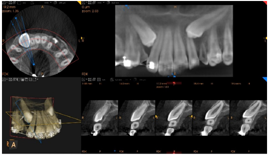
a) The tooth 13 impacted and the deciduous tooth 53 retained; the tooth 13 is
mesialized and facing the buccal side of the maxilla, as shown in Figure 1.
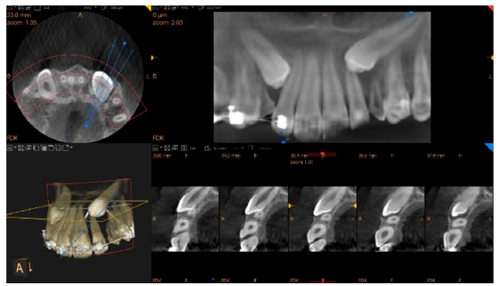
b) The tooth 23 impacted and the deciduous tooth 63 retained; the tooth 23 is
mesialized, over the apex of the tooth 22 and facing the buccal side of the maxilla; we still
observed a small radiolucency surrounding its crown, as shown in Figure 2.
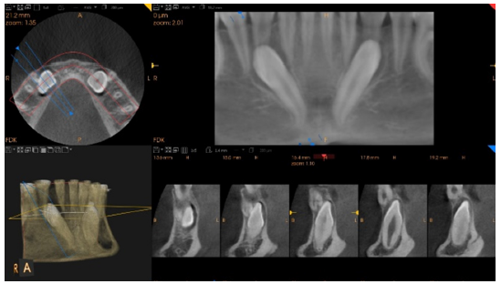
c) The tooth 33 impacted and the deciduous tooth 73 retained; the tooth 33 is
destabilized and facing the lingual side of the mandible, as shown in Figure 3.
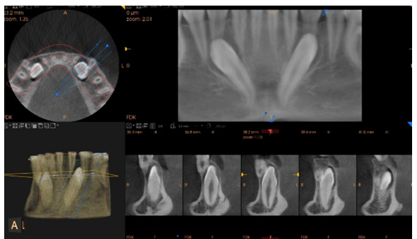
d) The tooth 43 impacted and the deciduous tooth 83 retained; the tooth 43 is distalized
and facing the lingual side of the mandible, as shown in Figure 4.
Figure 1:13 Impacted.

Figure 2:23 Impacted.

Figure 3:43 Impacted.

Figure 4:33 Impacted.

After the examination, the patient was released and returned to her orthodontist for treatment follow-up and planning for the 4 impacted canines.
Discussion
Despite the impaction of both maxillary and mandibular is rare, the impaction of the canines, mainly, the maxillary is one the most common impacted tooth [1,3]. The morphology of the maxilla plays an important role for the canine impaction [8,11]. Due to the importance of the canines in the occlusion and esthetics aspect of the patient, an early detection and treatment is much important [1,7]. The CBCT-Cone Beam Computed Tomography is the golden standard imaging examination for the most accurate location and position of these condition, for both maxillary and mandibular studies [3,4,7,8]. The case reported here shows a rare case where the 4 canines, maxillary and mandibular are impacted and the importance of the CBCT examination.
Conclusion
We concluded that a careful and meticulous clinical evaluation supported by a good CBCT examination are fundamental for the correct diagnosis and the therapeutic planning and treatment for the impacted maxillary and mandibular canines.
References
- Singh S, Parihar AV, Chaturvedi TP, Shukla N (2022) A case series of orthodontic traction of maxillary impacted canine. Natl J Maxillofac Surg 13(1): 147-152.
- Stabryła J, Plakwicz P, Kukuła K, Zadurska M, Czochrowska EM (2021) Comparisons of different treatment methods and their outcomes for impacted maxillary and mandibular canines: A retrospective study. J Am Dent Assoc 152(11): 919-926.
- Cacciatore G, Poletti L, Sforza C (2018) Early diagnosed impacted maxillary canines and the morphology of the maxilla: A three-dimensional study. Prog Orthod 19(1): 20.
- Alfaleh W, Thobiani SA (2021) Evaluation of impacted maxillary canine position using panoramic radiography and cone beam computed tomography. Saudi Dent J 33(7): 738-744.
- Bedoya KA, Escobar M, Juanito GP, Henriques B, Benfatti CA (2022) Multidisciplinary treatment of an impacted maxillary canine with immediate implant installation. J Indian Soc Periodontol 26(2): 192-196.
- Chen J, Lv D, Li M, Zhao W, He Y (2020) The correlation between the three-dimensional radiolucency area around the crown of impacted maxillary canines and dentigerous cysts. Dentomaxillofac Radiol 49(4): 20190402.
- Pico CL, Vale FJ, Caramelo FJ, Real AC, Pereira SM (2017) Comparative analysis of impacted upper canines: Panoramic radiograph Vs cone beam computed tomography. J Clin Exp Dent 9(10): e1176-e1182.
- Díaz ME, González AM, Navarro RA, Lorenzo AA, López NE, et al. (2022) Skeletal and dental morphological characteristics of the maxillary in patients with impacted canines using cone beam computed tomography: A retrospective clinical study. J Pers Med 12(1): 96.
- Xia Q, He Y, Jia L, Wang C, Wang W, et al. (2022) Assessment of labially impacted canines traction mode with clear aligners vs. fixed appliance: A comparative study based on 3D finite element analysis. Front Bioeng Biotechnol 10: 1004223.
- Goswami M, Johar S (2020) Surgical removal of odontoma: A case report. Int J Clin Pediatr Dent 13: S122-S124.
- Leonardi R, Muraglie S, Crimi S, Pirroni M, Musumeci G, et al. (2018) Morphology of palatally displaced canines and adjacent teeth, a 3-D evaluation from cone-beam computed tomographic images. BMC Oral Health 18(1): 156.
© 2023 Victor X Mosquera. This is an open access article distributed under the terms of the Creative Commons Attribution License , which permits unrestricted use, distribution, and build upon your work non-commercially.
 a Creative Commons Attribution 4.0 International License. Based on a work at www.crimsonpublishers.com.
Best viewed in


.jpg)


 Editorial Board Registrations
 Submit your Article
 Refer a Friend
 Advertise With Us
.jpg)


.jpg)


.bmp)
.jpg)
.png)
.jpg)


.jpg)


.png)

.png)


.png)


