- Submissions

Full Text
Surgical Medicine Open Access Journal
Post-Intubation Tracheal Tear in Myomectomy Patient
Aboud AlJa’bari MD MBBS DESA*
Consultant Cardiothoracic, Regional Anesthesia and Pain Management, Lausanne University Hospital, Switzerland
*Corresponding author: Aboud AlJa’bari MD MBBS DESA, Consultant Cardiothoracic, Regional Anesthesia and Pain Management, Lausanne University Hospital, Switzerland
Submission: June 28, 2021Published: October 06, 2021

ISSN 2578-0379 Volume4 Issue4
Abstract
Twelve hours after extubating, a 33-year-old woman developed extensive Subcutaneous (surgical) emphysema, involving the neck, and upper chest Extending from skull base down to the mediastinum left sided Pneumomediastinum and pneumothorax, was confirmed by neck and chest CT-scan. The location of the lesion and features of the patient favored conservative Treatment with antibiotic cover. The patient made a full and uncomplicated Recovery and was discharged ten days after the original injury.
Keywords: Anesthesia; Intubation; Myomectomy; Trachea; Tear
Introduction
Tracheal tear is a rare and life-threatening complication, that usually occur after blunt trauma to the chest, but which may complicate tracheal Intubation. We report this rare case of post-intubation tracheal tear after abdominal Myomectomy under general anesthesia.
Case report
A 33-year-old woman was scheduled for abdominal myomectomy under general
anesthesia. After induction of anesthesia with propofol 150mg, fentanyl 150mcg and
rocuronium 40mg, oral intubation was performed without any difficulty (Cormack lehane
view 1) using a 7.5 cuffed preformed orotracheal tube without stylet. The cuff was inflated
with 7ml of air and continuously monitored to sustain the pressure during the procedure.
Anesthesia was maintained with isoflurane. The entire surgery lasted approximately 60min.
Six hours after extubating the patient started to complain of shortness of breath and chest
discomfort. Consequently, ECG showed: Normal sinus rhythm, no significant changes, ABGS
“on room air”: PO2 80mmHg and 90% O2 sat. She responded positively as 96% improved on
O2 face mask and no shortness of breath anymore.
Twelve hours after extubating, the anesthesiologist attended because the patient had
suddenly developed subcutaneous emphysema of the facial, bilateral later cervical and upper
anterior chest, although she did not complain of chest pain or dyspnea. In addition, the chest
X-ray did not present any abnormal signs.
The results of the arterial blood gas on room air were pH=7.35 PCO2=43mmHg,
PO2=68mmHg HCO3= 22mmHg and SpO2=92%. A thoracic Computed Tomography (CT)
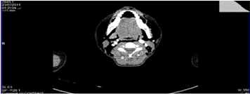
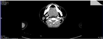
showed: Extensive subcutaneous (surgical) emphysema, involving the neck (Figure 1), and
upper chest (Figure 2), extending from skull base down to the mediastinum (Figure 1),
moderate left sided pneumomediastinum and pneumothorax, defect 40×10mm is seen in the
posterior (membranous) portion of dorsal trachea from the +1-D3.
Traumatic Tracheal tear was suspected, and the patient was transferred to surgical ICU.
Then a chest tube was inserted while the patient kept on simple face mask with daily ABGs.
According to the location and the features of the lesion, the conservative treatment with
antibiotic cover (cephalosporin and aminoglycoside) and monitoring in the intensive care
unit were the ideal treatment plan. Mechanical ventilation, bilateral endobronchial intubation
and surgery were available if necessary.
Figure 1: Thoracic CT showing diffuse soft tissues emphysema of the neck and skull base.

Figure 2: Thoracic CT showing diffuse soft tissues emphysema of the upper chest.

The patient improved, and five days later, the chest CT showed that the left tube is seen with almost complete resolution of left pneumothorax, and the surgical emphysema appears smaller in size at tracheal tear. There are no significant changes seen neither a fistula was noted. After three more days in a general medical ward, the patient was discharged home in good condition, ten days after the initial injury. One month later, the patient was scheduled to come back to surgical clinic for reevaluation; she was presented with good general condition.
Discussion
Tracheal tear following intubation is a rare complication [1].
Risk factors for this complication may be related to the patient, the
anesthesiologist, the endotracheal tube size, and the technique of
intubation [1,2]. Often, the cause is multifactorial [1]. Although, in
some cases, the main cause may remain iatrogenic [1]. Patient risk
factors include conditions affecting tracheal anatomy as the trachea
is more vulnerable in female, advanced age, short stature, chronic
obstructive pulmonary disease and corticosteroid therapy [2]. The
assumption that a membranous trachea is fewer firms in women
than in men [2].
There are multiple factors that contribute to the technique of
intubation, which may be related to mechanical factors as trauma
during intubation, or over inflation of the cuff, coughing etc., and
size of the tube [3-5]. Although, over inflation of endotracheal cuff
and sudden manipulation of the tube are two commonest reasons
but direct tear caused by the tube itself is extremely rare [6].
Multiple intubations attempt in difficult cases could result
tracheal laceration. Nevertheless, the operators involved are welltrained
anesthesiologists and the intubations were reported as easy
and uneventful as in our case. Other causes of iatrogenic tracheal
laceration are the use of a stylet inside the tube while the stylet
not being removed as soon as the tube tip passed the vocal cords,
or the use of a double-lumen tube with direct trauma of the tip or
the carina [2]. We can differentiate this depending on the level of
the tear, at cervical level (when using a single-lumen tube as in our
case) or at thoracic level (when using a double-lumen tube), may
indicate a cuff-induced rupture [2].
Finally, cuff over-inflation acts as a distension force and is
reported as cause in some cases [1]. Using nitrous oxide tends to
expand more the cuff, so monitoring cuff pressure during surgery is
recommended and essential. The possible explanation in our case
report is most probably due to large endotracheal tube and to lesser
extent the amount of air inflation in cuff. The clinical manifestations
of tracheal tear are subcutaneous emphysema, pneumothorax,
hemoptysis and respiratory failure [1]. Usually, they appear during
surgery or immediately in postoperative period [2]. Sometimes it
may delay hours later as in our case report.
Chest X-ray and chest CT can show soft tissues emphysema,
pneumomediastinum, pneumopericardium and/or pneumothorax
[2,4]. An emergency bronchoscope is necessary to confirm the
diagnosis and to determine the extension of the laceration [1,2,5].
Regarding treatment strategy of a post-intubation tracheal tear
depends on the size and the location of the rupture, clinical
presentation, and the condition of the patient [1].
Two therapeutic strategies are proposed: a surgical or nonsurgical
approach [3]. The current tendency is to decrease invasive
surgical treatment for the benefit of conservative management
[1,3] Surgical repair is the preferred when a transmural tear with
a 2cm length causing pneumothorax and/or pneumo-mediastinum
and if the lesion is at thoracic level.
Conservative treatment could be an alternative in some patients
if: small lacerations in the upper third of the trachea, without full
thickness of the tracheal wall, no gross air leak and patients who are
breathing spontaneously without any compromise [6]. Conservative
treatment in large complete tracheal rupture in old age with
critical general condition could be an option [3]. Other treatment
can include mechanical ventilation after tracheal or bilateral
endobronchial intubation (with the cuff inflated distal to the tear as
in our case), chest drain, continuous airway humidification, broadspectrum
antibiotics, and regular chest physiotherapy [3].
Good healing and the absence of stenosis could be verified with
tracheal fiber endoscopy one month after the initial injury [3]. Our
case report shows the effectiveness of the conservative therapeutic
strategy of a large tracheal injury. Selection of treatment for
post-intubation tracheal tear must be individualized. Airway
management is a fundamental aspect of anesthetic practice, and it is
essential for anesthesiologists to be vigilant to these complications,
and to have an effective strategy to prevent and manage these
complications when they happen.
References
- Carbognani P, Bobbio A, Cattelani L, Internullo E, Caporale D, et al. (2004) Management of postintubation membranous tracheal rupture. Ann Thorac Surg 77(2): 406-409.
- Ané MCH, Picard E, Jonquet O, Mary H (1995) Membranous tracheal rupture after endotracheal intubation. Ann Thorac Surg 60(5): 1367-1371.
- Jougon J, Ballester M, Choukroun E, Dubrez J, Reboul G, et al. (2000) Conservative treatment for postintubation tracheobronchial rupture. Ann Thorac Surg 69(1): 216-220.
- Gries CJ, Pierson DJ (2007) Tracheal rupture resulting in life-threaten subcutaneous emphysema. Respir Care 52(2): 191-195.
- Eipe N, Choudhrie A (2007) Tracheal rupture in a child with blunt chest injury. Pediatric Anesthesia 17(3): 273-277.
- Beiderlinden M, Adamzik M, Peters J (2005) Conservative treatment of tracheal injuries. Anesth Analg 100(1): 210-214.
© 2021 Aboud AlJa’bari. This is an open access article distributed under the terms of the Creative Commons Attribution License , which permits unrestricted use, distribution, and build upon your work non-commercially.
 a Creative Commons Attribution 4.0 International License. Based on a work at www.crimsonpublishers.com.
Best viewed in


.jpg)


 Editorial Board Registrations
 Submit your Article
 Refer a Friend
 Advertise With Us
.jpg)


.jpg)


.bmp)
.jpg)
.png)
.jpg)


.jpg)


.png)

.png)


.png)


