- Submissions

Full Text
Surgical Medicine Open Access Journal
Complicated Congenital Diaphragmatic Bochdalek Hernia in Adult
Elvia María SC1, Allan Fernando DM1* and José Daniel RB2
1Third-year Resident of General Surgery Residency, National Autonomous University of Honduras, Honduras
2Specialist in General Surgery, University School Hospital, Honduras
*Corresponding author: Allan Fernando DM, Third-year Resident of General Surgery Residency, National Autonomous University of Honduras, Honduras
Submission: June 16, 2021Published: June 29, 2021

ISSN 2578-0379 Volume4 Issue3
Abstract
Diaphragmatic hernia is an extremely rare entity in adults, it is defined as the transposition of abdominal organs to the thoracic cage, through defects of the phrenic muscle. They can be classified as congenital or acquired. The incidence is reported to be 0.17%, with most hernias occurring on the left side. In Honduras there are few cases reported in pediatrics and one case reported in adults. 53-year-old male patient, farmer, from Tegucigalpa, Honduras. Who attended the Surgery Emergency at the Hospital Escuela Universitario? With a single history of chronic alcoholism, she presented stopping of evacuation of 10 days of evolution which channeled gases, accompanied by abdominal distention in turn, abdominal pain of 1 week of colic-like evolution, in the left iliac fossa irradiated to the flank and right hypochondrium, progressive insidious onset, of mild to moderate intensity, sometimes it did not remit with analgesics, accompanied by dyspnea from small to medium efforts and paroxysmal episodes of cough, without cyanosis. Abdomen: globose at the expense of adipose tissue, distended, on auscultation with abolished bowel sounds, tympanic percussion, with pain on superficial and deep palpation with involuntary muscular resistance. The chest radiograph showed the presence of intestinal and stomach loops in the left hemithorax. Abdominal radiographs show hydroaerous levels, with an image of a pile of coins, with dilated loops of the large intestine. After laparotomy, the patient presented a satisfactory evolution, with subsequent hospital discharge.
Keywords: Diaphragmatic hernia; Bochdalek; Abdominal obstruction
Introduction
Diaphragmatic hernias represent an extremely rare entity especially when diagnosed in adulthood. Its causes can be congenital or generally acquired after a thoracic trauma. Congenital diaphragmatic hernias occur in 1 out of 2,200 to 5,000 live births, with high intrauterine and post-labor mortality, making it difficult to identify the true incidence of this pathology due to its high mortality prior to referral to a third-level center of attention [1]. The most common type of congenital diaphragmatic hernia is the Bochdalek hernia, which receives its name in honor of Victor Bochdalek who first described it in 1948, it is defined as a posterolateral [2] diaphragmatic defect that in most cases is left (85%) produced by a defect in the closure of the posterolateral foramen during the embryonic period between the 8th and 10th week [3]. Most of the cases that are asymptomatic until adulthood is diagnosed incidentally (14%) or present with abdominal (62%) respiratory (40%), obstructive (36%) symptoms [4].
Clinical Case
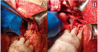
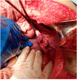
53-year-old male patient, farmer, from Tegucigalpa, Honduras. Who attended the Surgery emergency at the University School Hospital? With a single history of chronic alcoholism, he presented evacuation arrest of 10 days of evolution which channeled gases, accompanied by abdominal distension in turn, abdominal pain of 1 week of evolution type colic, in left iliac fossa irradiated to flank and right hypochondrium, Progressive insidious onset, of mild to moderate intensity, sometimes it did not subside with analgesics, accompanied by dyspnea from small to medium efforts and paroxysmal episodes of cough, without cyanosis. Physical examination revealed: conscious patient, blood pressure 130/90mmHg, heart rate 98 per minute, respiratory rate 20 per minute, oxygen saturation 89%, weight 80 kilograms, height 1.75 meters, body mass index 26.5kg/m3 Chest auscultation revealed: hypoventilation of the left lung base and bowel sounds in the left hemithorax. Abdomen: globose at the expense of adipose tissue, distended, on auscultation with abolished bowel sounds, tympanic percussion, with pain on superficial and deep palpation with involuntary muscular resistance. The chest X-ray showed the presence of bowel and stomach loops in the left hemithorax (Figure 1). X-rays of the abdomen present hydroaerous levels, with a coinpile image, with dilated loops of the large intestine. Hemogram: hemoglobin 12g/dl, Hematocrit 32%, platelets 270,000, white blood cells 16,000, Neutrophils 85% (Figure 2). The intraoperative findings were dilated small bowel and colon loops, diaphragmatic defect in the left posterior region of more or less 8 centimeters in length, stomach, spleen, splenic flexure of the colon and momentum in the left pleural cavity with multiple firm adhesions, without contamination of the pleural cavity, collapsed left lung, for which a chest tube was placed on the left side, 28 French (Figure 3).
Figure 1: Preoperative chest X-ray.

Figure 2: A. Manual reduction of the hernia.
B. Identification of diaphragmatic defect.

Figure 3: Diaphragmatic defect repair with interrupted suture of non-absorbable material.

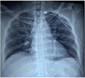
In the postoperative period, the patient presented hypoxemia and a decrease in blood pressure in blood gases, for which he was left under observation for 24hours. On the third postoperative day, in view of the fact that clinically hypoventilation areas were not auscultated, the vesicular murmur was present in both lung fields and the radiologically expanded lung, it was decided to remove the chest tube. On the fifth hospital day in view of the good clinical evolution, tolerating the oral route, environmental oxygen and chest X-rays, observing that there was no pleural effusion and presence of good lung expansion (Figure 4), without area of pneumothorax or atelectasis, it was decided to leave the healthcare center.
Figure 4: X-rays of the thorax in the third postoperative period.

Discussion
The Bochdalek hernia was described by the Czech anatomist Vincent Alexander Bochdalek in 1847 as a maldevelopment of pleuroperitoneal folds with septum transversum as well as absent migration of musculature during embryologic development, resulting in a posterolateral diaphragmatic defect [5]. Bochdalek hernias present in newborns with dramatic respiratory insufficiency due to lung hypoplasia, requiring immediate surgery [6]. Adult BH is an uncommon form of diaphragmatic hernia [7]. The incidence is reported to be 0.17%, with the majority of hernias occurring on the left side [8]. Symptoms in adults are chronic and variable. Emergency surgery is rare in adults [9,10]. Most hernias are asymptomatic and found incidentally. Patients may present with chronic symptoms such as recurrent chest or abdominal pain and postprandial fullness or vomiting [4,11]. Usually Bochdalek hernia is detected incidentally, but very rarely patients might present as an acute emergency due to the strangulation of herniated abdominal contents [6,8]. In contrast to the patient, a Japanese study adult BH has a female predominance, and 58 patients (60%) were female, with a mean age of 58 years (range 20-89 years) [4]. The hernia contents were the stomach (n=46), colon (n=40), momentum (n=27), small intestine (n=24), spleen (n=20), liver (n=6), pancreas (n=4), kidney (n=4), and retroperitoneal tissue (n=5). The most common initial presentations of adult BH were abdominal (pain, distension, discomfort; 67.7%), pulmonary (cough, chest pain, dyspnea; 29.1%), and obstructive symptoms (vomiting, nausea; 43.8%) [7]. Surgical reduction of the prolapsed organs and closure of the hernial orifice are recommended immediately following diagnosis/suspicion of adult Bochdalek hernia [4,8].
References
- Iqbal CW (2019) Congénital diaphragmatic hernia. In: Fischer JE, Jones DB, et al. (Eds.), Mastery of Surgery 7th (Edn), Lippincott Williams & amp, Wilkins, Philadelphia, USA, pp. 2420-2466.
- Deb SJ (2011) Massive right sided Bochdalek hernia with two unusual findings: A case report. J Med Case Rep 5: 519.
- Toral CI, Palacios PA, Castillo CR (2019) Bochdalek hernia in adult: An extremely rare entity. Journal of the Faculty of Medicine 62(3): 1-5.
- Machado NO (2016) Laparoscopic repair of Bochdalek diaphragmatic hernia in adults. N Am J Med Sci 8(2): 65-74.
- Longoni M, Pober BR, High FA (2006) Congenital diaphragmatic hernia overview. In: Adam MP, Ardinger HH, et al. (Eds.), Gene Reviews PubMed, USA.
- Miyasaka T, Matsutani T, Nomura, T (2020) Laparoscopic repair of a Bochdalek hernia in an elderly patient: A case report with a review from 1999 to 2019 in Japan. Surg Case Rep 6(1): 233.
- Akhtar K, Qurashi K, Rizvi A, Isla R (2009) Emergency laparoscopic repair of an obstructed Bochdalek hernia in an adult. Br J Hosp Med (Lond) 70(12): 718-719.
- DeAlwis K, Mitsunaga EM (2009) Sudden death due to nontraumatic diaphragmatic hernia in an adult. Am J Forensic Med Pathol 30(4): 366-368.
- Shah R, Reddy MM, Mascarenhas AF (1986) Bochdalek hernia in an adult presenting as an emergency. Indian J Chest Dis Allied Sci 28(4): 237-240.
- Laaksonen E, Silvasti S, Hakala T (2009) Right-sided Bochdalek hernia in an adult: A case report. J Med Case Rep 3(1): 109.
- Mullins ME, Stein J, Saini SS, Mueller PR (2001) Prevalence of incidental Bochdalek’s hernia in a large adult population. AJR Am J Roentgenol 177(2): 363-366.
© 2021 Allan Fernando DM. This is an open access article distributed under the terms of the Creative Commons Attribution License , which permits unrestricted use, distribution, and build upon your work non-commercially.
 a Creative Commons Attribution 4.0 International License. Based on a work at www.crimsonpublishers.com.
Best viewed in


.jpg)


 Editorial Board Registrations
 Submit your Article
 Refer a Friend
 Advertise With Us
.jpg)


.jpg)


.bmp)
.jpg)
.png)
.jpg)


.jpg)


.png)

.png)


.png)


