- Submissions

Full Text
Surgical Medicine Open Access Journal
Is H. Pylori the New Risk Factor for GB Cancer?
Shilpi B1* and Mathur AV2
1Resident, General surgery, SGRRIMS, Dehradun, India
2Professor, Dept of general surgery, SGRRIMS, Dehradun, India
*Corresponding author: Shilpi B, Resident, General Surgery, SGRRIMS, Dehradun, Uttarakhand, India
Submission: April 14, 2021Published: June 23, 2021

ISSN 2578-0379 Volume4 Issue3
Abstract
To find out the various clinicopathological presentations of gall stone disease with special relation to H. pylori infections of Gall Bladder. Institution based observational study in a tertiary care center. A total of 50 patients were selected who were admitted with the diagnosis of cholelithiasis and planned for cholecystectomy. Gallbladder specimen was then sent for routine H&E stain and additional Giemsa stain. Patients were grouped under two categories based on result of Giemsa stain. Out of which 10% showed the presence of H. pylori in the GB wall specimen. The presence of H. pylori in gall bladder wall was mostly seen in patients depicting chronic cholecystitis features on histopathological examination. Chronic cholecystitis is one of the most common risk factors for development of GBC. And inflammation being an important predisposing factor for development of cancer, could help us establish a relationship between GB cancer and H. pylori. Thus, in areas of high prevalence of GBC, like the gang belt H. pylori should further be evaluated as an etiological agent for the development of gall bladder cancer.
Keywords: H. pylori; Cholelithiasis; Gall bladder cancer (GBC); Cholecystitis
Introduction
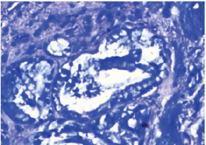
Figure 1: Chronic cholecystitis with positive H. pylori in mucosal glands.

India is a high incidence area for GBC. GBC is one of the three leading cancers among women of North and North-east India. The incidence has been steadily rising in India among women as well as men. India accounts for 10% of the global burden of GBC. In India, the incidence of GBC is 10 times higher in north India compared to the southern Indian states [8.9/100,000 population (Delhi) VS. 0.8/100,000 population (Chennai)] [1]. Gallbladder Cancer (GBC) arises from the epithelial lining of the Gall Bladder (GB) and the cystic duct (Figure 1). It is the most common biliary tract malignancy worldwide and manifests as either diffuse thickening of the GB wall or as a GB mass arising from the fundus, neck or body of the GB [2]. Helicobacter species has been associated with increased risk of GBC. H. pylori was first described in the gallbladder mucosa in patients with gallstones 1996, and a relationship between H. pylori and gallstone formation was reported [3]. All studies from India have shown a definite but small risk of H. pylori in the causation of GBC. It is hypothesized that a relationship like that of H. pylori and gastric carcinoma exists in the gall bladder (Figure 2). According to this, H. pylori is a causative agent of gallbladder cancer by causing chronic cholecystitis due to gall stones and later developing dysplasia, metaplasia and carcinoma. A history suggestive of chronic cholecystitis is present in approximately 50 % of the GBC patients. Recently, inflammation was distinguished as a hallmark of cancer. Increasing proof demonstrated that a large number of tumors originate from sites of chronic inflammation, which supports the perspective that chronic inflammation can incline to cancer [4]. Chronic inflammation plays a serious role in evoking cancer development in the gallbladder.
Figure 2: Gallbladder biopsy stained for H. pylori using Giemsa staining.

Aims and Objective
To study the presence of H. pylori in cholecystectomy specimen of patients of gall stone disease and its co-relationship with histopathological changes.
Material and Methods
A total of 50 patients who presented to surgery department
with diagnosis of gall stone disease from the month of April
2020 to October 2020 were included in the study. The study was
started after approval from the ethical committee of the medical
college (Table 1). These patients were selected randomly between
the age group of 18-60 years, with informed written consent and
a proforma was created and filled by every patient. Patient taken
treatment for H.pylori eradication in the past 6 months were
excluded from the study. Patient underwent cholecystectomy, Gall
bladder specimens was sent for histopathological examination in
10% buffered formaldehyde solution. Gross tissue processing and
routine H&E staining of the sections was done as per standard lab
protocol (Table 2). Additional sections were taken and subjected to
Giemsa staining. The results were studied and compared according
to the HPE findings. Based on HPE report, patients will be divided
into two categories H. Pylori positive and negative group. Further
the two groups shall be compared based on the histopathological
findings as follows:
A. Inflammation
B. Hyperplasia
C. Pyloric and intestinal metaplasia
D. Dysplasia
E. Cancerous changes.
Table 1.

Table 2.

Result
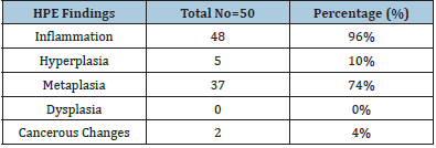
Among the 50 patients included in the study, 20% (10) were male and 80% (40) were females. Out of which 93% were symptomatic for gall stone disease and all of them were diagnosed for cholelithiasis based on USG findings (Table 3). The histopathological report depicted chronic inflammation/ cholecystitis in 90% (45) of the patients. 20% (10) patients showed acute and acute on chronic inflammation. Presence of inflammation of depicted in 48/50 patients, accounting for 96% patients .74% (37/50) patients showed metaplasia in GB wall on HPE. Hyperplasia was seen in 10% of the cases. Cancerous changes were evident in 4% (02) of evaluated cases. 10% patients showed presence of H. pylori with Giemsa staining in GB wall. Out of the 10%, every patient had chronic cholecystitis appearance on HPE.
Table 3.

Discussion
The true prevalence and relationship of different species of Helicobacter in the pathogenesis of these diseases is not known. There is currently insufficient evidence regarding the route whereby H. pylori settles in the gallbladder. It may reach the gallbladder via an ascending route from the duodenum or via the portal circulation system [5]. Researchers continue to discover new associations between H. pylori and idiopathic diseases, as well as potential benefits of H. pylori infection. Zhou 14 found H. pylori in 20% of chronic cholecystitis cases, and also emphasized that gallbladder metaplasia and pre-malignant lesions such as adenomyomatosis were more frequent in patients positive for gallbladder H. pylori [6]. In another study, Hassan [7] 19 reported that H. pylori infection aggravated potentially precancerous mucosal lesions. Helaly [8] in their study also showed the presence of H. pylori in almost 40.9% of samples in patients with chronic calcular cholecystitis. They also found a significant association between gastric and gall bladder H. pylori positivity. The source for gall bladder infection may be gastric colonization with H. pylori. They suggested that H. pylori may act as a lithogenic component, especially in presence of pure pigmented gallstones. Bansal, they concluded that there was a high prevalence of H. pylori infection in the gallbladder in northern Indian patients undergoing cholecystectomy for benign gallbladder disease which was detected only by PCR. Mishra [9] H. pylori may be endemic to the Varanasi region and may not play a significant role in the etiopathogenesis of gallbladder cancer in that region. They concluded that H. pylori colonizes areas of gastric metaplasia in gallbladder producing histological changes like those in gastric mucus. A recent study has shown that H. pylori can damage human gallbladder epithelial cells in vitro and could be the key factor that leads to clinical Cholecystitis. Histological changes, considered preneoplastic, were demonstrated in the mucosa of gallbladder, limited to mice infected with Helicobacter spp such as, intestinal metaplasia, hyperplasia, dysplasia in addition to eosinophilic inflammation and hyalinosis. Higher prevalence of cholesterol stones in the North Indian population and presence of heavy metals in the water and soil of the Indo Gangetic plains have been postulated as the reason for the same.
Conclusion
H. pylori was found in 10% of the patients included in this study. GBC being asymptomatic, is usually diagnosed in later stages. Therefore, any risk factor that could be evaluated earlier could be an added asset for the early diagnosis of GBC. It raises chances of potential prevention of this disease by eradication of H. pylori through medical therapy. Also, clinicians treating patients with gastric H. pylori infection need to be aware that associated gall bladder disease may need attention at the same time.
References
- (2012-14) National Cancer registry programme. Consolidated report of population-based cancer registries.
- Wistuba II, Gazdar AF (2004) Gallbladder cancer: Lessons from a rare tumour. Nat Rev Cancer 4(9): 695-706.
- Kawaguchi M, Saito T, Ohno H, S Midorikawa, T Sanji et al. (1996) Bacteria closely resembling Helicobacter pylori detected immunohistologically and genetically in resected gallbladder mucosa. J Gastroenterolgy 31(2): 294-298.
- Li Y, Zhang J, Ma H (2014) Chronic inflammation and gallbladder cancer. Cancer Lett 345(2): 242-248.
- Cen L, Pan J, Zhou B, Chaohui Yu, Youming Li et al. (2018) Helicobacter Pylori infection of the gallbladder and the risk of chronic cholecystitis and cholelithiasis: A systematic review and meta-analysis. Helicobacter 23(1):
- Zhou D, Guan WB, Wang JD, Zhang Y, Gong W, et al (2013) A comparative study of clinicopathological features between chronic cholecystitis patients with and without Helicobacter pylori infection in gallbladder mucosa. PLoS One 8(7): e70265.
- Hassan EH, Gerges SS, El-Atrebi KA, El-Bassyouni HT (2015) The role of pylori infection in gall bladder cancer: Clinicopathological study. Tumour Biol 36(9): 7093-7098.
- Helaly GF, Ghazzawi EF, Kazem AH, Dowidar NL, Anwar MM, et al (2014) Detection of Helicobacter pylori infection in Egyptian patients with chronic calcular cholecystitis. Br J Biomed Sci 71(1): 13-18.
- Mishra RR, Tewari M, Shukla HS (2013) Association of Helicobacter pylori infection with inflammatory cytokine expression in patients with gallbladder cancer. Indian J Gastroenterol 32(4): 232-235.
© 2021 Shilpi B. This is an open access article distributed under the terms of the Creative Commons Attribution License , which permits unrestricted use, distribution, and build upon your work non-commercially.
 a Creative Commons Attribution 4.0 International License. Based on a work at www.crimsonpublishers.com.
Best viewed in


.jpg)


 Editorial Board Registrations
 Submit your Article
 Refer a Friend
 Advertise With Us
.jpg)


.jpg)


.bmp)
.jpg)
.png)
.jpg)


.jpg)


.png)

.png)


.png)


