- Submissions

Full Text
Surgical Medicine Open Access Journal
Primary Torsion of Omentum-Case Report
Momcilo S* and Igor S
Department of Surgery Vranje, Vojvode Misica 17, 17500, Vranje Health Centre, Serbia
*Corresponding author: Momcilo S, Department of Surgery Vranje, Vojvode Misica 17, 17500, Vranje Health Centre, Serbia
Submission: March 24, 2021Published: May 21, 2021

ISSN 2578-0379 Volume4 Issue3
Abstract
The purpose of the study is to report a case of spontaneous, primary torsion of omentum. The analyses showed the signs of inflammation, and the main symptom was vomiting. Physical examination: high tenderness and guarding in the ileocecal region. It was decided to operate on the patient immediately since the pain was severe and it was treated as an urgent case of appendicitis. The study aims to present that the typical clinical presentation of appendicitis can have other causes, for example, the torsion of omentum. The torqued part was gangrenous, and it was resected. In the last 40 years, this has been the first registered case in our hospital. Having done the MEDLINE literature review, we are proposing a more elaborate classification of the torsion of omentum.
Keywords: Omentum; Torsion; Diagnosis
Introduction
The primary torsion of the omentum is a rare condition that can be diagnosed only if the twisted part of the omentum is not completely gangrenous. In the process of literature reviewing, we have not found a single study dealing with the suspicion of the torsion before doing CT scans, MRIs or rarely, an ultrasound. The total number of studies found by the PubMed search was 500 (and almost the same number of the cases for all ages). The prevention of the primary omental torsion is still unknown.
Case Report
A 56-year-old man (a flooring installer) felt severe pain in the ileocecal region while he was working, and he had to stop working. He was referred to the department of surgery where he was immediately operated on, after necessary preparation, as an urgent case suggestive of appendicitis, since he presented with tenderness in ileocecal region with localized guarding, and with signs of inflammation and forced positioning. A lower right pararectal incision was done. The exploration revealed a normal appendix, and therefore the incision was enlarged superiorly. Torqued omentum with gangrenous distal part was noticed above the cecum and it was removed by means of resection. Further exploration did not reveal any other pathological substratum. The patient’s recovery was rapid and easy, and he was discharged after 3 days.
Discussion
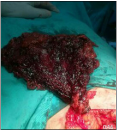
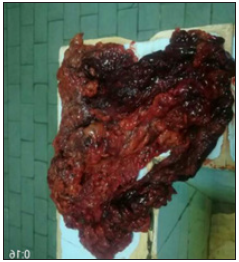
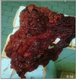
Torsion of the omentum is a rare pathological condition that can be primary and secondary, depending on the causes [1]. In the 40-year period of time, this has been the first case of primary gangrenous torsion in the hospital which is gravitated to by 300.000 people (Figure 1). Ratio is less than 4 cases per 1000 cases of appendicitis. The patient was immediately operated on as the case of appendicitis was suspected and which is the most frequent differential diagnostic dilemma. The surgery is easily done through open resection Eitel [2], and, since 1999. by laparoscopic surgery [3]. Torsion may occur with incomplete occlusion of blood vessels, and therefore there are 3 to 4 days left for further examination-CT, MRI, laparoscopic exploration [4,5]. In the presented case, the pain was sudden, severe and intraoperative the omentum was found to be twisted about 720 degrees on its axis, including distal gangrene (Figures 1-3). On Medline and PubMed, we found 635 cases of torsion by searching “torsion of omenti”. Including a filter “human”, the number was decreased to 576, and when adding a filter “English”, it was 286. After including a filter “adult”, the total number of the studies was 161. When the “non-English-language” filter was added, it still showed a rare number of cases. Having reviewed 161 studies published in English and presenting cases of adult patients, we noticed 10 studies at least which were related to ovarian torsion, without the omentum. A small number of studies contained more than 1 case, mostly 2 or 3, often for a longer period. Analysis of all cases clearly indicates that the causes of secondary torsion are not the same. Therefore, we are proposing a classification of torsion into primary and secondary, while the origin of the secondary causes can be:
Figure 1: Torsion and distal gangrene.

Figure 2: Resected part.

Figure 3: Resected part-the other side.

1. In pathological substrata in the omentum itself.
2. Outside the omentum.
The most frequent cause is a hernia [6], carcinoma or a polycystic
ovary [7], and appendicitis [8]. Pathological causes in the omentum
itself are (fibro)lipoma of the omentum [9], a cyst or a polycystic
omentum [10], fat necrosis [10], RY anastomosis [11]. In rare
cases, as ours is, clinical features do not allow further examination.
The pathogenesis of the omental torsion is more interesting than
the surgical procedure itself, which includes the resection of the
affected part and suturing the healthy edge. The most probable
cause is a sudden twist of the body with a predisposing greater
omentum or obesity/ pregnancy [10]. There is no explanation still
without a doubt. When ileocecal pain is present, torsion should be
considered although the surgical procedure is relatively easy, and
laparoscopy can also be done.
References
- Occhionorelli S, Zese M, Cappellari L, Stano R, Vasquez G (2014) Acute abdomen due to primary omental torsion and infarction. Case Reports in Surgery 2014: 208382.
- Eitel GG (1899) Rare omental torsion. NY Med Rec 55: 715-716.
- Sánchez J, Rosado R, Ramírez D, Medina P, Mezquita S, et al. (2002) Torsion of the greater omentum: treatment by laparoscopy. Surg Laparosc Endosc Percutan Tech 12(6): 443-445.
- Maeda T, Mori H, Cyujo M, Kikuchi N, Hori Y, et al. (1997) CT and MR findings of torsion of greater omentum: a case report. Abdom Imaging 22(1): 45-46.
- Puylaert JBCM (1992) Right-sided segmental infarction of the omentum: clinical, US, and CT findings. Radiology 185(1): 169-172.
- Hirano Y, Oyama K, Nozawa H, Hara D, Nakada K, et al. (2006) Left-sided omental torsion with inguinal hernia. World J Gastroenterol 12(4): 662-664.
- Dugalic V, Popovic N, Gojnic M, Arsenijevic Lj, Filimonovic D, et al. (2006) Torsion of carcinomatous ovarian cyst and polycystic omental diseases-case report. Eur J Gynaecol Oncol 27(6): 629-631.
- Kumar A, Shah J, Vaidia P (2016) Primary omental gangrene mimicking appendicular perforation-A case report. Int J Surg Case Rep 21: 67-69.
- Pérez NJV, Flores CA, Anaya PR, González IJ, Ramírez BEJ (2009) Angiofibrolipoma of the greater omentum: case report and literature review (esp). Cir Cir 77(3): 229-232.
- Karanikas M, Kofina K, Boz Ali F, Vamvakerou V, Effraemidou E, et al. (2018) Primary greater omental torsion as a cause of acute abdomen-A rare case report. J Surg Case Rep 8(8): 1-3.
- Descloux A, Basilicata G, Nocito A (2016) Omental torsion after laparoscopic Roux-en-Y gastric bypass mimicking appendicitis: A case report and review of the literature. Case Reports in Surgery, p. 2.
© 2021 Momcilo S. This is an open access article distributed under the terms of the Creative Commons Attribution License , which permits unrestricted use, distribution, and build upon your work non-commercially.
 a Creative Commons Attribution 4.0 International License. Based on a work at www.crimsonpublishers.com.
Best viewed in


.jpg)


 Editorial Board Registrations
 Submit your Article
 Refer a Friend
 Advertise With Us
.jpg)


.jpg)


.bmp)
.jpg)
.png)
.jpg)


.jpg)


.png)

.png)


.png)


