- Submissions

Full Text
Research in Pediatrics & Neonatology
Scalp Swelling: An Uncommon Etiology
Margarida Peixoto1*, Maria João Gaia1, Joana Brandão Silva1, Joana Reis1 and Sérgio Alves2
1Pediatric Service, Hospital Center of Vila Nova de Gaia/ Espinho, Portugal
2Pediatric Service, Porto University Hospital Center, Portugal
*Corresponding author: Margarida Peixoto,Pediatric Service, Hospital Center of Vila Nova de Gaia/ Espinho, Portugal
Submission: February 08, 2023; Published: February 16, 2023

ISSN: 2577-9200 Volume7 Issue3
Abstract
Langerhans cell histiocytosis (LCH) is a rare pediatric disease of unknown etiology, the skull being the most affected site. We present a healthy 4-year-old girl with a scalp swelling, without history of head trauma or other symptoms. Physical examination and bloodwork had no significant findings. The radiography and CT-scan revealed an osteolytic lesion, without intracranial involvement. Pathologic examination diagnosed a LCH. Being a single lesion in a low-risk location, she was put on clinical surveillance. The swelling regressed and she awaits imaging reevaluation. LCH can go unnoticed when symptoms resemble trauma or other conditions, but it should be considered when a mass arises from the skull in children.
Keywords:Langerhans cell histiocytosis; Osteolytic lesion; Scalp swelling; Multinucleated cells; CD1a; S-100 marker
Case Presentation
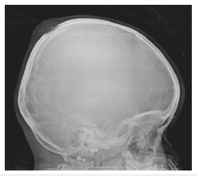
A 4-year-old previously healthy girl presented to the emergency department with scalp swelling detected the previous day. There was no history of head trauma or other accompanying symptoms. On examination she presented a focal well-defined area of scalp swelling in the right parieto-occipital bone transition, with 5cm diameter, with local tenderness and soft consistency. There were no skin changes, adenopathy or organomegalies. Cranial radiography (Figure 1) showed a lytic lesion in the parietal bone, with edema of the adjacent soft tissues. Bloodwork including hemogram and coagulation were unremarkable.
Figure 1: Skull radiograph - lytic lesion of the parietal bone, with surrounding edema of the adjacent soft tissues

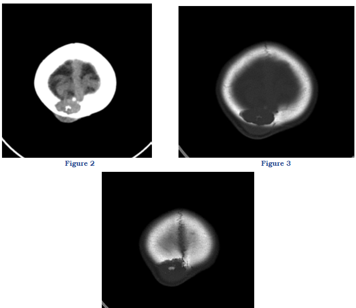
The CT scan (Figure 2-4) demonstrated an osteolytic lesion in the high convexity of the right parietal bone, about 25mm thick with slightly elevated densitometry, affecting both cranial boards, with an extra cranial component. No mass effect on the brain was observed, although a slight deviation of the sagittal sinus was apparent. The brain and the CSF pathways had normal shape and dimensions. These radiologic features suggested the diagnosis of eosinophilic granuloma.
Figure 2,3,4: Nonenhanced CT scan, showing osteolytic lesion in the right parietal bone, affecting internal and external boards. It is associated with soft tissue edema. No mas effect on the brain is observed.

Pathologic examination revealed numerous histiocytic cells, with irregular nucleus. Multinucleated and polynuclear giant cells were present, along with eosinophils. They expressed CD1a and S-100 markers. Langerhans cell histiocytosis (LCH) was then diagnosed. A subsequent scintigraphy and PET-CT scan revealed that there were no other organs affected. Being a single lesion in a low-risk location, she maintained only clinical surveillance. The swelling and the pain regressed. She awaits imaging reevaluation. LCH is a rare pediatric disease of unknown etiology. The clinical spectrum can go from a solitary eosinophilic granuloma to multisystem involvement. The most common site of involvement is the skull. LCH can go unnoticed in patients whose symptoms resemble trauma or other conditions. This diagnosis should always be considered when a mass arises from the skull in children. Involvement of the skeleton is best assessed with plain radiographs, and CT scans can better define osteolytic lesions, as well as the extra and intracranial soft-tissue components.
© 2023 Margarida Peixoto. This is an open access article distributed under the terms of the Creative Commons Attribution License , which permits unrestricted use, distribution, and build upon your work non-commercially.
 a Creative Commons Attribution 4.0 International License. Based on a work at www.crimsonpublishers.com.
Best viewed in


.jpg)


 Editorial Board Registrations
 Submit your Article
 Refer a Friend
 Advertise With Us
.jpg)


.jpg)


.bmp)
.jpg)
.png)
.jpg)


.jpg)


.png)

.png)


.png)


