- Submissions

Full Text
Research in Pediatrics & Neonatology
FATCO Syndrome with Infant of Diabetic Mother
Gulfer Akca1 And Unal Akca2*
1Samsun University Faculty of Medicine, Department of Pediatrics, Samsun, Turkey
2Samsun University Faculty of Medicine, Department of Pediatric Neurology, Samsun, Turkey
*Corresponding author: Unal Akca, Samsun University Faculty of Medicine, Department of Pediatric Neurology, Samsun, Turkey
Submission: November 11 , 2022; Published: December 07, 2022

ISSN: 2577-9200 Volume7 Issue1
Abstract
Fibular aplasia, tibial campomelia and oligosyndactyly (FATCO) syndrome (OMIM 246570) is an extremely rare syndrome first described by Hecht and Scott [1]. The etiology of the syndrome is currently unknown. This syndrome is commonly sporadic, but autosomal dominant inheritance has also been proposed. Although fibular aplasia is among the congenital malformations that develop due to diabetes, FATCO syndrome is a separate entity. In addition to the unknown genetic cause of this syndrome, which has typical findings, the mother’s pregnancy and diabetes history should also be taken well. Corrective operations, physical therapy and regular development follow-up very important in these patients who do not have mental, cardiac and facial dysmorphism, requires multidisciplinary care.
Keywords: Campomelia; Fibular aplasia; FATCO syndrome; Infant of diabetic mother; Oligosyndactyly
Introduction
Fibular aplasia, tibial campomelia and oligosyndactyly (FATCO) syndrome (OMIM 246570) is an extremely rare syndrome first described by Hecht and Scott [1]. Courtens et al. [2], reported a further case and compare it with earlier four reports of similar conditions [1,3,4]. They proposed the name FATCO as all cases had fibular aplasia, tibial campomelia and oligosyndactyly in common. Individuals with FATCO showed shortening and anterior bowing of the lower limb at the distal third of the tibia with overlying soft tissue dimpling, oligodactyly of the foot, and oligosyndactyly of the hand.
The etiology of the syndrome is currently unknown. This syndrome is commonly sporadic, but autosomal dominant inheritance has also been proposed [5]. Previous reports have already excluded WNT7A as a potential FATCO candidate gene [6,7]. Mutations in WNT7A cause Fuhrmann syndrome (OMIM 228930) and the Al-Awadi/ Raas-Rothschild syndrome (OMIM 276820), characterized by various degrees of limb aplasia/hypoplasia and joint dysplasia [8]. Conflicting reports have been published concerning inheritance of fibular aplasia with ectrodactyly [9]. We report a 3 years old boy in the FATCO syndrome clinic born to mother with diabetes.
Case
We report a 3 year-old -boy with congenital lower limb deficiency. This deficiency consists of shortness of left leg, anterior bowing at the tibia, with associated overlying soft tissue dimpling, together with oligosyndactyly (4 toes) of the left foot. Both upper limbs and right leg are completely normal. He has neither dysmorphic facial features nor other associated anomalies. The patient was born at full term by elective Cesarean section after an uneventful pregnancy and delivery. He was the third-birth of a mother with diabetes. Birth weight, length, and occipital frontal circumference were 2520g. (<10th percentile), 49cm. (25th-50th percentile), and 35cm. (50th–75th percentile), respectively. Apgar scores were normal. The umbilical cord contained two arteries and a single vein. The first pregnancy of the mother had been a spontaneous quadruplet pregnancy that had resulted in intrauterine death. A child born after the mother’s second pregnancy was a 5-year-old girl in good health [10-17]. The father and mother were 33 and 32 years old, respectively, at the birth. There was no history of consanguinity, although the parents were born in the same small town, which has only about 20000 inhabitants. Family history was unremarkable. The mother had been using insulin for 7 years with the diagnosis of diabetes mellitus and had no history of alcohol, tobacco and exposure to radiation. The pregnant woman who has a follow-up did not have postnatal respiratory distress.
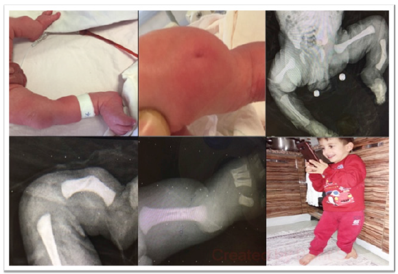
In the newborn physical examination, a difference in length was detected between two legs. Right leg: 18cm (right femur 8cm, right tibia 9cm), left leg: 13cm (left femur 7cm, left tibia 6cm) measured. Left leg was curved and there was a tibial dimple in the 1/3 proximal front side. There were 4 toes on the left foot, and cutaneous syndactyly was present in the second and third toes. Radiographic examination revealed complete absence of left fibula (fibular aplasia, FA type II), anterior bowing and shortness of left tibia (tibial campomelia) and absence of lateral rays of the foot (4 toes only-oligosyndactyly). No other abnormalities were detected on skeletal survey. External genital were normal. He had neither facial dysmorphism nor other associated anomalies. Systemic examination was normal, including fundal examination, auditory evoked potentials, cranial ultrasound examination and abdominal, renal, and cardiac examinations. There were no other associated anomalies. The second hour blood glucose was 46mg/dl. Insulin: 10μIU/mL. c- peptide: 2, 86. Mothers’ HbA1c level: 8,33 and BMI was 37.1. Body blood sugar regulation of the patient was achieved with glucose infusion and increased oral intake. All values returned to normal in following controls.
Psychomotor development of the patient was appropriate for his age. He had head control at 2 months, sat up at 6 months, talk at 12 months, and walk at 14 months. The first correction surgery was performed at 21 months of age. The Denver developmental screening test was normal when he was 2 years old. Chromosomal analysis was (46,XY) normal. Other follow-up investigations were all normal, including laboratory examinations, eye fundus, auditory evoked potentials and abdominal renal and cardiac ultrasound examinations. The child is now 3 years, with normal mental development. The second operation was planned at the age of 4 years. The patient still does not have any active health problems other than short legs (Figure 1).
Figure 1:Distinct length difference between two legs(A). left tibial dimple(B). X- ray: Congenital total absence of left fibula (C). X ray: Curved left tibia (campomelic) (D). X-ray: Agenesis of finger2 on the left foot (E). The patient still does not have any active health problems other than short legs (F).

Discussion
Congenital limb deficiencies are common birth defects, occurs in 1 in 2000 neonates and characterized by the aplasia or hypoplasia of bones of the limbs [18]. Fibula hemimelia (FH) is a rare congenital anomaly and was described by Gollier in 1698 [2,19]. This term encompasses a spectrum of disease from mild fibular hypoplasia to fibular aplasia. It has been estimated that there are 5.7 to 20 cases per one million births [20]. FH commonly occurs unilaterally, isolated. However it may be a part of a malformation syndrome. These components may include femur and tibia shortening, clubfoot, valgus deformity, flexion contracture, instability of knee and ankle, tarsal coalition with deficiency of lateral rays of the foot. Even though fibular hemimelia is rare among long bone deficiency disorders, it is the most common malformation [19].
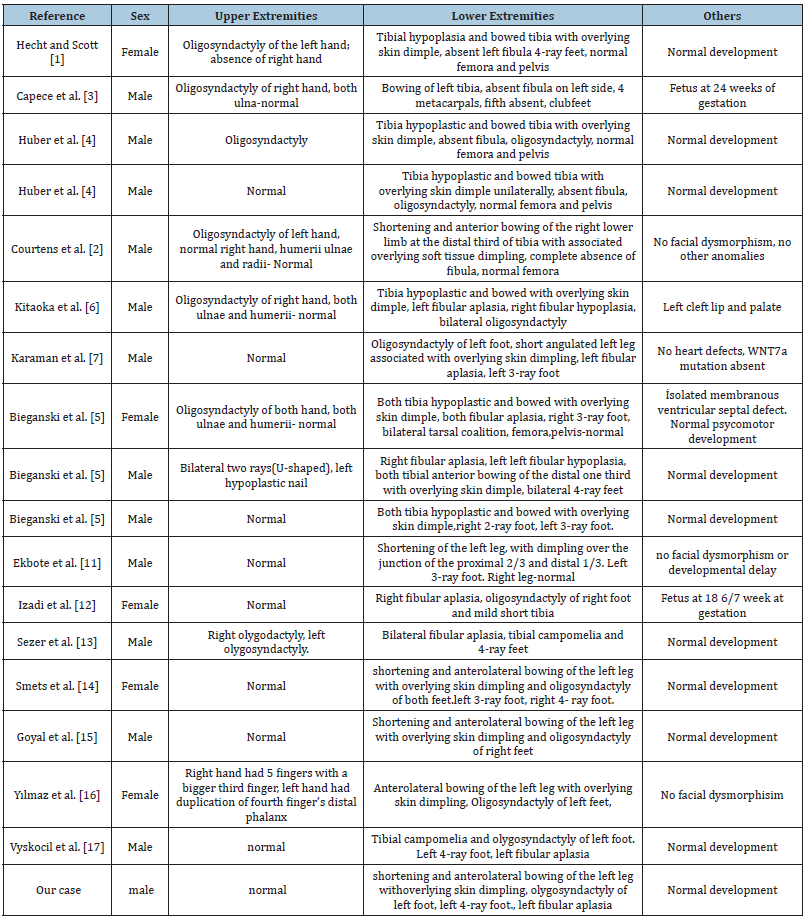
A rare congenital limb malformation syndrome characterized by the left fibular hypoplasia, the right fibular aplasia, tibial campomelia, and lower limb oligosyndactyly involving the lateral rays that was first defined by Hecht and Scott in [1]. Courtens et al. [2] reported on a male infant with oligosyndactyly of the left hand and the right foot, absence of right fibula, and anterior bowing of the ipsilateral tibia with associated overlying soft tissue dimpling and rewieved four other cases [1-4]. All of five cases had same three major findings that fibular agenesis, tibial campomelia and oligo-syndactyly, they proposed to name it Fibular Aplasia- Tibial Campomelia-Olygosyndactytly (FATCO) syndrome [6]. The cases previously reported by various authors had a great deal of phenotypic heterogeneity. We are presenting a phenotypic review of all the previously reported cases to date. A total of 18 cases have been reported to date (Table 1).
Table 1:A total of 18 cases have been reported to date.

Upper extremity involvement was present in 50% of patients, and 44% of those affected were unilateral. Skin dimpling and syndactyly in the lower extremity was present in all patients. There was unilateral involvement in the lower extremity at a rate of 55%. Fibular hypoplasia was detected in half of 18 patients. Except for the patient with cleft palate, none of them had facial dysmorphism [6]. The femur, pelvis, ulna, and radius were preserved in all patients. Mental and cardiac effects were not observed in any of the patients. The gene or genes responsible for the FATCO syndrome are not known yet. Most of the cases are sporadic. There was a definitive male predominance (14/18). There was no evidence of consanguinity in any of cases. Bieganski et al. [5] declined that it may be autosomal dominant and inherited X-linked inheritance because of male preponderance [5].
Single or double-sided fibular bone aplasia with tibial bone anomalies syndromes; Acheiropody, Chondrodysplasiacromelic- Genital anomalies Schinzel phocomelia and FATCO syndrome. Of the major components of FATCO syndrome: oligo syndactyly and tibial campomelia are not seen in Acheiropody and Chondrodysplasia Akromelik-Genital anomalies syndrome. Again, Fuhrmann syndrome and Al-Awadi syndrome are diseases with similar clinical features with two alleles and are differentiated by the involvement of pelvis, femur, radius and ulna, and nail deformities with oligo/ polydactyly [21]. Fibula aplasia or hypoplasia is the most common long bone developmental defect seen often isolated. In etiology, non-genetic reasons are in place. These; exposure to radiation during pregnancy, cytotoxic busulfan drug use, retinoic acid use, and maternal diabetes mellitus [19].
Diabetes is an important medical condition affecting pregnancy. Gestational diabetes, which comprises approximately 80% of cases of diabetes in pregnancy [22]. Women with Type-1 diabetes who receive optimum pre-conception and antepartum care through a multidisciplinary antenatal clinic achieve a perinatal mortality rate equivalent to that observed in women who do not have diabetes in pregnancy [23]. In a recent study, an increased HbA1c level above 6.5% causes an increase in the prevalence of congenital anomalies, glycaemia and BMI are the key modifiable risk factors. There was no significant difference between type-1 and type-2 diabetes. In our case the mother had high HbA1c ( 8.82-8.33) and high BMI [24].
Our case is an infant of Diabetic Mother (IDM) but this is the first study with FATCO syndrome with IDM. Although fibular aplasia is among the congenital malformations that develop due to diabetes, FATCO syndrome is a separate entity. The goal of treatment for FATCO syndrome is to correct the leg length discrepancy and to functionally stabilize knee and ankle joints. Commonly accepted avenues of treatment include the use of an orthoses, epiphysiodesis, limb lengthening, and amputation with prosthesis [25]. In our case, he had first corrective surgery at the age of 21 months and his second operation was planned.
Conclusion
The FATCO syndrome is a rare genetic development limb disorder of the long bones with proposed autosomal dominant inheritance and so far has an unknown molecular basis. Chromosomal analysis should be performed in addition to the other investigations in patients with deformity and dysmorphism in selected cases where specific molecular diagnosis is not possible.
We know that diabetes causes congenital joint deformities, but this syndrome has been reported for the first time. In addition to the unknown genetic cause of this syndrome, which has typical findings, the mother’s pregnancy and diabetes history should also be taken well. Corrective operations, physical therapy and regular development follow-up very important in these patients who do not have mental, cardiac and facial dysmorphism, requires multidisciplinary care.
References
- Hecht JT, Scott CI (1981) Limb deficiency syndrome in half-sibs. Clin Genet 20(6): 432-437.
- Courtens W, Jespers A, Harrewijn I, Puylaert D, Vanhoenacker F (2005) Fibular aplasia, tibial campomelia, and oligosyndactyly in a male newborn infant: A case report and review of the literature. Am J Med Genet A 134(3): 321-325.
- Capece G, Fasolino A, Monica DM, Lonardo F, Scarano G, et al. (1994) Prenatal diagnosis of femur-fibula-ulna complex by ultrasonography in a male fetus at 24 weeks of gestation. Prenatal Diagn 14(6): 502-505.
- Huber J, Volpon JB, Ramos ES (2003) Fuhrmann Syndrome: Two Brazilian cases. Clin Dysmorphol 12(2): 85-88.
- Bieganski T, Jamsheer A, Sowinska A, Baranska D, Niedzielski K, et al. (2012) Three new patients with FATCO: Fibular agenesis with ectrodactyly. Am J Med Genet A 158A(7): 1542-1550.
- Kitaoka T, Namba N, Kim JY, Kubota T, Miura K, et al. (2009) A Japanese male patient with ‘fibular aplasia, tibial campomelia and oligodactyly’: An additional case report. Clin Pediatr Endocrinol 18(3): 81-86.
- Karaman A, Kahveci H (2010) A male newborn infant with fatco syndrome [fibular aplasia, tibial campomelia and oligodactyly]: A case report. Genet Couns 21(3): 285-88.
- Woods CG, Stricker S, Seemann P, Stern R, Cox J, et al. (2006) Mutations in WNT7A cause a range of limb malformations, including Fuhrmann syndrome and Al-Awadi/Raas-Rothschild/Schinzel phocomelia syndrome. Am J Hum Genet 79(2): 402-408.
- Evans JA, Elliot AM (2006) Letter re: fibula aplasia, tibial campomelia, and oligodactyly. Am J Med Genet A 140(10): 1127.
- Menke LA, Bijlsma EK, Essen AJV, Boogaard MJHVD, Rijn VRR, et al. (2008) Ectrodactyly with fibular aplasia: A separate entity? Eur J Med Genet 51(5): 488-496.
- Ekbote AV, Danda S (2012) A case report of fibular aplasia, tibial campomelia, and oligosyndactyly [FATCO] syndrome associated with Klinefelter syndrome and review of the literature. Foot Ankle Specialist 5(1): 37-40.
- Izadi M, salehnia N (2020) Prenatal diagnosis of FATCO syndrome (Fibular Aplasia, Tibial Campomelia, and Oligosyndactyly) with 2D/3D Ultrasonography. Ultrasound Int Open 6(2): E44-E47.
- Sezer O, Gebesoglu I, Yuan B, Karaca E, Gökçe E, et al. (2014) Fibular aplasia, tibial campomelia, and oligosyndactyly: A further patient with a 2-year follow-up. Clin Dysmorphology 23(4): 121-126.
- Smets G, Vankan Y, Demeyere A (2016) A female newborn infant with FATCO syndrome variant (Fibular Hypoplasia, Tibial Campomelia, Oligosyndactyly)- A Case Report. J Belg Soc Radiol 100(1): 41.
- Goyal N, Kaur R, Gupta M, Bhatty S, Paul R (2014) FATCO Syndrome Variant-Fibular Hypoplasia, Tibial Campomelia and Oligosyndactyly--A Case Report J. Clin Diagn Res 8(9): LD01-2.
- Yılmaz H, Topak D, Yilmaz O, Çakmakli S (2019) A Turkish female twin sister patient with fibular aplasia, congenital tibia pseudoarthrosis, oligosyndactyly, and negative WNT7A gene mutation. J Pediatr Genet 8(2): 95-99.
- Vyskocil V, Dortova E, Dort J, Chudacek Z (2011) FATCO syndrome: Fibular aplasia, tibial campomelia and oligo-syndactyly. Joint Bone Spine 78: 217-218.
- Espinasse HM, Devisme L, Thomas D, Boute O, Vaast P, et al. (2004) Pre- and postnatal diagnosis of limb anomalies: A series of 107 cases. Am J med genet A 124A(4): 417-422.
- Lewin S, Opitz JM (1986) Fibular a/hypoplasia: Review and documentation of the fibular development field. Am J Med Genet Suppl 2: 215-238.
- Geipel A, Berg C, Germer U, Krokowski M, Smrcek J, et al. (2003) Prenatal diagnosis of femur-fibula-ulna complex by ultrasound examination at 20 weeks of gestation. Ultrasound Obstet Gynecol 22(1): 79-81.
- Kantaputra PN, Mundlos S, Sripathomsawat W (2010) A novel homozygous arg222trp missense mutation in WNT7A in two sisters with severe Al Awadi/Raas Rothschild/Schinzel phocomelia syndrome. Am J Med Genet 152A(11): 2832-2837.
- Miller E, Hare JW, Cloherty JP, Dunn PJ, Gleason RE, et al. (1981) Elevated maternal hemoglobin A1c in early pregnancy and major congenital anomalies in infants of diabetic mothers. N Engl J Med 304(22): 1331-1334.
- Evers IM, Valk, HWD, Visser GHA (2004) Risk of complications of pregnancy in women with Type 1 diabetes: Nationwide prospective study in the Netherlands. BMJ 328(7445): 915-920.
- Murphy HR, Howgate C, O'Keefe J, Myers J, Morgan M, et al. (2021) Characteristics and outcomes of pregnant women with type 1 or type 2 diabetes: A 5-year national population-based cohort study. Lancet Diabetes Endocrinol 9(3): 153-164.
- Hamdy RC, Makhdom AM, Saran N, Birch J (2014) Congenital fibular deficiency. J Am Acad Orthop Surg 22(4): 246-255.
© 2022 Unal Akca. This is an open access article distributed under the terms of the Creative Commons Attribution License , which permits unrestricted use, distribution, and build upon your work non-commercially.
 a Creative Commons Attribution 4.0 International License. Based on a work at www.crimsonpublishers.com.
Best viewed in


.jpg)


 Editorial Board Registrations
 Submit your Article
 Refer a Friend
 Advertise With Us
.jpg)


.jpg)


.bmp)
.jpg)
.png)
.jpg)


.jpg)


.png)

.png)


.png)


