- Submissions

Full Text
Research in Pediatrics & Neonatology
A Novel Procedure for the Removal of Supernumerary or Accessory Tragus on Neonates
Roig JC1*, Leibovici A2, Taylor K3, Major E1 and Roig SM3
1Department of Pediatrics, Division of Neonatology, University of Florida College of Medicine, Florida, USA
2Advent Health Orlando, Florida, USA
3East Carolina University, Greenville, North Carolina, USA
*Corresponding author: Department of Pediatrics, Division of Neonatology, University of Florida College of Medicine, Florida, USA
Submission: June 04, 2020; Published: June 25, 2020

ISSN: 2577-9200 Volume4 Issue4
Abstract
The majority of supernumerary or accessory tragus in humans are noted soon after birth, and are generally benign isolated lesions not associated with other genetic abnormalities. When present, these lesions are typically managed by the primary care provider, but occasionally the caretakers opt to refer the patient to a surgeon to have the lesion resected surgically as an outpatient. This practice may place an unnecessary financial burden on the patient’s family, and may pose added difficulty due to the availability of the subspecialist. The current literature lacks other practical and effective methods for dealing with these lesions despite the incidence of up to 1.5% of the population [1]. Traditionally, however, these lesions are managed by pediatricians or the PCP by placing a suture ligature at its base so that the distal portion of the tragus will fall off after the ischemic necrosis has occurred [2]. This approach is the current standard of care, and is the method being taught at most pediatric training programs. When successful, this process can take days if not weeks to run its course. Another approach may be to refer these patients to a Plastic Surgeon or a Pediatric Surgeon for care which may be to have the lesions managed by means of application of surgical clips [3] at their base thus achieving a similar effect as a ligature. Alternatively, the lesions can be permanently surgically excised later when the patient is older.
At the University of Florida we have been successfully excising these lesions when devoid of cartilage prior to the patient’s discharge using the Digiclamp® device. We report 7 lesions which were permanently removed using this method; the clamp was placed at their base flush with the skin, and the accessory tragus was excised. This novel minimally invasive procedure does not require suturing, and has proven to be safe and poses minimal risk to the patient when performed correctly. All of these excisions took place prior to the patient’s discharge and uniformly required only minimal care thereafter. Among the advantages of utilizing this procedure are: the time needed to perform the procedure is brief, on average requires only 10 minutes or less to perform; the procedure has consistently been well tolerated by all of the patients; and although all of the excisions took place in the patient’s center of birth prior to their discharge, it can easily be performed in the outpatient setting since it requires minimal time, equipment, and is relatively simple to perform.
Keywords: Accessory Tragus; Supernumerary Tragus; Accessory Auricle; Ear Tag; Removal Procedure; Digiclamp®
Background
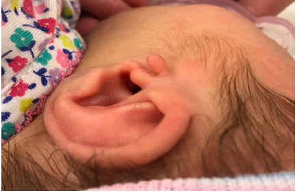
The supernumerary or accessory tragus, first reported by Burkett in 1858 [4], is a benign congenital abnormality that typically is unilaterally present at birth (Figure 1). The embryological derivation of the tragus is the first branchial arch which also gives rise to the mandible in humans [5]. When present, the most typical appearance for these lesions is that of a nodular skin colored protrusion located in the pre-auricular region on either side of the head. Less commonly, these nodules can also be located along the line from the tragus to the angle of the mouth; along the anterior aspect of sternocleidomastoid muscle; in the cheeks; the upper sternum or on the glabella [6-10]. Additionally, other clinical conditions may resemble the accessory tragus, such as acrochordons or “skin tags”, auricular fistulas, fibromas, polyps, epidermoid cysts, and wattles [7,11,12]. These lesions are more frequently present unilaterally, but may also be present bilaterally, and may be pedunculated or sessile [4]. These lesions may or may not contain cartilage, the presence of which is accurately confirmed with a careful physical exam. Generally these lesions are 10 mm or less in size at their base and are typically removed for cosmetic reasons only [13]. In cases where the lesions contain a central core of elastic cartilage, surgical excision is generally recommended.
Figure 1: Example of a left sided pedunculated supernumerary tragus.
Method for the Procedure
Figure 2: Disposable Digiclamp® device.
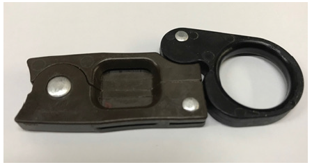
Figure 3: Site of local block for the Digiclamp® application and excision.
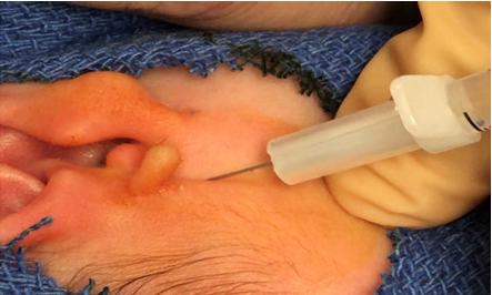
A careful manual examination of each of the accessory or supernumerary tragi is initially performed to confirm that no cartilage is contained within the lesion. Those patients with cartilage were excluded from the Digiclamp® excision procedure (Figure 2) and referred for surgical resections. All of our patient’s families had previously been offered and declined options the suture ligature application and outpatient surgical referral prior to giving us their consent. After consent was obtained and “timeout” was observed, the site of the lesion was prepped in sterile fashion using either povidone iodine and 70% Isopropyl alcohol or Hibistat. The patients were then offered oral Sucrose solution for analgesia after which, the pre-auricular area was infiltrated subcutaneously just below and anterior to the pinna using a 27 French needle and injected with approximately 0.25mL of 1% lidocaine solution without Epinephrine (Figure 3). After allowing several minutes for the anesthetic to take effect, the Digiclamp® was placed at the base of the accessory tragus applying gentle upward traction to the distal part of the lesion prior to closing the device (Figure 4).
Figure 4: Digiclamp application to the base of the lesion.
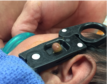
Once the instrument is applied to the lesion and closed, the Digiclamp® is left in place compressing the base of the lesion for approximately 5 minutes. During this time, the opposing edges of skin at the base of the accessory tragus are being fused together. After the recommended period of time has elapsed, the lesion is safe to be excised using a scalpel. This is done easily by passing the scalpel through the side ports of the clamp, while the instrument remains closed over the base of the lesion (Figure 5). Attention should be given towards resecting any redundant which may be protruding above the facet of the clamp (Figure 5 & 6) so that there is no skin projecting above the clamp. After excision has taken place, the instrument is removed and the site is carefully cleaned with alcohol. There should be a translucent flap of fused skin apparent where the base of the accessory auricle had previously been and no bleeding from the lesion should be apparent. A skin flap approximately 2mm in height should be present where the lesion had previously been (Figure 7). A small band aid should be placed to cover the site, and the family should be instructed to allow the band-aid to spontaneously fall off the lesions after discharge. Prior to discharge the family receives anticipatory guidance to observe and report the presence of any erythema, bleeding, discharge or foul smell to their primary care provider should any of these be apparent after the resection at the operative site.
Figure 5: Example of the scalpel excision of the accessory auricle.
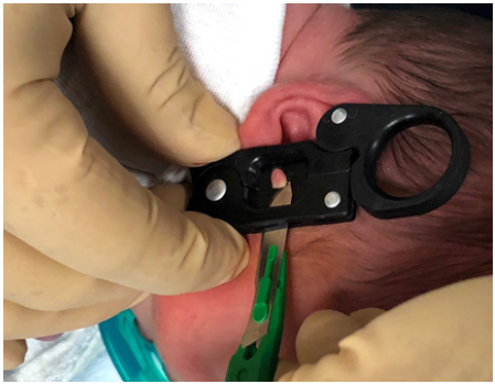
Figure 6: Example of the excision site after the Accessory Auricle has been trimmed.
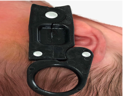
Figure 7: View of the skin flap apparent after the Digiclamp® is removed showing the compressed skin of the base of the accessory auricle.
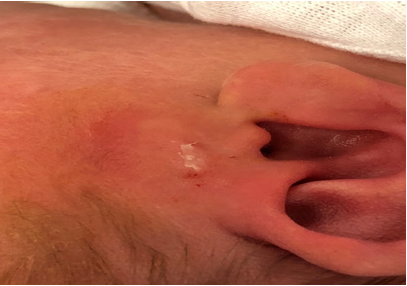
Follow up for these patients typically consisted of a phone call several weeks later to ask about any subsequent complications, recurrence of the lesion, and their overall satisfaction with the procedure and the results. The parents were also asked about any post-operative bleeding, or any ER related to our procedure, site infections as well as the cosmetic appearance of the site of the lesion. The feedback received from all of the patient’s parents were extremely satisfactory, and all of the patients’ parents expressed that they would opt to have our procedure done again in the event that subsequent births presented with the same lesion.
Discussion
The Accessory tragus of the ear is a limited deformity, when associated with a syndrome it is typically associated with defects that extend to involve both the first and second branchial arches such as in Goldenhar syndrome. Less frequently, accessory tragus may be present with Townes-Brocks syndrome, Treacher-Collins syndrome, VACTERL association, and Wolf-Hirschhorn syndrome, but when present, the accessory tragus is generally not associated with other malformations [14]. Although suture ligature or surgical excision of these lesions are viable options for management, these options are not necessarily the most practical. In the case of the former, ligature success is operator dependent and may fail if the ligature is not tight enough to cause ischemia. In the cases when ligature is successful, the lesions may take days or weeks to fall off. Additionally there is also the likelihood that a residual nubbin of tissue may be present at the site afterwards because there is a tendency for the ligature itself to migrate upwards and away from the lesion’s base. In these instances, the parents may be forced to explore surgical options for a cosmetic repair which can be costly, may require the use of general anesthesia and need for hospitalization [7,15]. In instances where primary surgical repair is preferred, the centers need to have pediatric or cosmetic surgeon on staff to perform either a surgical clip application or a primary resection.
Our success rate and patient satisfaction using the Digiclamp® device and this method has been excellent. This suggests that our device, or one that is similar, as well as this methodology may be superior to the ligature method currently used to manage these lesions in newborns. Moreover, we feel that formal studies including long term follow up to document the evolution of the lesions after their resection, and a larger number of patients is warranted, since this method is significantly less costly than surgery, was very well received by parents, and when appropriately performed poses a small risk for complications.
Conflict of Interest
Roig JC Is a listed inventor on U.S. patent Serial No. 15/030,054 related to Digiclamp licensed to XDG Technologies.
References
- Jones S, Alvi R, Burton D (1996) Accessory auricles: Unusual sites and the preferred treatment option. Arch Pediatr Adolesc Med 150(7): 769-770.
- Mehmi M, Balasubramian P, Bhat J (2007) Accessory tragus-beware of the preauricular ‘skin tag. Journal of the American Academy of Dermatology 56(Suppl 2): AB54.
- Wong PY, Laing T, Milroy C (2014) One stop outpatient management of accessory auricle in children with titanium clip. Plastic Surgery International 2014: 1-4.
- Shin MS, Choi YJ, Lee JY (2010) A case of accessory tragus on the nasal vestibule. Ann Dermatol 22: 61-62.
- Bendet E (1999) A wattle (cervical accessory tragus). Otolaryngol Head Neck Surg 121(4): 508-509.
- Beder LB, Kemaloglu YK, Maral I, Serdaroğlu A, Bumin MA (2002) A study on the prevalence of accessory auricle in Turkey. Int J Pediatr Otorhinolaryngol 63(1): 25-27.
- Sebben JE (1989) The accessory tragus--no ordinary skin tag. J Dermatol Surg Oncol 15(3): 304-307.
- Hodges FR, Sahouria JJ, Wood AJ (2006) Accessory tragus: A report of 2 cases. J Dent Child 73(1): 42-44.
- Sayama S, Tagami H (1982) Cartilagenous nevus on the glabella. Acta Derm Veneorol 62(2): 180-181.
- Kim SW, Moon SE, Kim JA (1997) Bilateral accessory tragus on the suprasternal region. J Dermatol 24(8): 543-545.
- Cohen PR, Barness EG (1993) Pathological cases of the month: Accessory tragus. Am J Dis Child 147(10): 1123-1124.
- Christensen P, Barr RJ (1985) Wattle: An unusual congenital anomaly. Arch Dermatol 121(1): 22-23.
- Han HH, Kim HY, Oh DY (2016) A novel surgical method using two triangular flaps for accessory tragus. Archives of Aesthetic Plastic Surg 22(2): 63-67.
- Bahrani B, Khachemoune A (2014) Review of accessory tragus with highlights of its associated syndromes. Int Journal of Dermatology 53(12): 1442-1446.
- Frieden IJ, Chang MW, Lee I (1995) Suture ligation of supernumerary digits and “tags”: An outmoded practice? Archives of Pediatrics and Adolescent Medicine 149(11): 1284.
© 2020 Roig JC. This is an open access article distributed under the terms of the Creative Commons Attribution License , which permits unrestricted use, distribution, and build upon your work non-commercially.
 a Creative Commons Attribution 4.0 International License. Based on a work at www.crimsonpublishers.com.
Best viewed in


.jpg)


 Editorial Board Registrations
 Submit your Article
 Refer a Friend
 Advertise With Us
.jpg)


.jpg)


.bmp)
.jpg)
.png)
.jpg)


.jpg)


.png)

.png)


.png)


