- Submissions

Full Text
Research & Investigations in Sports Medicine
Angle of Spinal Curvature and SEMG Frequency of the Erector Spinae in Young School-Children
Jacek Wilczyński1*, Przemysław Karolak2, Sylwia Janecka2 and Natalia Habik2
1 Department of Posturology, Poland
2 Faculty of Medicine and Health Sciences, Poland
*Corresponding author:Jacek Wilczyński, Department of Posturology, Poland
Submission: June 11, 2019; Published: July 26, 2019

ISSN: 2577-1914 Volume5 Issue1
Abstract
The aim of the study was to analyse the relationship between the angle of spinal curvature and the SEMG frequency of the erector spinae in young school-children. 251 children aged 7-8 participated in the study. The analysis involved 103 (41%) children with scoliosis, 141 (56.17%) children with scoliotic posture and 7 (3.0%) with correct body posture. The Diers formetric III 4D optoelectronic method was used to evaluate body posture. Analysis of the SEMG frequency of the erector spinae was performed with the Noraxon TeleMyo DTS. Significant differences were only found in the boys' group regarding the frequency of SEMG of the erector spinae between groups with scoliosis, scoliotic posture and those within the norm regarding the test: prone position, trunk up, thoracic segment, right side. The most important predictor for body posture and the frequency of the erector spinae in the scoliosis group were the variables: prone position, trunk up, thoracic segment, left side; standing position, lumbar section, right side; prone position, thoracic segment, left side; standing position, thoracic segment, right side. In the group with scoliotic posture, the most important model predictor was: prone position, trunk up, thoracic segment, right side.
A significant relationship was found between the angle of spinal curvature and the SEMG frequency of the erector spinae. An important model predictor for the variables: SEMG frequency of the erector spinae and the angle of curvature in the scoliosis group, turned out to be the test: prone position, lumbar section, left side. In the scoliotic posture group, an important predictor of the model was: prone position, lumbar section, right side. The asymmetrical activity of the erector spinae visible in the SEMG frequency record is associated with an increase in the angle of spinal curvature. The largest generalised SEMG frequency of the erector spinae in girls was found in the case of scoliotic posture, while in boys, in the scoliosis group.
Keywords: Scoliosis, Scoliotic posture, Surface electromyography, Diers formetric III 4D
Introduction
The formation and development of scoliosis depends on etiological and biomechanical factors. The first can be very diverse and initiate the curvature, the latter is common for all curvatures regardless of etiology and works in accordance with the laws of gravity and laws of growth (Delpech-Wolff’s law) [1]. This factor controls the development of scoliosis. The course of scoliosis can be presented in 3 aspects referred to as I, II or III order symptoms. First order symptoms (direct) concern the spine and the sacral bone, the second order (intermediate, close) include the chest and the pelvis, and the third (intermediate, distant from the spine) include further parts of the musculoskeletal system [2].
First order symptoms primarily include the lateral curves of the primary and secondary spine (compensatory), rotation and lateral displacement of the vertebrae, as well as changes in their shape (torsion, wedging, flattening and widening) [3]. These changes are also accompanied by disorders of the anterior-posterior curvature of the spine. Lateral deflection can be full- or half-arches (half-arches in the end sections of the spine) of "return to straight"-type. These arches have a specific length, height and angular size. They are usually determined using a radiograph, after eliminating the Cobb angle. The angle of curvature is the basic criterion for the division of scoliosis. Studies on the pathomechanics of scoliosis are carried out as experimental work on animals, experimental research on mechanical models, as theoretical analysis of ways of creating distortions on the basis of changes found in clinical and radiological studies and sections of deceased corpses with scoliosis [4].
In recent years, research has also been carried out using computer three-dimensional imaging of scolioses [5]. The complete reconstruction of the pathomechanics of the formation and development of idiopathic scolioses is possible only on the basis of the results of research carried out with all the aforementioned methods. This task is particularly difficult, because many of the observations made so far about scoliosis are definitely contradictory. The results of experimental studies require close confrontation with clinical and anatomopathological observations, and after appropriate selection, they may form the basis for further pathomechanical considerations [6]. Studies on the SEMG frequency of the erector spinae are related to the issue of the etiology and pathogenesis of idiopathic scoliosis.
In the literature, the relationship between SEMG amplitude and scoliotic changes is generally analysed [7]. Some light on the pathogenesis and development of scoliosis, in conjunction with the theory of disturbances in the balance of muscle tone, is also shed by the examination of erector spinae SEMG frequency [8]. Modern computer technology enables simple application of Fast Fourier Transformations (FFT) for analysis and estimation of SEMG frequency components. In this model, the imposed SEMG signal can be treated as the sum of sinusoidal waves at different frequencies [9]. The FFT algorithm can be described as the partition of the SEMG signal into sinusoidal components. The most dominant sinusoidal SEMG component is the 80Hz one, because the most SEMG recording power falls on this frequency.
After performing such an analysis of the power distribution in a continuous manner at a given frequency range, the frequency of distribution or the total power spectrum is created [10]. The distribution of power indicates its different size for particular frequencies. Peak power is the maximal value of the total power spectrum curve, which is used to describe the characteristics of the SEMG frequency [11]. The most important parameters in SEMG frequency analysis are the mean and the median frequencies and their changes in the time domain of the performed contractions.
An alternative to calculations based on the FFT algorithm is the simple calculation of the zero-point intersection with the SEMG signal. The zero-point intersection coefficient correlates well with the average frequency and can be used as an alternative to FFT calculations that require time and specific computer equipment. At present, calculations of the zero-point intersection have lost their importance and those based on the FFT algorithm are preferred [12]. Movement and generation of muscle power are associated with muscle contraction, which occurs under the influence of impulses coming from the nervous system [9]. As a result of muscle contraction, electrical phenomena occur, associated with an increase or decrease in muscle tone, which can be recorded using an electromyograph.
Thus, the SEMG signal expresses the electrical activity of the muscle associated with its contraction and the generation of force. The SEMG signal is stochastic in nature and is contained in the frequency band of approximately 5÷1000Hz. However, it is assumed that the upper limit is the value of 450Hz [13]. The frequency of the recorded SEMG signal depends on the type of muscle fibres, the frequency of contractions and the developed power. The SEMG signal is the source of much information about the processes taking place in the muscle, including muscle stress and fatigue. However, an unprocessed SEMG signal carries only qualitative information, which can help to determine whether the muscle is active and generated. The graphs show a marked increase in the signal frequency resulting from the increase in muscle strength. Based on the record of the raw SEMG signal [14], only qualitative assessment is possible indicating whether the muscle works with greater or lesser force.
To obtain quantitative information, mathematical processing of the signal should be performed. On its basis, it is possible to extract parameters characterising the SEMG signal which indicate the processes occurring in the muscle. The electromyographic signal can be recorded from the muscle using needle electrode punctures or, as in our research, surface electrodes. Needle electrodes are used in the diagnosis of muscle diseases. In most cases, kinesiology tests use electrodes that collect the signal from the surface of the skin (surface electrodes), making it possible to measure the SEMG signal in a non-invasive manner [15].
Surface electromyography is a very useful tool, especially when measuring signals from large muscles, allowing easy placement of test electrodes. Analysis of SEMG frequency of the erector spinae can be effectively used to recognise the correct muscle work patterns helpful in the treatment of scoliosis. It was found that asymmetric muscle activity is associated with increased axial rotation, which, in turn, is associated with an increase in the angle of the curvature.
The aim of the study was to analyse the relationship between the angle of the spinal curvature and the SEMG frequency of the erector spinae in young school-children.
Material and Methods
251 children took part in the study, including 113 (45.02%) girls and 138 (54.98%) boys. There were 130, 7-year-old children (51.79%). Among them, there were 63 girls (48.46%) and 67 boys (51.54%). There were 121, 8-year-old children (48.21%). Among them, there were 50 girls (41,32%) and 71 boys (58,68%). The selection of subjects was mixed. The research was carried out in 2017 at the Posturology Laboratory, Faculty of Medicine and Health Sciences, UJK in Kielce. All research procedures were carried out in accordance with the 1964 Declaration of Helsinki and with the consent of the University Bioethics Commission of Jan Kochanowski University in Kielce (Resolution No. 5/2015).
Body posture was examined via the optoelectronic method - Diers formetric III 4D - using raster stereography. Three-dimensional analysis of the spine is a combination of the newest techniques of optical imaging and digital data processing. It is a quick and non-contact 4D photogrammetry measurement and analysis of the patient's back and spine. The measurement results are very precise, and thanks to the fast image transfer to the computer, the data analysis takes place immediately after the test. Because of the measurement, precise diagnosis is achieved, which enables selection of the best possible individual therapy (59,61,62). The room in which the measurements were performed was darkened in a way that prevents direct sunlight on the patient's body. At a distance of about 3m from the optical tripod, a dark background was mounted. During the measurement, the patient undressed down to shorts and placed his/her back to the apparatus at a distance of 2m. The subject assumed usual posture and the feet were placed in front of the line attached to the floor. The projector emitted about 1cm wide horizontal stripes onto the patient's back. According to the Diers formetric III 4D manufacturer guidelines, body posture examination was carried out via the DiCAM programme using the ‘Average’ measurement option. It consisted in making a sequence of 12 film frames, which by creating an average value, reduced the variance of the posture and thus, improved the clinical value of the study. The computer programme analysed the data and determined the digital, photogrammetric image of body posture [16]. Body posture examination was performed twice. The researcher (a specialist in postural re-education) decided which test was more reflective of the postural habits and only this test was further analysed. Body posture testing with the Diers formetric III 4D device took about 15 minutes.
However, it was preceded by clinical examination, the Adams test was performed, and the length of the lower limbs was measured. According to the guidelines of the Diers formetric III 4D manufacturer, the presence of scoliosis and scoliotic posture was determined considering the values of 3 variables: pelvic obliquity expressed in millimetres, lateral deviation expressed in millimetres and surface rotation expressed in degrees. Scoliotic posture occurred when pelvic tilt totalled 1-5mm, and at the same time, when lateral deviation was 1-5°, while surface rotation was 1-5°. Scoliosis occurred when pelvic tilt and lateral deviation were greater than 5mm (>5mm), and surface rotation was greater than 5 degrees (>5°) [17]. If three requirements were not met, it was assumed that scoliosis or scoliotic posture did not occur.
After examining the posture of the body, the children went to another room, where under the supervision of caregivers, they rested for about 1 hour. Then, the SEMG examination of the erector spinae was performed, which lasted about 30 minutes. Electromyographic analysis was performed using the Noraxon TeleMyo DTS 12 channel camera. The device was certified by the EC (Certificate Production Quality Assurance Directive 93/42/EEC Medical Devices Annex V). This apparatus complies with the requirements of the 93/42/EEC Medical Devices Directive for class I products. It has the appropriate CE mark and corresponds to the definition of Class I devices and standards of the product. Safety: IEC 60601-1 (1988) and EMC: IEC 60601-1-2 [18]. Pre-gel electrodes with a diameter of 3cm were used for the tests. The skin of the subjects was cleaned with abrasive liquid at the electrode application site in order to obtain resistance between the skin and the electrode below 2kΩ [19]. The electrodes were located parallel to the direction of the tested muscle fibres. The distance between them was about 2 centimetres.
The erector spinae muscle was examined in the thoracic and lumbar segments, both on the left and right sides. Each of the trials lasted 10 seconds. After connecting the Noraxon device to the computer and the electrodes to the erector spinae muscle, each of the tests was preceded by entering data for the examined person. Then, via the Noraxon TeleMyo DTS programme, the erector spinae were selected for analysis. Following, a raw signal lasting about 15 seconds appeared on the computer screen, preceding the target recording of the potential of the muscle. Next, the measurement of the raw signal was recorded and analysed. The researcher chose a signal modification programme consisting in cleaning the raw record from extreme, maximum and minimum deflections. Then, the option of processing the raw signal calculation of the average SEMG frequency was selected. Furthermore, the examiner wrote down the processed mean SEMG frequency of the erector spinae muscle expressed in Hertz (Hz). The results of the test considered the scale of the voltage intensity in the time interval, which was 100 milliseconds.
The study used the continuous trace recording mode. Functional potentials were recorded from the erector spinae in the thoracic and lumbar sections at the top of the curvature arches:
1. In a habitual standing position (Frankfurt plane),
2. In a resting position: prone position (lower limbs straightened in the knee joints, upper limbs placed along the trunk),
3. Under isometric contraction conditions:
i. In prone position (lower limbs straightened in the knee joints, upper limbs placed along the trunk, pelvis stabilised), the subject lifts the trunk within the limits of the mobility of the spine, and then maintains this position for 10 seconds,
ii. In prone position, with the upper trunk stabilised (shoulders and chest, limbs positioned as before), the subject lifts both lower limbs to the maximum possible height and maintains this position for 10 seconds.
The SEMG frequency of the erector spinae was analysed. This variable is used to assess the degree of activity and muscle fatigue. SEMG measurement was in line with SENIAM (Surface ElectroMyoGraphy for the Non-Invasive Assessment of Muscles) recommendations, a European research programme containing a number of guidelines on the selection of electrode types, their location, anatomy and muscle function, muscle group tests and signal processing as well as equipment conditions [20]. Both the postural examination and SEMG frequency analysis were painless and non-invasive.
To determine normality of distribution regarding the variables, the Kolmogorov-Smirnov test was used. In order to assess the differences in the size of the spinal curvature, between girls and boys in scoliosis groups, in those with scoliotic posture and with normal posture, and SEMG frequency of the erector spinae in different study positions, one-way ANOVA analysis of variance was used. In order to determine dependence and the predictors between dependent variables being the types of posture and scoliosis angle and the independent variable, that is the SEMG frequency of the erector spinae, multiple, stepwise and progressive regression models were used. Prior to regression analysis, k-fold cross validation was performed. The verification parameter used to assess the models was the coefficient of determination, the corrected coefficient of determination and the test statistic and the level of statistical significance, which clearly allowed to select models with the assumed level of statistical significance, p<0.05 [21].
Results
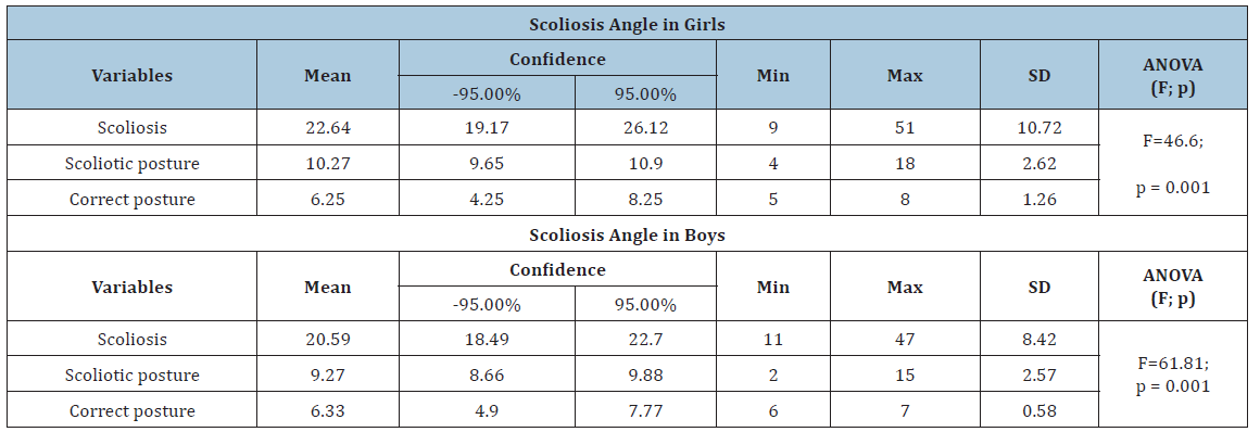
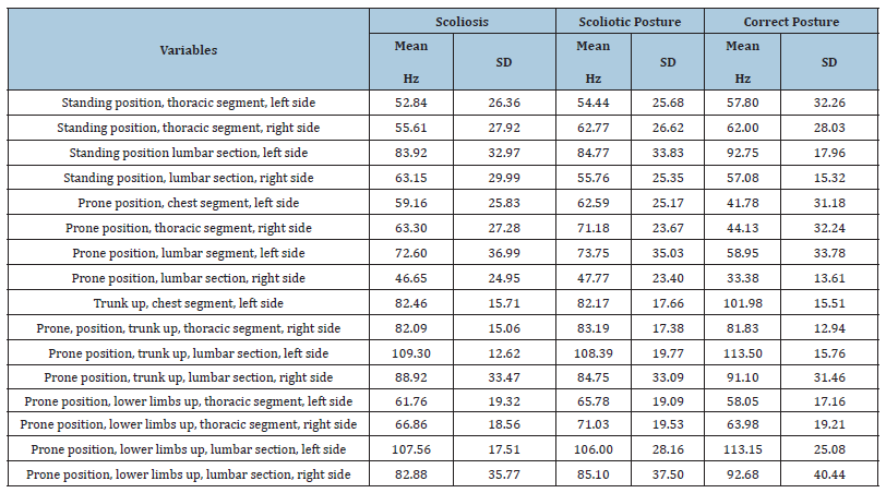
In the study group, there were 103 (41%) children with scoliosis, 141 (56.17%) with scoliotic posture and 7 (3.0%) with normal posture. The values of the position and dispersion for curvature angles were characterised by differentiation in variable distributions for girls and boys in the scoliotic, scoliotic posture and the norm groups (Table 1). The highest absolute differences in the angle of curvature occurred in the scoliosis group both in the case of girls (S=10.72) and boys (S=8.42). One-way analysis of variance demonstrated significant differences between groups in the angle of curvature both in girls (F=46.6, p=0.001) and boys (F=61.81, p=0.001). This means that the values of the curves were significantly different between the scoliosis, scoliotic and norm groups (Table 1). Also, the values of position and dispersion measurements for the SEMG frequency of the erector spinae had different variable distributions in girls and boys from the scoliosis, scoliotic and correct posture groups (Table 1). The highest absolute differentiation of SEMG frequency among girls from the scoliosis group occurred for the variable: prone position, lumbar section, left side (S=36.99) (Table 2).
Table 1:Analysis of differences in ANOVA regarding scoliosis angle in boy and girls.

Table 2:Frequency of erector spinae SEMG in girls.

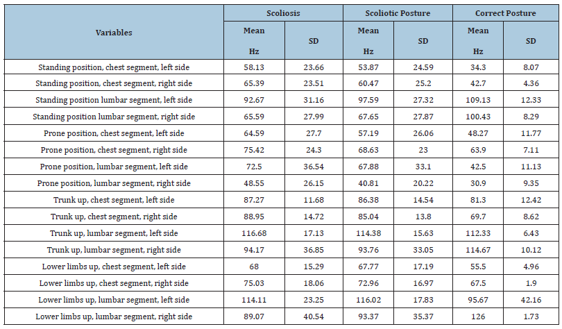
In turn, the SEMG frequency in the lower limb test of the right lumbar section was the most absolutely differentiated among girls in the scoliotic posture group (S=37.5) and in the group within the norm (S=40.44) (Table 2), as well as among boys in the scoliosis (S=40.54) and scoliotic posture (S = 35.37) groups (Table 3). In boys in the group within the norm, the greatest absolute differentiation of the SEMG frequency for the lower limb variable was observed: lumbar section, left side (S=42.16) (Table 3).
Table 3:Frequency of erector spinae SEMG in boys.

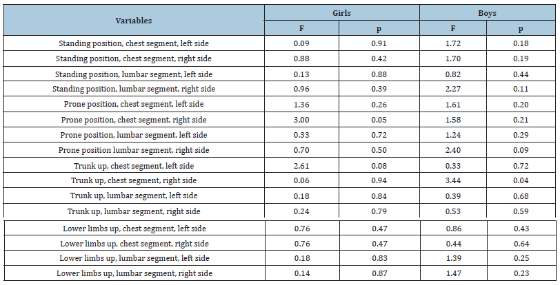
In girls, the highest generalised frequency occurred in scoliotic postures (x=73.69Hz), while in boys, this occurred in the scoliosis group (x=79.75Hz). One-way analysis of variance only showed significant differences in the group of boys between the scoliosis, scoliotic posture and the normal posture groups regarding SEMG frequency of the erector spinae in measurements for the prone position, trunk up, thoracic segment, right side (F=0.04; p=0.04) (Table 4).
Table 4:Analysis of differences in ANOVA for the SEMG frequency of the erector spinae between the scoliosis, scoliotic posture and the group within the norm among boys and girls

The relationship between body posture and EMG frequency of the erector spinae
Independent variables were the SEMG frequency of the erector spinae, while the dependent variables were scoliosis, scoliotic posture and correct posture. The regression model for the group within the norm did not reach the assumed statistical significance, therefore, it was not shown and was not considered proper in modelling the relationship between body posture and the frequency of the erector spinae examined in different positions. The most important and statistically significant predictors for body posture and the frequency of the erector spinae examined in different positions in the scoliosis group were the variables: prone position. trunk up, thoracic segment, left side (p=0.02), standing position, lumbar section, right side (p=0.03), prone position, thoracic segment, left side (p=0.02), standing position, thoracic segment, right side (p=0.04). The variance explained by the independent variables adopted in the model is 42% of the total variability (R^2=0.42), which indicates average adjustment to the data, but the assumed level of statistical significance (p=0.01) was met and additionally, the appropriate value for the statistical F was obtained, F=2.87. In the group with scoliotic posture, the most important and statistically significant model predictor turned out to be the trunk up position -chest segment, right side (p=0.04). The model was only explained 25% (R^2=0.25), which is low, but the level of statistical significance (p=0.03) was also met, as well as the appropriate value for the statistical test, F=2.97 (Table 5).
The angle of curvature of the spine and the EMG frequency of the erector spinae
The regression model for the group within the norm did not reach the assumed level of statistical significance, therefore, it was not demonstrated and was not considered as proper in modelling the relationship between SEMG frequency and the curvature angle. The most important and statistically significant model predictor for the SEMG frequency of the erector spinae and the angle of the spinal curvature in the scoliosis group was prone position, lumbar section, left side (p=0.03). The variance explained by the independent variables adopted in the model constitutes 29% of the total variability (R^2=0.29), which indicates small adjustment to the data, however, the statistical significance level (p=0.03) was met and the corresponding statistical test value was obtained, F=3.14. In the group with scoliotic posture, the most important and statistically significant model predictor was prone position, lumbar section, right side (p=0.03). The model was only explained in 29% (R^2=0.29), i.e. the low but assumed level of statistical significance (p=0.04) was also met, and also an appropriate value for the statistical test was obtained, F=3.14 (Table 5).
Discussion
Tylman [6] divided the concepts of the formation and development of idiopathic scoliosis into: the theory of congenital changes, the theory of physiological curvatures, the theory of curvature changes, the anatomical-functional theory, osteopathic theory, mechanical-static-dynamic theory, theory of growth disorders, the theory of disturbances of transformation processes, the theory of primary disorders of joint processes, the theory of hereditary changes. Among them, the theory of disturbances in muscle tension balance occupies a special place. This concept is considered among the oldest, as it was already described in 1741 by Nicolas Andry de Bois-Regard [6].
In 1865, Huetter carried out experiments on animals, which consisted of unilaterally cutting out the back muscles and obtaining the curvatures of the spine, directed by convexity towards the damaged one [6]. Similar experiments were carried out on rabbits by Arnd (1903) and Pusch (1928) and on dogs by Plagemann (1927) [6]. Not all of the subjects demonstrated curvature with convexity towards the damaged muscles. Pusch, for example, obtained curvatures in the opposite direction [6]. Experimental studies on animals, consisting of unilateral excision of the back muscles, were continued in the 1940s [22]. The supporters of the theory of muscular disorders were Scott and Morgan [23]. In Poland, Wejsflog [24] and Gruca [25] were advocates of the theory of muscular disorders. Most researchers did not see the primary etiologies of scoliosis-induced disorders in the muscles. In the opinion of these authors, the disturbances in balance of muscle tone are secondary as a result of not yet examined changes occurring in the central nervous system. There were also proponents of primary metabolic muscle disorders [26]. The theory of muscular tension disorders recognises the problem of the formation and development of scoliosis in an etiopathomechanical manner, i.e., without seeing the possibility of detecting primary etiological causes, aiming to reproduce the mechanism of distortion [6].
The starting point of the considerations was the statement that disturbances in muscle tension balance can be caused by a set of changes that create a clinical and anatomopathological image of the curvature of the spine. For this purpose, in addition to direct muscle damage, indirect damage was induced by excision of the peripheral and spinal nerves. Lesser, Hoffa and Frey [6] performed one-sided excision of the diaphragmatic nerve, and in this manner, they sought to create asymmetry of the chest. The results of these experiments were negative. Liszka's experiments [27] conducted on young rabbits consisted of one-sided intradural dissection of four consecutive dorsal roots of the spinal nerves. This experimental damage caused the development of severe structural scoliosis. All rabbits developed curvatures of the spine with convexity towards the cut roots. According to Liszka [27], the cause of spinal curvature is the interruption of sensory roads and damage to the spinal cord reflex activities related to the gamma system, the so-called core ataxia. These concepts coincide with the clinical observations of Boldrey et al. [28], who found developmental malformations, cystic lesions, hemangiomas, varicose veins, epidural and intradural adhesions as well as intramedullary tumours in the cervical and thoracic spinal cords of scoliosis patients. Stilwell's experiments in 1962 were particularly valuable for the theory of muscle tone disorders [29]. These experiments were performed on young monkeys age 1 to 1.5 years and consisted of one-sided excision of the erector spinae, starting from the sacrum to the upper part of the thoracic segment, and one-sided excision of the interspinous and intertransverse ligaments in 3-6 intervertebral spaces. After four weeks, Stilwel found distinct lateral-posterior curvatures that were transformed into structural changes during the 5th-8th week [29].
The above experiments and histological studies have explained the pathogenesis of growth disorders in the vertebral bodies. Increased pressure on the concave side of the vertebrae, and their extension on the convex side - in accordance with the law by Huetter-Volkmann and Wolff-Delpech - led to the distortion of the vertebral bodies in the shape of wedges in monkeys. Stilwell stated that these changes were the consequence of disturbances in the growth of the cartilage and connective tissue of the vertebral bodies. Stilwell's research showed that changes in the muscular ligament apparatus may induce secondary bone growth disorders [29]. The experiments by Goryński and Bojkowa delivered significant data contributing to the theory of muscle tone disturbances [30]. These experiments were carried out on rabbits divided into five groups. In subsequent experimental groups, the functioning of the illiocostalis muscles and the longest one of thorax were weakened unilaterally, the muscles of the transversospinales were unilaterally cut out, part of the erector spinae muscle was unilaterally removed, in the intervertebral area, one-sided dissection of the intercostal nerves over the course of 6 successive episodes was performed and one-sided costotransverse ligaments were cut in 6 intervertebral spaces in order to obtain intercostal and abdominal muscle paralysis. The observations from the above experiments allowed the author to conclude that the unilateral weakening or excision of the erector spinae muscle, as well as the weakening of the illiocostalis muscles and the longest one of the thorax over the 6 sections, led to spinal curvatures in rabbits. The curvature convexity was in the direction of the damaged muscles. Isolated damage of the intercostal and abdominal muscles of rabbits did not cause spinal curvature. The difference in muscle tone was apparently too small to disturb the four-legged animal's balance of the spine supported by the damaged back muscles. The unilateral destruction of the lateral costotransverse ligaments over the 6 segments did not cause the development of spinal curvatures.
These studies confirmed the possibility of developing scoliosis caused in experimental animals by asymmetry of muscle tone in the back. These studies, however, cannot be literally equated with the changes observed in humans. The most important obstacle in comparative studies is the upright posture of human beings and the related differences in the axial load on the spine and its balance apparatus. Since the trunk muscles do not have identical functions in humans and rabbits, the validity of the quadrupedal animal model for studying scoliosis may be questioned. The evolution of bipedal gait is accompanied by a different function of the trunk muscles in humans than animals [31,32]. Besides the functional differences between the species, there are also obvious anatomical differences between humans and quadrupeds. The vertebral bodies in rabbits are higher, the cephalic endplates are convex and the caudal flat, whereas the human endplates are biconcave, which may influence the kinematics of the spine in the two species. On the other hand, the anatomy of the muscles with regard to location, shape and relative size, is similar.
An important argument of the proponents of the muscle balance disorder theory is paralysis. From a pathomechanical point of view, they arise as a result of one-sided weakening or paralysis of the erector spinae muscles. In histopathological examination of the erector spinae muscles in people with idiopathic spinal curvatures during surgical procedures, morphological differences on the convex and concave sides were found [6], but it was not possible to indefinitely establish whether the changes in muscles on the convex side of the curvature are primary or secondary, caused by their mechanical overload. Scoliosis-related muscular balance disorders, and in particular, the erector spinae muscles, undoubtedly have an effect on the progressive nature of the curvature. SEMG tests have shed some light on muscle changes in the curvature of the spine, but the differences in the interpretation of the obtained results are very vast and even contradictory. Riddle & Roaf [33] and Bayer [34], on the basis of electromyographic tests, concluded that in idiopathic scoliosis, stronger muscles are always at the top of the original curvature. EMG and chronaxiometric examinations carried out by Żuk & Tokarowski [35,36], Weiss et al. [37] are interpreted in a different way.
Żuk, on the basis of electrodiagnostic tests, introduced the division of idiopathic scolioses dividing them into four groups:
a. Paretic
b. Dystonic
c. Atactic
d. Of other origins
The paretic group covers approx. 46% cases of idiopathic scoliosis. These curves have a long arch with simultaneous retroversion of the spine. In the electromyographic image, decrements and hypertrophy of motor units can be observed. The dystonic group includes about 39% of idiopathic scoliosis cases. Clinically, they are the most common type of scoliosis with co-existing anteflexion. In electromyographic tests, these curves are characterised by an increased response of the back muscles. The clinical, radiological and electrodiagnostic image makes the above-mentioned idiopathic scoliosis similar to spastic curvatures. According to Żuk, the atactic group (spinal ataxia) includes about 11% of idiopathic scolioses. These are clinically, long-arch curvatures with clearly increased retroversion of the spine. Clinically and electromyographically, deep sensory disturbances are found (no response to the stretching of muscles of the convex side under axial pressure). The fourth group does not show significant deviations in electromyographic image except for the typical image associated with spinal axis disorder and increased speed of muscle reaction and activation during movement on the convex side of the curve. Żuk stated that in idiopathic scoliosis, there are numerous secondary disorders of muscle function associated with their hypertrophy on the convex side of the curve, which were misinterpreted by some authors as primary lesions [36].
The studies by Doménech, Tormos, Barrios and Pascual-Leone have also indicated some developments in the theory of muscle tone balance disorders in recent years [38]. According to these authors, the aetiology of idiopathic scoliosis (IS) remains unknown; however, there is a growing body of evidence suggesting that spine deformity could be the expression of a subclinical nervous system disorder. Defective sensory input or anomalous sensorimotor integration may lead to abnormal postural tone and therefore, the development of spinal deformity. Inhibition of the motor cortico-cortical excitability is abnormal in dystonia. Consequently, the study of cortico-cortical inhibition may shed some insight into the dystonia hypothesis regarding the pathophysiology of IS. Paired pulse transcranial magnetic stimulation was used to study cortico-cortical inhibition and facilitation in 9 adolescents with IS, 5 teenagers with congenital scoliosis (CS) and 8 healthy age-matched controls. The effect of a previous conditioning stimulus (80% intensity of resting motor threshold) on the amplitude of the motor-evoked potential induced by the test stimulus (120% of resting motor threshold) was examined at various inter-stimulus intervals (ISIs) in both abductor pollicis brevis muscles. The results for healthy adolescents and those with CS showed a marked inhibitory effect of the conditioning stimulus as a response to the test stimulus at inter-stimulus intervals shorter than 6ms. These findings do not differ from those reported for normal adults. However, children with IS revealed abnormally reduced cortico-cortical inhibition at short ISIs. Cortico-cortical inhibition was practically normal on the side of the scoliotic convexity, while it was significantly reduced on the side of scoliotic concavity. In conclusion, these findings support the hypothesis that dystonic dysfunction underlies IS. Asymmetrical cortical hyperexcitability may play an important role in the pathogenesis of IS and represents an objective neurophysiological finding that may be used clinically [38].
The presented research indicates the validity of the concept regarding the formation and development of idiopathic scoliosis as a consequence of the asymmetry of muscle tone in the back. Since the beginning of research on the electromyography of the spine muscle, it has been noticed that there is a relationship between spinal curvature and the unequal muscle tension of the spine extensors. Therefore, the aim of scoliosis therapy should be to restore proper connections between the vertebrae so that the distribution of pressing and shearing burdens the vertebral bodies, not the joint processes. Passive stabilising structures, such as: intervertebral discs, interstitial joint capsules, ligaments, are not able to ensure correct segmental stabilisation. Muscle dystonia, which occurs in children with scoliosis, causes destabilisation and additional burden on these structures. It is only the introduction of forces of neuro-muscular origin that brings reduction of displacements in three planes and correction of scoliosis.
Conclusions
Only in boys in the prone position, trunk up, thoracic segment, right side, were there significant differences in the SEMG frequency of the erector spinae between the scoliosis, scoliotic and correct posture groups. The most important model predictors for the body posture and the frequency of the erector spinae in the scoliosis group were the variables: prone position, trunk up, thoracic segment, left side; standing position, lumbar section, right side; prone position, thoracic segment, left side; standing position, thoracic segment, right side. In the group with scoliotic posture, an important model predictor was prone position, trunk up, thoracic segment, right side. A significant relationship was found between the angle of curvature of the spine and the SEMG frequency of the erector spinae. An important model predictor for the SEMG frequency of the erector spinae and the angle of curvature in the scoliosis group turned out to be the test: prone position, lumbar section, left side. In the group with scoliotic posture, an important model predictor was prone position, lumbar section, right side. The largest, generalised SEMG frequency of the erector spinae in girls was found in scoliotic postures, while in boys, in the scoliosis group. The asymmetrical activity of the erector spinae visible in the SEMG frequency records is associated with an increase in the angle of curvature of the spine.
References
6. Tylman D (1995) Pathomechanics of lateral spinal curvatures. Severus, Warsaw, Poland.
16. Hierholzer E, Drerup B (2001) Influence of length discrepancy on ratserstereographic back shape parameters. Orthopade 30: 242-250.
22. Schwartzmann JR, Miles M (1945) Experimental production of scoliosis in rats and mice. J Bone Joint Surg (Am) 27: 59-69.
25. Gruca A (1974) On the pathology and treatment of idiopathic scolioses. Acta Med Pol 15(3): 139-156.
31. MacEwen G (1973) Experimental scoliosis. Clin Orthop Relat Res 93: 69-74.
35. Żuk T, Tokarowski A (1973) Pathomechanics of experimental scoliosis in rabbits as determined by electromyography. Chir Narzadow Ruchu Ortop Pol 38(1): 39-44.
36. Żuk T (1965) Etiology and pathogenesis of idiopathic scoliosis from the viewpoint of electromyographic studies. Beitr Orthop Traumatol 12: 138-141.
37. Weiss M, Milkowska A, Kozinska M (1957) Conservative treatment of scoliosis in the light of electromyographic data. Chir Narzadow Ruchu Ortop Pol 22(2): 197-209.
© 2019 Jacek Wilczyński. This is an open access article distributed under the terms of the Creative Commons Attribution License , which permits unrestricted use, distribution, and build upon your work non-commercially.
 a Creative Commons Attribution 4.0 International License. Based on a work at www.crimsonpublishers.com.
Best viewed in


.jpg)


 Editorial Board Registrations
 Submit your Article
 Refer a Friend
 Advertise With Us
.jpg)


.jpg)


.bmp)
.jpg)
.png)
.jpg)


.jpg)


.png)

.png)


.png)


