- Submissions

Full Text
Research & Investigations in Sports Medicine
New Tools for Medical Surgery Using Digital Image Processing
MI Mario A González Torres
Psychiatrist in San Juan, Mexico
*Corresponding author:MI Mario A González Torres, Psychiatrist in San Juan, Mexico
Submission: March 15, 2019; Published: March 27, 2019

ISSN: 2577-1914 Volume4 Issue5
Abstract
In this article we present a tool that can play a vital role for the patient’s life when operating, using digital image processing is proposed a new model in three dimensions of the patient for easy examination and exploration with non-invasive studies such as magnetic resonance nuclear
Introduction
Since the emergence of modern surgery as we know it, it has evolved constantly, the tools and everything that has an important role for these processes have already been analyzed arduously, for this reason, in this article will not analyze forms al-ready studied, but the proposition of a new tool that can play a new role in modern surgery.
The preparation for a surgery, is related until the ancient times of Egyptian surgery. There are papyri that chronicle clinical cases with a lot of emphasis on prognosis Dubois ND [1] and although the medical area was gradually gaining strength, as was a notable example in ancient Greece during the time of Hippocrates that not only took this branch very hard of knowledge, but also, a high degree of perfectionism. It is reported that not only surgery was improved, but also the diagnosis it implied before being able to proceed for a patient Cortés ND [2] and Dubois ND [1]. This character had so much impact in the medical area that today many of the doctors who profess these activities are categorized as Hippocratic doctors. Antralgo PL [3] As the years passed, with the help of other branches of knowledge, innovative ways of facilitating medical diagnosis were emerging, or at least to give another approach to the diagnosis and thus be able to help humanity. For example, when humanity acquires new knowledge, it seeks to apply them, and military and aerospace projects are a notable example of this [4].
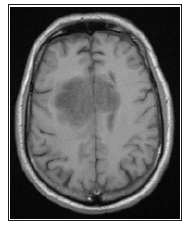
Currently, medical instrumentation is present in almost all procedures, varying from diagnosis to treatment. For the case of this article, we are considering medical imaging and, even more precisely, nuclear resonance imaging. The advantages that it brings with it will be taken from the fact that it produces a series of images presented as succession to one another that together can resemble a three-dimensional shape to perceive the bodies. Although this method is classified as a non-invasive form of instrumentation, many researchers believe otherwise due to the passive effects of nuclear magnetic resonance and what it entails, however, it has not been proven to present any risk to the patient , unlike radiation-based studies such as X-rays that can modify the structure of the atoms ionizing them, causing severe dam-age to health [5]. These devices can read our body in a magnetic way, changing strongly the resonance frequencies, aligning their Larmor precession [6]. To cause strong changes in the magnetic field, the hydrogen atoms cause momentous changes in frequency and large transceivers can capture these fluctuations thus representing the visual that is conceivable as shown in Figure 1.
Methodology
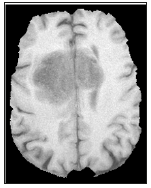
The idea that is proposed is one where can be given a new angle for diagnosis, the proposal is to have in your hands an exact replica of the patient to be able to explore without some scruple and without some consequence, again and again, both for diagnosis and prior to surgery if is that it is required. For this, a study of an anonymous patient consisting of 128 sectional cuts of the skull to the base of the chin was taken using a Siemens Resonator with a magnetic field of 4.5 Tesla; will begin with the digital processing of images with the help of a mathematical image processor and, therefore, they begin to remove sections of the skull and other regions that are not of interest as in this case the head of a patient, as It shows in the following Figure 2.
Figure 1:Image of an MRI with a Siemens resonator in a 5 Tesla magnetic field.

Figure 2:Same post processing resonance image.

This processing is a series of simple steps to reach the end, these successions of steps are shown in the following Figure 3. As you can see, the process is carried out from the indexing of the images that make up the study, these images are read and put into an array of three dimensions, width, length and number of images is built (therefore show that way in the diagram). Meticulously chosen filters are applied for this case by removing only bone structure and periosteum, this processing is carried out to remove sections that are not of interest, likewise, it is worth mentioning that this processing does not remove blood vessels. After an algorithm is applied to reconstruct three-dimensional spatial arrangement.
Figure 3:Flow diagram for point cloud recovery using magnetic resonances.

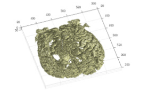
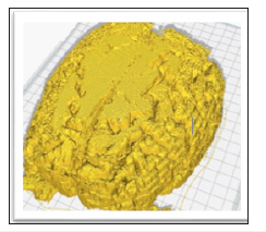
Once it has been processed, it can be exported to a standardized file so that it can be viewed in a spatial point editor and, finally, if desired, printing in 3d for a better analysis. Then, once the images are processed, we can apply the spatial point reconstruction algorithm and we can obtain the following Figure 4. Once the data has been processed, it can be Exported and prepared them to be printed in 3d. The following Figure 5 shows the brain ready to be printed in open source software.
Figure 4:Three-dimensional model made mathematically from a study of magnetic resonances.

Figure 5:Mathematically generated model represented in the Cura software ready to be printed.

In the same way this process for generating digitized models can be applied without previous processing for isolation, applying directly to the algorithm for reconstruction in three dimensions as shown in the following Figure 6.
Figure 6:Digitization of a patient using magnetic resonances.

Conclusion
This method can be implemented in some hospitals, both in the private and in the public sector, to reduce the mortality rate in surgeries and to reduce the time to make a real diagnosis. This method can play a very important role in the exploration not only cerebral. When an anomaly appears in the body and it is necessary to see with imaging, anomalies can be characterized and even touched outside the human body with the help of new 3D printing technologies and materials. From identifying tumors, to identifying veins in the body.
Biomedical engineers must be used to innovate, create and improve the processes for diagnosis and prior, during and after operation, that the technology used in day-to-day can serve to pose new challenges and help the humanity.
References
- Dubois MS. History of surgery. Fascicle primero: Preoperatorio, pp. 1-15.
- Cortés ST. Evolution of surgery.
- Antralgo PL (1981) The origins of the medical diagnosis. Gelfand doctor- blade science and history Dynamis 1: 3-15.
- Anonymous. Introduction to biomedical instrumentation. pp. 5-21.
- Physics and Society (2017) Ionizing radiations.
- Anonymous (2016) IDC-online
© 2019 MI Mario A González Torres. This is an open access article distributed under the terms of the Creative Commons Attribution License , which permits unrestricted use, distribution, and build upon your work non-commercially.
 a Creative Commons Attribution 4.0 International License. Based on a work at www.crimsonpublishers.com.
Best viewed in


.jpg)


 Editorial Board Registrations
 Submit your Article
 Refer a Friend
 Advertise With Us
.jpg)


.jpg)


.bmp)
.jpg)
.png)
.jpg)


.jpg)


.png)

.png)


.png)


