- Submissions

Full Text
Orthopedic Research Online Journal
Simultaneous Bado Type II Monteggia Fracture Dislocation with Ipsilateral Acute Osteochondral Fracture of the Capitellum in an Adult: A Case Report
Mohamed Elkabbani1*, Volkhardt Studtmann2 and Michael Schulte3
1Associate Professor Orthopedic Surgery and Traumatology, Faculty of Medicine, Mansoura University, Egypt
2Senior Physician at the Clinic for Trauma Surgery and Orthopedics, Pediatric Orthopedics, Agaplesion Diakoniekllnikum Rotenburg. Dr. Med, Specialist in Surgery, Focus on Trauma Surgery, Emergency Medicine, Senior Emergency Physician, Egypt
3Head Physician at the Clinic for Trauma Surgery and Orthopedics, Pediatric Orthopedics, Agaplesion Diakoniekllnikum Rotenburg. Prof. Dr. Med, Specialist in Surgery, Focus Accident Surgery, Specialist in Orthopedics and Accident Surgery, Special Accident Surgery, Physical Therapy, Egypt
*Corresponding author:Mohamed Elkabbani, Associate Professor Orthopedic Surgery and Traumatology, Faculty of Medicine, Mansoura University, Egypt
Submission: September 09, 2023;Published: October 06, 2023

ISSN: 2576-8875 Volume10 Issue4
Abstract
Background: Simultaneous Monteggia fracture dislocations and ipsilateral fracture of the Capitellum is an extremely rare injury in adults.
Materials and methods:A 43-year-old male patient was presented with open grade I Monteggia fracture dislocation Bado type II with ipsilateral osteochondral fracture of the Capitellum which was not apparent on initial radiographs. We describe the diagnosis and management of these extremely rare coexisting fractures.
Results and conclusion: this case emphasizes the importance of exclusion of any associated ipsilateral Capitellar fracture in any case of Monteggia fracture dislocation. Careful clinical and radiological examination are essential for a satisfactory outcome in such complex injuries.
Keywords:Monteggia; Capitellum; Osteochondral fracture
Introduction
Giovanni Battista Monteggia described in 1814 “a traumatic lesion distinguished by a fracture of the proximal third of the ulna and an anterior dislocation of the proximal epiphysis of the radius” [1,2].
In 1967, Bado classified the Monteggia fracture into four types, depending on the direction of the dislocation of the radial head and the angulation of the ulnar fracture [3].
Bado classification system is the most commonly used to describe Monteggia fractures. Currently the term Monteggia fracture generally refers to fractures of the proximal ulna associated with dislocation of the radial head. They account for approximately 1% to 2% of all forearm fractures [4].
Bado type 1 (anterior radial head dislocations) are more common in the pediatric age groups while Bado type 2 (posterior radial head dislocations) are more common in adults. Several authors have described the predominance of Bado type II fractures in adults [5-8].
Jupiter et al further subclassified type II Monteggia fracture dislocations into four groups depending on the position of the ulna fracture: [9]
Monteggia fracture dislocation associated with ipsilateral capitellar fractures are extremely rare injuries in adults. Careful assessment of the fracture and exclusion of associated injury are essential for favourable outcome.
In spite of advances in surgical management and a better understanding of the biomechanical principles, a Monteggia fracture is still frequently associated with complications, poor functional results and further operations [5,6].
Case Report
A 43 yeras old male patient was presented in the trauma department at late night with severe pain and swelling after a fall on the right elbow after being stumbled over a rowing machine. Examination revealed tenderness, deformity and swelling of the upper third of the right forarm and elbow with painful limitaion of movement in all directions and a skin wound below the olecranon which was less than 1cm. No associated neurovascular deficit. Ipsilateral wrist joint was free.
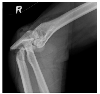
Only an immediate lateral x-ray view of the elbow and forearm was possible because of pain and deformity, and it showed fracture of the proximal third of the ulna with posterior dislocation of the radial head (Figure 1). Accordingly, the patient was diagnosed to have a Grade 1 open monteggia fracture dislocation Bado type II.
Figure 1:Pre-operative radiograph of the right forearm showing fracture of the proximal ulna and posterior dislocation of the radial head.
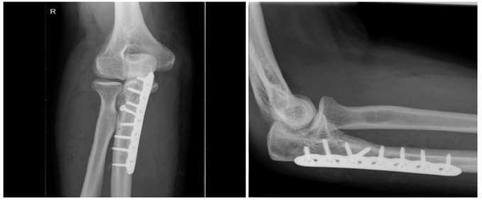
The patient was immediately transferred to the operative theatre for open reduction and internal fixation of the monteggia fracture dislocation after antibiotic administation and wound debridment under general anathesia. The fracture was rigidly fixed through a posterior approach with 3.5mm 7 holes locking compression plate and 3.5mm cortical screws with lag screw fixation. The radial head was spontaneously reduced as being confirmed by intra operative fluoroscopy. The elbow and forearm were placed in an elastic bandage and the patient was instructed for unloading of the limb for 6 weeks with early active mobilization after removal of the drainage. Postoperative antibiotic therapy was given in the form of cefazolin for 3 days.
Immediate postoperative x-ray views revealed proper reduction of the fracture and proper alignment of the radio capitellar joint (Figure 2).
Figure 2:Radiographs of the elbow showing operative fixation of the posterior Monteggia lesion. a) anteroposterior and b) lateral views.
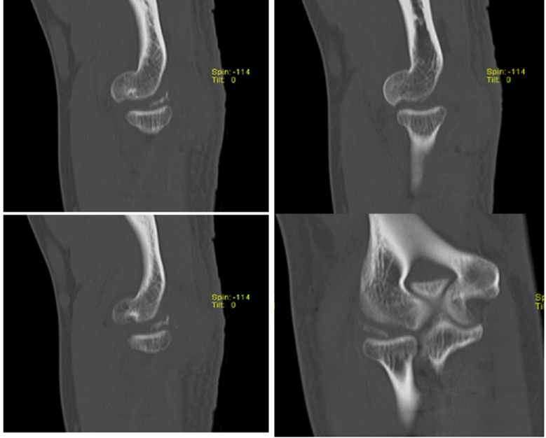
However, the patient continued to suffer from significant limitation in elbow motion and pain with elbow movement. CT scan of the elbow was indicated in the fourth postoperative day, and it revealed the presence of defective capitulum and a loose bony fragment in the elbow joint suggesting the diagnosis of associated traumatic osteochondral fracture of the capitullum (Figure 3).
Figure 3:Post-operative sagittal and coronal CT cuts showing defective posterolateral aspect of the capitellum and loose osteochondral elbow joint fragment.
Accordingly, a second operation was decided to deal with this loose body in the elbow joint and osteochondral capitellar fracture. Elbow arthroscopy was performed one week after the first operation. It revealed intact humeroulnar articulation and superficial injury of the articular cartlige covering the radial head.
However, because of difficult access to the dorsolateral aspect of the radio capitellar articulation arthroscopically, elbow arthrotomy was performed using the proximal part of the previous posterior approach which was used during the first operative intervention and splitting the anconeus to approach the elbow joint around the medial epicondyle.
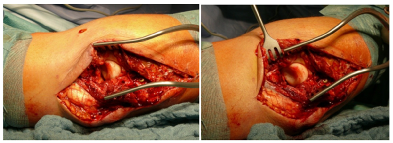
Surgical assessment of the elbow joint revealed defective posterolateral part the capitellum in addition to a loose osteochonral joint fragment. The radial head dislocates posteriorly with increasing elbow flexion and on extension it jams on the remaining anterior portion of the capitellum (Figure 4).
Figure 4:Photographs of the operated right elbow showing the radial head and the defective fractured capitellum.
The loose bony fragment was removed and the sharp edges of the fractured capitullum were smoothened with subchondral drilling and the elbow was immbolized in mild flexion. The necessity of a third operation for restoration of the radio capitellar joint stability by osteocartligenous grafting was discussed with the patient who accepted a third operation and grafting from the knee.
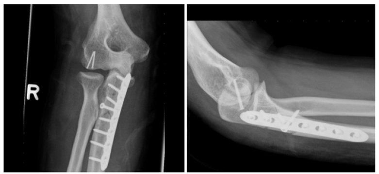
One week later osteocartligenous grafting was performed using Osteochondral Autograft Transfer System (OATS) to take 1 cm osteochondral fragment from the lateral femoral condyle to be re-inserted in the defective capitellum and fixed with two mini fragment screws with proper restoration of the radio capitellar joint stability as being proved intra-operatively (Figure 5).
Figure 5:Immediate postoperative elbow anteroposterior and lateral radiographs showing the osteochondral autogenous graft being fixed in position by two mini fragment screws.
Postoperatively the elbow was immobilized with a long arm splint for 6 weeks in midway flexion. After the third week the splint can be temporary removed with beginning of progressive rangeof- movement exercises only between 50 and 110 degrees of elbow flexion.
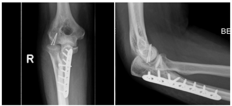
The first follow up x-rays were done 2 weeks postoperatively after removal of the sutures. They revealed slight bulge of the two mini fragment screws which may require further readjustment, but the patient refused the idea of being operated for a fourth time and was lost in the next follow up (Figure 6).
Figure 6:Two weeks postoperative elbow radiographs showing loosening and slight bulge of the two mini fragment capitellar screws.
Discussion
The reported incidence of misdiagnosis of Monteggia fractures varies from 16% to 52 % [10]. Accordingly, these fractures represent a challenging clinical phenomenon in spite of increased understanding of these fractures and advances in surgical management in the Orthopaedic community.
With respect to adult patients, good results can only be obtained by early diagnosis and stable internal fixation [9,11]. The posterior Bado type 2 fracture is the most common type of Monteggia fractures in adult [5-8,11]. Many authors attributed the poor functional outcome of the posterior Monteggia lesion to the commonly associated radial head or neck and coronoid fractures [5,6,9,12,13].
Paval et al. [7] considered a triangular shaped chip fracture of the radial head as one of the three major components of a posterior Monteggia lesion. He assumed that the radial head fracture probably occurred as the dislocated head is forced against the capitellum and this may also produce a cartligenous or osteochondral fracture of the capitellum.
Ring et al. [5] noted that associated fracture of the head of radius occurred in 68% of type II injuries. He considered elbow problems related to coronoid and radial head fractures as the most challenging element in treatment of these injuries.
Furthermore, Konrad et al. [13] found that Patients with Bado type II fracture, Jupiter type IIa fracture, fracture of the radial head, coronoid fracture, and complications requiring further surgery should be informed about the potential risk of functional deficits and the possible need for further surgery.
In a series of seven patients, Penrose et al. [8] detected obivious damage to the articular surface of the capitellum intraoperatively in two patients. He described the mechanism of injury by axial forces acting on the forearm with a semi flexed elbow, fracturing the posterior cortex of the ulna and leading to posterior radial head dislocation and in which excessive rotation in either direction does not enter in the mechanism of injury [8].
This mechanism differs from Evans theory who postulated hyper pronation as the possible mechanism of injury [1]. Furthermore, Penrose postulated that the posterior Monteggia fracture is simply a variation of dislocated elbow in which the ligamentous attachment of the elbow proves stronger than the shaft of ulna and the shaft therefore fractures [8].
Osborne and Cotterill first described in 1966 an osteochondral fracture in the posterolateral margin of the capitellum with or without a crater or shovel like defect in radial head in recurrent elbow dislocation as similar to the glenoid defect (bony bankart lesions) observed in in recurrent shoulder dislocation. They attributed recurrence of the posterolateral dislocation of the elbow to collateral ligament laxity with secondary damage to the capitellum and head radius [14].
Jeon et al. [15] called this lesion Osborne and cotterill lesion and described its importance in the diagnosis of posterolateral rotatory instability of the elbow on normal x-rays. To our Knowledge, the coexistence of ipsilateral capitullum fracture with posterior Monteggia lesion is rarely described in the literature with no previous reported cases describing such simultaneous lesions.
This case highlights the need for a high index of suspicion in Monteggia fracture dislocation in adults even if the cause is low energy trauma and in spite of no evidence of assosciated elbow fractures in initial radiographs.
We suggested considering this lesion as a Monteggia type II equivalent.
The lesion found in this case is considered a traumatic Osborne and Cotterill lesion in the posterolateral margin of the capitellum and it affects the radio capittillar joint stability markedly thus suggesting a similar mechanism in the common posterolateral instability of the elbow.
The true incidence of capitellar fracture with the posterior Montegia lesion is still to be investigated. Many of the cases are missed because there is either little bone attached to the articular cartilage fragment or a purely cartligenous lesion, thereby resulting in poor visualization on standard radiographs and requiring further imaging.
Capitellum fracture or at least articular cartlige injury is to be expected in every Monteggia posterior lesion. However, whether it needs intervention or not, depends on the degree of impairment of elbow range of motion and radio capitellar joint stability. The focus in the literature is always on the associated radial head or coronoid fracture. However, the possibility of associated capitellar fracture or just articular injury must be always taken into consideration, not because of its frequency, but because of its hazardous effect on the prognosis of such complicated injuries..
This is to the best of our knowledge the first case report of an adult with an occult osteochondral fracture with posterior Monteggia lesion in spite of normal postoperative radio capitellar joint on radiographs.
Early and proper diagnosis of such lesions associated with posterior Monteggia lesions is critical for proper management. Currently the accepted treatment option for adult Monteggia fracture dislocations is open reduction and rigid anatomical fixation of the ulnar fracture. Once this has been performed the radial head will either spontaneously reduce or will be reducible by closed manipulation [4].
This must be confirmed intraoperatively with a true lateral radiograph. Associated lesions must be dealt with in the same setting. Treatment options for associated osteochondral fractures of the capitellum include debridement of loose bodies, subchondral drilling, bone grafting with or without internal fixation, and reattachment. The choice of the appropriate surgical procedure depends on the size and location of the lesion, stability of the fragment, and its effect on the radio capitellar joint stability. They can be done either arthroscopically, or by open arthrotomy or combined.
Conclusion
Our case report highlights a rare combination of injuries. While it is true that such injuries occur rarely, but they dramatically affect the prognosis. Good-quality radiographs are essential in the assessment of Monteggia fracture dislocations. If any doubt exists as to the extent of the patient’s injury a CT scan must be indicated in order to clearly define the anatomy of the radio capitellar joint and to exclude not only the commonly associated radial head or coronoid fractures but also fractures of the capitellum as well.
Compliance with Ethical Standards
Funding
No funding was received for this study.
Conflict of interest
The authors declare that they have no conflict of interest.
Ethical approval
All procedures performed in this study involving human participants were in accordance with the ethical standards of the institutional and/or national research committee and with the 1964 Helsinki declaration and its later amendments or comparable ethical standards.
Informed consent
Informed consent was obtained from all individual participants included in the study (one patient for this case report).
References
- Evans EM (1949) Pronation injuries of the forearm, with special reference to the anterior Monteggia J Bone Joint Surg Br 31(4): 578-588.
- Monteggia GB (1814) Surgical Institutions. Milan, Maspero, Italy.
- Bado JL (1967) The Monteggia Clin Orthop Relat Res 50: 71-86.
- Reckling FW (1982) Unstable fracture-dislocations of the forearm (Monteggia and Galeazzi lesions). J Bone Joint Surg Am 64(6): 857-863.
- Ring D, Jupiter JB, Simpson NS (1998) Monteggia fractures in adults. J Bone Joint Surg [Am] 80-A: 1733-1744.
- Egol KA, Tejwani NC, Bazzi J, Susarla A, Koval KJ (2005) Does a Monteggia variant lesion result in a poor functional outcome? A retrospective Clin Orthop 438: 233-238.
- Pavel A, Pitman JM, Lance EM, Wade PA (1965) The posterior monteggia fracture: A clinical J Trauma 5: 185-199.
- Penrose JH (1951) The monteggia fracture with posterior dislocation of the radial head. J Bone Joint Surg [Br] 33-B: 65-73.
- Jupiter JB, Leibovic SJ, Ribbans W, Wilk RM (1991) The posterior monteggia lesion. J Orthop Trauma 5: 395-402.
- Speed JS, Boyd HB (1940) Treatment of fractures of ulna with dislocation of head of radius (Monteggia fracture). JAMA 115(20): 1699-705.
- Ring D, Jupiter JB, Waters PM (1998) Monteggia fractures in children and J Am Acad Orthop Surg 6: 215-24.
- Givon U, Pritsch M, Levy O, Yosepovich A, Horoszowski H (1997) Monteggia and equivalent lesions: a study of 41 Clin Orthop 337: 208-215.
- Konrad GG, Kundel K, Kreuz PC, Oberst M, Sudkamp NP (2007) Monteggia fractures in adults: Long-term results and prognostic factors. J Bone Joint Surg [Br] 89B: 354-360.
- Osborne G, Cotterill P (1966) Recurrent dislocation of the elbow. J Bone Joint Surg Br 48: 340-346.
- In-Ho Jeon IH, Micic ID, Yamamoto N, Morrey BF (2008) Osborne-cotterill lesion: An osseous defect of the capitellum associated with instability of the elbow. AJR Am J Roentgenol 191(3): 727-729.
© 2023 Mohamed Elkabbani. This is an open access article distributed under the terms of the Creative Commons Attribution License , which permits unrestricted use, distribution, and build upon your work non-commercially.
 a Creative Commons Attribution 4.0 International License. Based on a work at www.crimsonpublishers.com.
Best viewed in


.jpg)


 Editorial Board Registrations
 Submit your Article
 Refer a Friend
 Advertise With Us
.jpg)


.jpg)


.bmp)
.jpg)
.png)
.jpg)


.jpg)


.png)

.png)


.png)


