- Submissions

Full Text
Orthopedic Research Online Journal
Deep Learning - A Rising Curve in the Field of Orthopaedics
Jyotsna*
University of Delhi, India
*Corresponding author:Jyotsna, University of Delhi, India
Submission: September 30, 2022;Published: October 13, 2022

ISSN: 2576-8875 Volume10 Issue2
Introduction
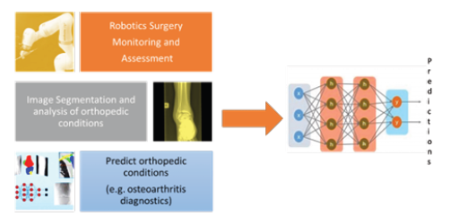
Figure 1: Deep learning in orthopaedics.

In recent years, Artificial Intelligence (AI) has revolutionized the medical domain. There has been an insurgence of the availability of medical data with Electronic Health Record (EHR) systems in place to track patients’ records. There have been attempts to visualize medical data with deep learning algorithms, making it more interpretable. Due to the advancement in deep learning algorithms and computational power, radiological assessment in the diseased condition of orthopaedics is gaining new clinical solutions. Orthopedics deal with musculoskeletal conditions such as joint pain from arthritis, bone fractures, injuries affecting muscles and ligaments, back pain, neck pain, shoulder pain, etc. Medical analysis of such conditions involves image analysis providing decision support to medical practitioners and improving various diagnostic and treatment processes. Deep learning has been successfully used in such applications relating to the medical analysis of orthopaedic conditions [1]. The American Academy of Orthopaedic Surgeons emphasized that X-rays are the most common and widely available diagnostic imaging technique allowing clinicians to diagnose certain diseased conditions. Deep learning has been successfully used in analyzing X-Rays to determine any fracture condition, osteoarthritis, knee or hip implant detection, any kind of bone tumor diagnosis, etc. [2]. Deep learning modelling for X-ray image analysis is inspired by the human brain connecting neurons. Deep learning includes two predominant algorithms based on neural modelling- Convolutional Neural Network (CNN) and Recurrent Neural Network (RNN) [3]. These algorithms use filters learned on a small subset of a larger image to detect certain features useful for analysis. Neural network trains over and combines all the high-level features from the deepest convolution layer and uses them to output the classification of regions on X-Ray images of orthopaedic conditions. Through the iterative training process, the CNN is able to update its learnable parameters and make important [4]. The current research curve suggests the development of deep learning models focused on classifying, diagnosing, and risk predicting orthopedic conditions from medical images (Figure 1). Some of the interesting studies highlighted how machine learning with deep models could help in predicting orthopaedic medical conditions and improving treatments with advanced deep models. Liao et al., recently conducted a study on improving orthopedics radiographic fracture classification. In an interesting work [5] deep models have been proposed to detect orthopaedic diseases in medical images efficiently.
In an interesting study by Murphy et al., it is highlighted when should orthopedic surgeon use deep learning. On contrary, some researchers also have highlighted the use of deep learning as a “black box model” with hidden functionality, However, further research work to develop “Explainable Artificial Intelligence (XAI)” [6] has explained and addressed such black box problems [7-9].
References
- Lee J, Chung SW (2022) Deep learning for orthopedic disease based on medical image analysis: present and future. Applied Sciences 12(2): 681.
- Hill BG, Krogue JD, Jevsevar DS, Schilling PL (2022) Deep learning and imaging for the orthopaedic surgeon: How machines “read” radiographs. JBJS 104(18): 1675-1686.
- Mahajan R, Parihar N, Kale I, Thakur M, Raut S (2021) Infrared thermography for orthopaedic disorders by image processing. International Journal of Research in Engineering and Science (IJRES) 9(7): 66-69.
- Lalehzarian SP, Gowd AK, Liu JN (2021) Machine learning in orthopaedic surgery. World Journal of Orthopedics 12(9): 685.
- Xiu F, Rong G, Zhang T (2022) Construction of a computer-aided analysis system for orthopedic diseases based on high-frequency ultrasound images. Computational and Mathematical Methods in Medicine.
- Yang G, Ye Q, Xia J (2022) Unbox the black-box for the medical explainable AI via multi-modal and multi-centre data fusion: A mini-review, two showcases and beyond. Information Fusion 77: 29-52.
- Chan HP, Samala RK, Hadjiiski LM, Zhou C (2020) Deep learning in medical image analysis. Adv Exp Med Biol 1213: 3-21.
- Liao Z, Liao K, Shen H, Van Boxel MF, Prijs J, et al. (2022) CNN attention guidance for improved orthopedics radiographic fracture classification. IEEE Journal of Biomedical and Health Informatics 26(7): 3139-3150.
- Holzinger A, Saranti A, Molnar C, Biecek P, Samek W (2022) Explainable AI methods-a brief overview. In International Workshop on Extending Explainable AI Beyond Deep Models and Classifiers. Springer, Cham, pp: 13-38.
© 2022 Jyotsna. This is an open access article distributed under the terms of the Creative Commons Attribution License , which permits unrestricted use, distribution, and build upon your work non-commercially.
 a Creative Commons Attribution 4.0 International License. Based on a work at www.crimsonpublishers.com.
Best viewed in


.jpg)


 Editorial Board Registrations
 Submit your Article
 Refer a Friend
 Advertise With Us
.jpg)


.jpg)


.bmp)
.jpg)
.png)
.jpg)


.jpg)


.png)

.png)


.png)


