- Submissions

Full Text
Orthopedic Research Online Journal
Knee Dislocation with Tibial Shaft Fracture
Nesa Milan1, Alireza Nezami2*, Aref Daneshi2, Yousef Fallah3, Paniz Nezami4 and Hossein Nematian1
1Research Assistant at Center of Orthopedic Trans-Disciplinary Applied Research (COTAR), Tehran University of Medical Sciences, Iran
2Orthopedic Resident, Tehran University of Medical Sciences, Iran
3Assistant Professor at Orthopedic Department, Tehran University of Medical Sciences, Iran
4Cardiology Resident, Tehran University of Medical Sciences, Iran
*Corresponding author:Alireza Nezami, Orthopedic Resident, Tehran University of Medical Sciences, Iran
Submission: July 23, 2021;Published: August 06, 2021

ISSN: 2576-8875 Volume8 Issue4
Abstract
We present a case of anterior dislocation of the knee combined with tibial shaft fracture and multi-ligament injury. To our knowledge, a complex case like this has rarely been documented. A 53-year-old male was involved in a car crash. Radiographies showed a fracture in the shaft of the tibia and fibula, anterior dislocation of the knee, and an avulsion fracture of the fibula. In MRI scan, ACL was torn entirely, and PCL had a high-grade intra-sheath tearing. The surgery was performed in two steps with 4-day interval. At first step, we fixed the tibial shaft fracture with a plate and screw, and in the second step, we reconstructed the ligament injury. All components of this lesion were quickly diagnosed and treated appropriately. Three months after treatment, the patient had achieved adequate daily functioning following the onset of early rehabilitation exercises.
Keywords: Knee dislocation; Tibial fracture; Ligament; ACL; PFL; Avulsion fracture
Introduction
Acute knee dislocation is a limb threatening emergency due to complications such as extensive soft tissue injury and disruption of regional arteries [1]. Since 50% of knee dislocations reduce spontaneously [2], they make it difficult to diagnose damage to the ligaments as well as the accompanying vascular injury, and if missed, can cause significant dysfunction in the limb [3]. This partly explains why, despite the low incidence of knee dislocations, the injury is associated with a high rate of complications such as amputation [4].
There is still debate about how to manage a traumatic complex, multiple ligamentous knee injury, and choosing between surgical treatment options or closed immobility is a controversy [5]. Concomitant fractures, including ipsilateral tibial diaphysis fractures, often challenge immediate ligament repair. Simultaneous occurrence of tibial diaphysis fracture and an anterior knee dislocation, leading to multiple ligamentous injury, is uncommon which occurs in only 2% of tibial fractures [6,7].
The treatment of choice for tibial shaft fractures is intramedullary nailing (IMN) [8]. Recent studies, however, show that transtibial tunnels implantation is difficult and challenging in the event of simultaneous occurrence of knee ligament injury and tibial shaft fracture and approach to both injuries at the same time should be avoided, and repair of ligament damage should be delayed until bone damage has healed [9]. We report a case of traumatic close left tibial shaft fracture and an anterior knee dislocation associated with extensive injuries in lateral, anterior, and posterior compartments of the knee.
Case Report
A 53-year-old man was admitted to the emergency department of Sina Hospital following a car accident causing his left leg trapped between two vehicles. The principles and protocols of Advanced Trauma Life Support (ATLS) was applied on admission. After the initial examination and stabilization of the patient, he had severe pain, deformity and swelling on his left knee. There were skin abrasions on the left knee, left leg, and left medial malleolus. Physical examination was not possible, because of the severe pain. Neurovascular examinations of the limb were normal. There was no numbness or weakness in the limb, and pulsation was normal. Plain radiography and Magnetic Resonance Imaging (MRI) of left lower limbs were taken.
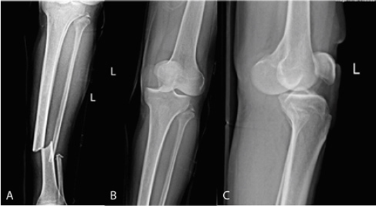
Radiographies revealed an anterior dislocation of the knee, a spiral-oblique fracture through the one-third mid-distal diaphysis of the tibia and fibula, and an avulsion fracture of the fibula (Figure 1). A closed reduction of dislocation was performed under sedation in operation room, and neurovascular examinations were rechecked. Then the limb was immobilized with a splint.
Figure 1: Initial radiographs showing the dislocated knee with simultaneous fracture of the tibial shaft. (A) Tibial shaft fracture. (B) Anterior dislocation of the knee combined with LCL and PFL avulsion. (C) Anterior dislocation of the knee.

MRI scan of the patient demonstrated a longitudinal meniscal tear, extending to the posterior horn and meniscofemoral ligament and a complete tearing of Lateral Collateral Ligament (LCL). Anterior Cruciate Ligament (ACL) was entirely torn, and Posterior Cruciate Ligament (PCL) had a high-grade intra-sheath tearing. Medial Patellofemoral Ligament (MPFL) showed a grade 2 sprain along with a partial avulsion from femoral insertion. Arcuate fracture at the fibular tip along with subjacent edema was also noticed as a result of Popliteofibular Ligament (PFL) avulsion (Figure 2).
According to the MRI report, it was a severe injury. 3 out of 4 compartments of the knee were severely injured. To detect any neurovascular injury The patient was carefully examined.
The surgery was performed after stabilizing the patient’s condition which was two days after the accident. Tibial shaft fracture was fixed using plates and screws to obtain proper stability and facilitate a later, second stage ligament reconstruction. Four days after initial surgery, the patient was taken back to operation room to repair the ligament injury. ACL and LCL tearing were fixed using suture anchors and PFL tearing was fixed with a screw (Figure 3). Hinged knee brace was used for immobilizing the limb for six weeks. After six weeks, gradual rehabilitative exercises began, and three month later the patient was able to do daily activities. In the case of knee joint capsule sprain and acute phase edema, PCL injury was managed non-operatively.
Figure 2: The preoperative MRI images. (A) ACL is completely torn. (B) LCL is completely torn. (C) MCL is severely injured.

Figure 3: Immediate postoperative radiographs. (A) and (B) Tibial shaft fracture fixation with plate and screw. (C) and (D) Suture anchor used for fixation of LCL avulsion.

References
- Kennedy J (1963) Complete dislocation of the knee joint. JBJS 45(5): 889-904.
- Wascher DC, Dvirnak PC, DeCoster TA (1997) Knee dislocation: initial assessment and implications for treatment. J Orthop Trauma 11(7): 525-529.
- Schenck RC, Richter DL, Wascher DC (2014) Knee dislocations: Lessons learned from 20-year follow-up. Orthop J Sports Med 2(5).
- Aydın A, Atmaca H, Müezzinoğlu ÜS (2013) Anterior knee dislocation with ipsilateral open tibial shaft fracture: a 5-year clinical follow-up of a professional athlete. Musculoskelet Surg 97(2): 165-168.
- Levy BA, Fanelli GC, Whelan DB, Stannard JP, MacDonald PA, et al. (2009) Controversies in the treatment of knee dislocations and multiligament reconstruction. J Am Acad Orthop Surg 17(4): 197-206.
- Templeman DC, Marder RA (1989) Injuries of the knee associated with fractures of the tibial shaft. Detection by examination under anesthesia: a prospective study. J Bone Joint Surg Am 71(9): 1392-1395.
- Thiagarajan P, Ang KC, Das De S, Bose K (1997) Ipsilateral knee ligament injuries and open tibial diaphyseal fractures: incidence and nature of knee ligament injuries sustained. Injury 28(2): 87-90.
- Ahmed N, Khan MS, Afridi SA, Awan AS, Afridi SK, et al. (2016) Efficacy and safety of interlocked intramedullary nailing for open fracture shaft of tibia. Journal of Ayub Medical College Abbottabad 28(2): 341-344.
- Campos TVO, Moraes MN, Andrade MAP, Schenck RC, Donell ST (2020) Knee dislocation with ipsilateral tibial fracture treated with an intramedullary locked nail and simultaneous transtibial tunnel knee ligament reconstruction: A Case Report of autografts and limited resources. Surg J (NY) 6(3): e160-e163.
- Richter M, Lobenhoffer P, Tscherne H (1999) Knee dislocation. Long-term results after operative treatment. Chirurg 70(11): 1294-1301.
- Meyers MH, Moore TM, Harvey JP (1975) Traumatic dislocation of the knee joint. J Bone Joint Surg Am 57(3): 430-433.
- Schenck RC, Hunter RE, Ostrum RF, Perry CR (1999) Knee dislocations. Instr Course Lect 48: 515-522.
- Brautigan B, Johnson DL (2000) The epidemiology of knee dislocations. Clin Sports Med 19(3): 387-397.
- Green NE, Allen BL (1977) Vascular injuries associated with dislocation of the knee. The Journal of Bone and Joint Surgery American Volume 59(2): 236-239.
- Robertson A, Nutton R, Keating J (2006) Dislocation of the knee. The Journal of Bone and Joint Surgery British Volume 88(6): 706-711.
- Gaski IA, Martinussen BT, Engebretsen L, Johansen S, Ludvigsen TC (2004) Knee luxation--follow-up after surgery. Tidsskr Nor Laegeforen 124(8): 1078-1080.
- Harner CD, Waltrip RL, Bennett CH, Francis KA, Cole B, et al. (2004) Surgical management of knee dislocations. JBJS 86(2).
- Liow RY, McNicholas MJ, Keating JF, Nutton RW (2003) Ligament repair and reconstruction in traumatic dislocation of the knee. J Bone Joint Surg Br 85(6): 845-851.
- Kinney MC, Nagle D, Bastrom T, Linn MS, Schwartz AK, et al. (2016) Operative versus conservative management of displaced tibial shaft fracture in adolescents. Journal of Pediatric Orthopaedics 36(7): 661-666.
- Shindell R, Walsh W, Connolly J (1984) Avulsion fracture of the fibula: The 'arcuate sign' of posterolateral knee instability. The Nebraska Medical Journal 69(11): 369-371.
- Birnie MF, van Schilt KL, Sanders FR, Kloen P, Schepers T (2019) Anterior inferior tibiofibular ligament avulsion fractures in operatively treated ankle fractures: a retrospective analysis. Archives of Orthopaedic and Trauma Surgery 139(6): 787-793.
- Gottsegen CJ, Eyer BA, White EA, Learch TJ, Forrester D (2008) Avulsion fractures of the knee: imaging findings and clinical significance. Radiographics 28(6): 1755-1770.
© 2021 Alireza Nezami. This is an open access article distributed under the terms of the Creative Commons Attribution License , which permits unrestricted use, distribution, and build upon your work non-commercially.
 a Creative Commons Attribution 4.0 International License. Based on a work at www.crimsonpublishers.com.
Best viewed in


.jpg)


 Editorial Board Registrations
 Submit your Article
 Refer a Friend
 Advertise With Us
.jpg)


.jpg)


.bmp)
.jpg)
.png)
.jpg)


.jpg)


.png)

.png)


.png)


