- Submissions

Full Text
Orthopedic Research Online Journal
External Fixator Distraction Idea to Reduce Complex Digital Fractures and Preserve Digital Joints
Ahmed Mokhtar Mahmoud Elbayer*, Iqbal Wani, Habib Al-Basti, Iqbal Wani, Sohail Quazi and Ahmed Mofeed Mokhallalati
Hamad Medical Corporation, Doha
*Corresponding author:Ahmed Mokhtar Mahmoud Elbayer, Doha
Submission: May 06, 2019;Published: May 09, 2019

ISSN: 2576-8875 Volume5 Issue4
Abstract
Complex digital fractures are so challenging to treat sometimes. Especially those involve digital joints surface, in this work we will illustrate how to tackle such fractures with minimal steps
Keywords: Digit; Digital; Fractures; External; Fixator; Distraction; Complex; Fracture; Joint; Joints; Reduction; Fixation; K wire; K wires; Kirschner wire; Kirschner wires; Hand; Thumb; Index; Middle; Long; Ring; Little; Metacarpal; Plate; Screws; Prosthesis
Abbreviations: K wires: Kirschner Wires; AP: Anteroposterior
Introduction
Digital Fractures are challenging to treat especially when it involves more than one joint surface, more than one phalanx and a lot of small pieces. These fractures usually happen with massive compression force, setting varies between work, sport injury or even road traffic accidents.
They are a lot of modalities to treat such fractures, eg. splinting without surgical intervention due to the complexity of the fracture with a later joint prosthesis if needed. Other modalities include open reduction with trial of fixing small pieces together which is impractical [1].
Case Presentation
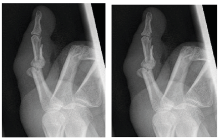
17-year-old male had a rollover car accident, presented with isolated little finger comminuted fractures proximal and middle phalanx involving the joint surface for each (Figure 1)
Figure 1:AP and Lateral views for little finger proximal and middle phalanx comminuted fracture.

Intraoperative
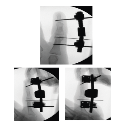
We decided to use External fixator to distract the fracture pieces with manual compression of the fractured phalanx to reduce the pieces under fluoroscopy.
a) Inserted the proximal pin first in the proximal phalanx shaft
b) Insertion of the distal pin in the distal phalanx (away from the fractured middle phalanx)
c) Xrays done to adjust the position and the depth of the pins
d) Insertion of the external fixator device on both pins.
e) Before tightening the clamps, application of distraction force done between the two pins by pulling the distal pin as maximum as possible with one hand and fixing the clamps using clamp drivers in the new position.
f) Xray is done shows great reduction of the fractured pieces
g) Reduction of the bicondylar proximal phalanx done using bone holder clamp
h) Insertion of transverse. 8mm k wire
i) Xray taken (below) (Figure 2)
Figure 2:

Discussion
Traction power can play a big role in complex comminuted fractures reduction. It should always be considered especially when other options are not that effective or have high morbidity.
References
© 2019 Ahmed Mokhtar Mahmoud Elbayer. This is an open access article distributed under the terms of the Creative Commons Attribution License , which permits unrestricted use, distribution, and build upon your work non-commercially.
 a Creative Commons Attribution 4.0 International License. Based on a work at www.crimsonpublishers.com.
Best viewed in


.jpg)


 Editorial Board Registrations
 Submit your Article
 Refer a Friend
 Advertise With Us
.jpg)


.jpg)


.bmp)
.jpg)
.png)
.jpg)


.jpg)


.png)

.png)


.png)


