- Submissions

Full Text
Orthopedic Research Online Journal
Metacarpal Swelling-Chondrosarcoma: A Rare Case Report
Kamudin NAF* and Abdul Yazid K
Department of Orthopedic, Pusat Perubatan Universiti Kebangsaan, Malaysia
*Corresponding author: Kamudin NAF, Department of Orthopedic, Pusat Perubatan Universiti Kebangsaan, Malaysia
Submission: November 03, 2017;Published: June 12, 2018

ISSN 2576-8875Volume3 Issue1
Introduction
Chondrosarcoma, a malignant bone tumour constitutes about 9% of primary bone malignancies [1]. Common in locations like proximal femur, proximal humerus, and pelvis, rarely it can occur in hand. We describe a patient with chondrosarcoma of the 5th metacarpal, a rare presentation of this tumour. To the best of our knowledge, only 13 cases (primary & secondary) are reported in literature so far [2].
Case Report
A 60-year-old male presented to our clinic with chief complaint of left hand swelling and pain for past 3 months. The swelling was gradually increasing in size, without any initial history of trauma. Patient had no constitutional symptoms. Clinically there was swelling over left hand over 5th metacarpal region with size of 3cm x 2cm, hard in consistency, non-mobile, not attached to skin and non-tender. Skin overlying was normal without any changes. Movement of the 5th metacarpophalengeal joint was full.
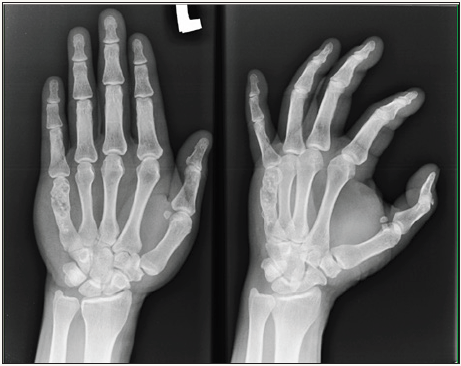
Radiograph of the left hand showed expansile sclerotic lesion over distal 3rd of 5th metacarpal with cortical destruction and increased soft tissue shadow, but no periosteal reaction. Magnetic resonance imaging (MRI) scan was done showed thinning cortex and endosteal scalloping of infection over distal aspect of 5th metacarpal with measurement of 0.3.5 x 1.1 x 1.5cm (AP x W x CC). The rest of bones show normal marrow signal. The visualized hand muscle and neurovascular bundle were normal. Computer Tomography Thoracic Abdomen Pelvis (CT TAP) revealed no evidence of metastasis elsewhere in the body (Figure 1).
Figure 1: Left hand x ray findings: lesion over proximal 5th metacarpal.

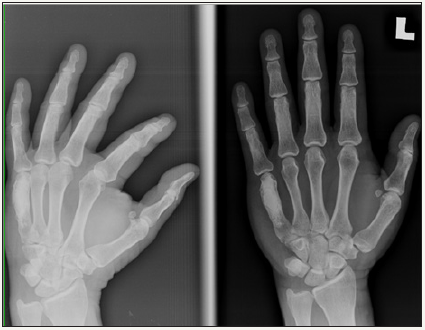
We proceed with bone curretage and cement of left 5th metacarpal. Bone biopsy was sent with findings showed atypical chondrocytes with nuclear pleomorphism and hyperchromatism, suggestive of low grade chondrosarcoma (Figure 2).
Figure 2: Left hand x ray after bone curettage and bone cement of left 5th metacarpal.

Patient then was discharge well and appointment was given to see for wound post operative. 6 months post-operative, MRI surveillance was done of left hand suggestive of residual lesion over proximal 5th metacarpal bone. However, clinically patient did not complaint of any pain and increasing in size of swelling. Thus, we decided for observation. Another MRI surveillance of left hand was done a year after showed no change of residual lesion over left proximal 5th metacarpal.
Discussion
Chondrosarcoma is a malignancy of mesenchymal origin with neoplastic cells in cartilaginous matrix [3]. It can be low-grade tumour of cartilage to high grade, it can be low risk of metastatic to aggressive tumour respectively [2]. Few types of chondrosarcoma have been reported such as conventional chondrosarcoma, dedifferentiated chondrosarcoma, clear cell chondrosarcoma, mesenchymal chondrosarcoma and juxtacortical chondrosarcoma. All examples above referred to primary chondrosarcoma, which a rise de-novo. Other type is secondary chondrosarcoma, which originate from pre-existing lesion of the cartilage [4].
Another way to classify this disease is by histological grading: grade 1(low grade), grade 2(intermediate) and grade 3 (high grade). The higher the grade the higher the risk of metastasis, with grade 2 10-15% and grade 3 more than 50% risk of metastasize [5]. It is difficult to differentiate between benign (enchondroma or osteochondroma) and malignant type (grade 1 chondrosarcoma) based on radiological and histological, as mentioned by Geirnaedt et al. [6] and Mirra et al. [7], thus depending more on interobserver variability.
Osseous chondrosarcoma commonly found in femur, humerus and pelvic. As in hand it is rare presentation. According to Wirbel et al. [5], chondrosarcoma of hand concluded of 0.5% of all cases related to chondrosarcoma, from these percentage 60% cases are located in phalanges and 40% are located in metacarpals. While Thanigai et al. [2] describe the radiological findings, lesion usually over metaphyseal region. The destruction of cortex usually unicortical and some periosteal reaction and intralesionalcalcification.
Surgical treatment can be intralesionalcurretage or wide local excision. Surgical treatment in low grade lesion of chondrosarcoma was choice of treatment in 60 to 85% cases as recorded by Ogose et al. [8], although recurrence might occur. In our case of low grade 1 of chondrosarcoma, intralesionalcurretage and bone cement was done. MRI surveillance repeated after 6 months and 1 year showed no new lesion.
Conclusion
Chondrosarcoma is common tumour that occurs over proximal long bones [2]. In our case reports we highlighted a rare presentation of left 5th metacarpal low-grade chondrosarcoma. This patient was treated with bone curettage and bone cement, and no recurrent of lesion on subsequent follow-up. This case report is to create awareness among doctors that such presentation of chondrosarcoma could happen and treatment with bone curettage with cement would be one of choices.
References
- Canale ST, Beaty J (2007) Campbell’s operative orthopaedics, (11th edn), 22: 907-910.
- Thanigai S, Douraiswami B (2012) Chondrosarcoma of metacarpal -A rare presentation. J Clin Orthop Trauma 4(2): 85-88.
- Stomeo D, Tulli A, Ziranu A, Mariotti F, Maccauro G (2014) Chondrosarcoma of the hand: A literature review. Journal of Cancer Therapy 5: 403-409.
- Gelderblom H, Hogendoorn PC, Dijkstra SD, Rijswijk CS, Krol AD, et al. (2008) The clinical approach towards Chondrosarcoma. Oncologist 13(3): 320-329.
- Wirbel RJ, Remberger K (1999) Conservative surgery for chondrosarcoma of the first metacarpal bone. Acta Orthop Belg 65(2): 226-229.
- Geirnaerdt MJA, Hermans J, Bloem JL, Kroon HM, Pope TL, et al. (1997) Usefulness of radiography in differentiating enchondroma from central grade 1 chondrosarcoma. AJR Am J Roentgenol 169: 1097-1104.
- Mirra JM, Gold R, Downs J, Eckardt JJ (1985) A new histologic approach to the differentiation of enchondroma and chondrosarcoma of the bones. A clinicopathologic analysis of 51 cases. Clin Orthop Relat Res (201): 214-237.
- Ogose A, Unni KK, Swee RG, May GK, Rowland CM, et al. (1997) Chondrosaroma of small bones of hands and feet. Cancer 80(1): 50-59.
© 2018 Kamudin NAF. This is an open access article distributed under the terms of the Creative Commons Attribution License , which permits unrestricted use, distribution, and build upon your work non-commercially.
 a Creative Commons Attribution 4.0 International License. Based on a work at www.crimsonpublishers.com.
Best viewed in


.jpg)


 Editorial Board Registrations
 Submit your Article
 Refer a Friend
 Advertise With Us
.jpg)


.jpg)


.bmp)
.jpg)
.png)
.jpg)


.jpg)


.png)

.png)


.png)


