- Submissions

Full Text
Orthoplastic Surgery & Orthopedic Care International Journal
Atraumatic Isolated Peroneal Compartment Syndrome
Mohamed Nagy1,2*, Neil Ashwood3,4, Veda Amara5 and Amit Kotecha6
1Trauma and Orthopedics Lecturer, Orthopaedic Surgery Department, Cairo University Kasr Alainy Faculty of Medicine, Egypt
2Trauma and Orthopaedics Department, Queens Burton Hospital, University Hospitals of Derby and Burton NHS Foundation Trust, UK
Honorary Professor, Research institute, University of Wolverhampton, UK
4Trauma and Orthopaedics Department, Trauma and Orthopaedics-Upper Limb Consultant, Queens Burton Hospital, University Hospitals of Derby and Burton NHS Foundation Trust, UK
5Senior Clinical Fellow, Trauma and Orthopaedics Department, University Hospitals Sussex NHS Foundation Trust, UK
6Trauma and Orthopaedics-Lower limb reconstruction Consultant, University Hospitals Birmingham, UK
*Corresponding author: Mohamed Nagy, Trauma and Orthopedics Lecturer, Cairo University Kasr Alainy Faculty of Medicine, Orthopaedic Surgery Department, Egypt and Trauma and Orthopaedics Department, Queens Burton Hospital, University Hospitals of Derby and Burton NHS Foundation Trust, UK
Submission: November 1, 2022;Published: November 11, 2022

ISSN 2578-0069Volume3 Issue1
Abstract
26-year-old male presented with leg lateral aspect pain with numbness over foot dorsum lateral aspect for 6 hours after rugby training with no trauma. Slight peroneal compartment tightness with negative stretch test. Creatine kinase 4659U/L. Peroneal compartment discomfort was worsening, fasciotomy of all leg compartments was done with only lateral peroneal compartment affected. Sensory changes in presentation highlights the importance of having a high index of suspicion. One could use biochemical markers aiding decision making on borderline situations, however we advise decompression in these cases. Although the patient had uneventful postoperative recovery, having ICP monitoring equipment or MRI would prevent overzealous opening of posterior compartment.
Abbreviations: Atraumatic; Isolated peroneal compartment; Compartment syndrome
Introduction
Acute Compartment Syndrome (CS) is a possibly overwhelming condition in which the pressure within an Osseo-fascial compartment rises to a level that diminishes the perfusion gradient across tissue capillary beds, driving to cellular anoxia, muscle ischemia, and necrosis. Diagnosis is essentially clinical, supplemented by compartment pressure measurements [1]. The clinical diagnosis of acute compartment syndrome is ordinarily made based on a classically known pentad of symptoms: pain, paresthesia, pallor, paralysis, and pulselessness [2]. Among the “5 P’s” of compartment syndrome, pallor, paralysis, and pulselessness are late findings [2]. It is known that pain with passive stretch and pain that is out of proportion to the injury are the earliest, most sensitive, and dependable findings in diagnosing compartment syndrome [3].
Compartment syndrome of the leg regularly influences whole leg compartments or the anterior compartment and can be traumatic or exertional (chronic) in nature [3]. Isolated lateral (peroneal) compartment syndrome of the leg is a less typical and lesser-known condition that can potentially miss the diagnosis if not considered in the workup of a painful leg [4]. This case report represents a case of atraumatic leg isolated lateral compartment syndrome. Consent for publishing is obtained from the patient.
Case Report
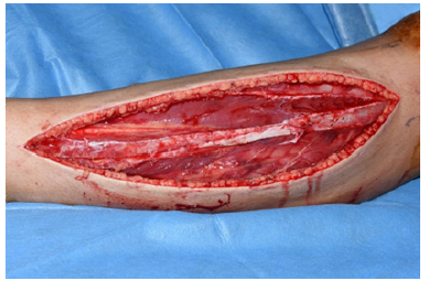
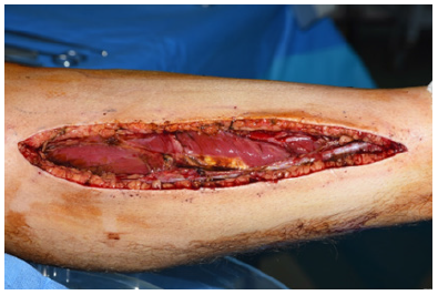
A 26-year-old male presented with niggle pain in his left leg lateral aspect over the last 6 hours to the Queens Burton Hospital Emergency Department. That started during rugby training giving no significant history of blunt injury or trauma. The pain was associated with numbness over the dorsum of the lateral aspect of the left foot. A week before, he had the same discomfort sensation, but it could be managed by NSAIDs, and he was able to carry on for the training. The patient had no significant past medical, surgical or allergy history. On presentation to hospital, the pain score was 5/10. That was associated with slight swelling and tightness of the peroneal compartment. The stretch test of different leg compartments was negative. The range of motion of the knee and ankle was normal, painless and full power against resistance. Distally, the limb was neurovascularly intact with no signs of infection. The radiographs of the leg were normal. Laboratory results were insignificant apart from obviously high Creatine Kinase (CK) level 4056U/L (trust lab normal range between 22 to 198U/L). Initially, in the emergency department, the patient was managed by analgesics, leg elevation and ice packs then admitted for close observation. The pain started to improve scoring 3. The decision was taken to hold analgesics in order not to mask the pain and other suggestive signs of compartment syndrome. Two hours late, the patient started to complain from increasing of the pain scoring 6/10. Also, the numbness included the distal lateral aspect of the leg. Examination showed that the planter-flexion with inversion, as a stretch test for the peroneal compartment, became positive. The CK increased to 4659U/L. Unfortunately, in our trust we didn’t have instruments to measure compartment pressure. The second consultant opinion was taken, and the decision for the surgical fasciotomy was taken. According to the BOAST guidelines, the patient was consented for 4 compartment decompression [5]. Intraoperatively, the anterolateral incision showed the muscles of the peroneal lateral compartments were dark, bruised and swollen. The muscles were bleeding and contracting on pinching indicating still viable. After a few minutes, the colour of the peroneal muscles improved (Figure 1). Muscles of the anterior compartment were normal (Figure 1). Posteromedial incision given to check for the posterior superficial and deep compartment muscles which were normal (Figure 2). The wounds were left open but approximated by shoelace technique. Postoperatively, on the day of surgery, the pain improved, the numbness subsided and the CK dropped to 2200U/L the day after. Re-exploration was done after 48 hours when the posteromedial skin incision was closed, but anterolateral skin incision closure seemed to be under tension and left approximated. Two days late, closure of the anterolateral wound was done free off tension.
Figure 1:Clinical photography of the antero-lateral incision showing the muscles of the peroneal lateral compartments after few minutes, the colour of the peroneal muscles improved with some residual bruises*. The peroneal muscles were dark, bruised and swollen once the incision was made, but unfortunately that photo is not available. Muscles of the anterior compartment were normal**.

Figure 2:Clinical photography of the posteromedial incision showed the posterior superficial and deep compartment muscles which were normal.

During hospital stay, the patient did well with gradual range of motion and partial weight bearing commenced. Sensations returned to normal after 48 hours post fasciotomy. CK level dropped to near normal 223U/L after closure of the wounds. He was discharged the next day of complete wound closure. The wounds were followed-up in 2 weeks for removal of sutures and were clean. A professional physiotherapist has taken over for gradual rehabilitation leading to complete return to his normal sports activities in 9 months.
Discussion
Compartment syndrome is an orthopedic emergency that can result from an assortment of causes, the foremost common being trauma [6]. Once in a while, it develops without any history of trauma, and several etiologies for atraumatic compartment syndrome have been portrayed; noteworthy strenuous activity, medical history of anticoagulants, bleeding disorders, vascular obstruction, medication injection, reported case of injection of insecticides for suicide and lengthy cardiac surgery [7]. The most common etiology for an atraumatic compartment syndrome is strenuous activity that may result in Chronic Exertional Compartment Syndrome (CECS). In CECS of the leg, the anterior and deep posterior compartments are mainly affected [4]. Conversely, there have been a few cases reported in the literature in which only the lateral compartment was involved. In most of these cases, there was significant strenuous activity or a concomitant traumatic event, such as an inversion injury or peroneal muscle rupture [8]. Our patient presented with a rare scenario in which symptoms and signs were isolated to the peroneal compartment without traumatic event. This combination of an atraumatic, possible exertional compartment syndrome isolated to the peroneal compartment is unusual and has been rarely reported..
Compartment syndrome with an unusual presentation can result in a delay in diagnosis and treatment; consequently, a high index of suspicion needs to be maintained in these cases [9]. Hope and McQueen verified that in cases without a fracture, there was a considerably greater delay to fasciotomy compared with those with a fracture. At the time of fasciotomy, 20% of patients without a fracture had muscle necrosis necessitating debridement compared with 8% of patients with a fracture [10]. Different methods of Intra- Compartmental Pressure (ICP) checking for the suspected acute compartment syndrome have been described in the literature. A review of 850 patients with acute CS showed that the sensitivity of ICP was 94%, with a valued specificity of 98%, an estimated positive predictive value of 93%, and an estimated negative predictive value of 99% [11]. While the compartment syndrome is a diagnosis of clinical suspension, some biomarker changes were found to be associated with the development of CS. In as study of 97 patients, 100% correlation was found between the diagnosis of CS and the CK level greater than 4,000U/L, maximal chloride level greater than 104 mg/dL, and minimal BUN level less than 10 mg/dL [12]. In our case, it was noticeable that the trend of the CK was rising from 4056 to 4659 preoperatively, then drop to 2200 the day after surgery and finally 223U/L at discharge. In addition, preoperatively, the chloride level was 104mg/dL, and the BUN was 1.85mg/dL which went with the above-mentioned correlation. Atraumatic bilateral acute compartment syndrome should always be included in the differential diagnosis for patients presenting with bilateral leg pain with no history of trauma, especially with cognitive impairment or mental health conditions. An elevated CK level can be a useful adjunct to guide the diagnosis in case of doubt, although this is not currently a part of UK guidelines. In addition, the use of ICP monitoring may avoid delayed diagnosis and treatment in cases where clinical signs are equivocal [13].
Radiology may play a role in such atypical cases of CS. MRI picture of acute compartment syndrome includes isolated involvement of the compartment and loss of normal muscle striations, micro-haemorrhages, and oedema of muscles within the affected compartment with peripheral convex bowing of the compartment margins [14]. In our case, the patient was admitted over the weekend with unfortunately no availability of MRI to be done. Early fasciotomy not only improves patient outcome but also is linked to decreased indemnity risk [2]. Management of compartment syndrome in the current era involves not only preventing the sequelae of a missed diagnosis but also lessening the risk of malpractice claima. Bhattacharyya and Vrahas described that increasing time from the start of symptoms to the fasciotomy was linearly coupled with an increased indemnity expense. A fasciotomy performed within 8 hours after the first presentation of symptoms was consistently associated with a successful defense [15].
Conclusion
This is rare case of isolated compartment involvement without trauma and sensory changes on presentation highlights the importance of having high index of suspicion to diagnose atypical cases of compartment syndrome. High level of creatine Kinase (more than 4000U/L) showed positive correlation with the diagnosis of compartment syndrome, so one could use biochemical markers to aid decision making on borderline situations. However, we would advise decompression in these cases, having the intracompartmental pressure monitoring equipment or MRI would prevent the overzealous opening of posterior compartment and prolong stay.
References
- Rubinstein AJ, Ahmed IH, Vosbikian MM (2018) Hand compartment syndrome. Hand Clin 34: 41-52.
- Cone J, Inaba K (2017) Lower extremity compartment syndrome. Trauma Surg Acute Care Open 2: e000094.
- Olson SA, Glasgow RR (2005) Acute compartment syndrome in lower extremity musculoskeletal trauma. JAAOS - Journal of the American Academy of Orthopaedic Surgeons 13(7): 436-44.
- Van Zantvoort APM, Hundscheid HPH, DeBruijn JA, Hoogeveen AR, Teijink JAW, et al. (2019) Isolated lateral chronic exertional compartment syndrome of the leg: A new entity? Orthop J Sports Med 7(12): 2325967119890105.
- BOAST diagnosis and management of compartment syndrome of the limbs.
- Bango J, Zhang E, Aaron DL, Diwan A (2020) Two cases of acute anterolateral compartment syndrome following inversion ankle injuries. Trauma Case Rep 30: 100371.
- Aydin A, Atakan MD, Topalan M, Tümerdem B, Erer M (2004) An unusual case of atraumatic compartment syndrome due to subcutaneous injection of insecticide for suicide attempt. Plast Reconstr Surg 113: 1302-1303.
- Zachariah S, Taylor L, Kealey D (2005) Isolated lateral compartment syndrome after weber C fracture dislocation of the ankle: a case report and literature review. Injury 36(2): 345-346.
- Livingston K, Glotzbecker M, Miller PE, Hresko MT, Hedequist D, et al. (2016) Pediatric nonfracture acute compartment syndrome: A review of 39 cases. Journal of Pediatric Orthopaedics 36(7): 685-690.
- Hope MJ, McQueen MM (2004) Acute compartment syndrome in the absence of fracture. Journal of Orthopaedic Trauma 18(4): 220-224.
- McQueen MM, Duckworth AD, Aitken SA, Court-Brown CM (2013) The estimated sensitivity and specificity of compartment pressure monitoring for acute compartment syndrome. Journal of Bone and Joint Surgery Am 95(8): 673–677.
- Valdez C, Schroeder E, Amdur R, Pascual J, Sarani B, (2013) Serum creatine kinase levels are associated with extremity compartment syndrome. Journal of Trauma and Acute Care Surgery 74(2): 441-447.
- Warren M, Dhillon G, Muscat J, Abdulkarim A (2021) Atraumatic bilateral acute compartment syndrome of the lower legs: A review of the literature. Cureus 13(12): e20256.
- Yılmaz TF, Toprak H, Bilsel K, Ozdemir H, Aralasmak A, et al. (2014) MRI findings in crural compartment syndrome: A case series. Emergency Radiology 21(1): 93–97.
- Bhattacharyya T, Vrahas MS (2004) The medical-legal aspects of compartment syndrome. J Bone Joint Surg Am 86(4): 864-868.
© 2022 Mohamed Nagy. This is an open access article distributed under the terms of the Creative Commons Attribution License , which permits unrestricted use, distribution, and build upon your work non-commercially.
 a Creative Commons Attribution 4.0 International License. Based on a work at www.crimsonpublishers.com.
Best viewed in


.jpg)


 Editorial Board Registrations
 Submit your Article
 Refer a Friend
 Advertise With Us
.jpg)


.jpg)


.bmp)
.jpg)
.png)
.jpg)


.jpg)


.png)

.png)


.png)


