- Submissions

Full Text
Orthoplastic Surgery & Orthopedic Care International Journal
Radial Club Hand, a Rare Congenital Abnormality: Report of Two Cases
Ashima Mahajan1* and Anshu Mahajan2
1 KIMS Karad, India
2 Department of neurosciences, Medanta the Medicity, India
*Corresponding author: Ashima Mahajan MBBS, MD Resident, Radiodiagnosis, KIMS Karad, India
Submission: March 16, 2018; Published: june 01, 2018

ISSN: 2578-0069Volume1 Issue5
Abstract
Congenital radial deficiency is an extremely rare congenital anomaly and its presentation may vary from mild hypoplasia to complete absence of radius. Club hand deformities are classified into two main categories radial and ulnar. Ulnar club hand is much less frequent than radial club hand. It is also known as radial club hand or radial dysplasia. We report two cases of rare congenital abnormality which can be helpful to the literature.
keywords Congenital radial deficiency (CRD); Limb anomaly; Radial club hand
Introduction
Congenital radial deficiency also known as radial club hand is a relatively rare congenital anomaly. Patient may present with variable degree of deficiency along the radial (or preaxial) side of the limb and is usually associated with absent thumb, thumb hypoplasia, thin first metacarpal and absent radius [1]. Ulnar club hand is much less frequent than radial club hand and ranges from mild deviation of hand on the ulnar side of forearm to complete absence of ulna. The estimated incidence of CRD is found to be 1 in 30,000 to 1 in 100,000 live births and most of the cases occurs sporadically with no known cause or associated with the syndrome [2-4].
Case 1
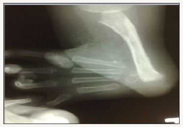
A 4-year-old male child was brought to the OPD with complaints of cough and fever for 2 days. There was a history of a deformed right upper limb since birth. He was a product of a non-consanguineous marriage and his perinatal history was uneventful (Figure 1). There was no family history of a similar deformity in the past two generations and his developmental history was normal for his age. There was no history of blood transfusion. The physical examination of the child revealed a shortened right forearm which was atrophied as compared to the opposite normal limb and he had a single forearm bone. There was limitation of movements of the right elbow flexion and extension including the rotatory movements of the forearm and the wrist. The systemic examination was normal. Evaluation of the radiographs of both the upper limbs revealed complete absence of the right radius and right first metacarpal bone. Chest X-ray, echocardiogram, hemogram including the platelet counts and ultrasound of the abdomen were normal.
Figure 1: X-ray right forearm with hand revealed complete absence of radius and thumb (type IV congenital radial deficiency).

Case 2
A 1year-old-male was referred from a peripheral health centre for evaluation of the congenital deformity of right upper limb. The baby was born to a 25-year-old primipara mother at term and his perinatal history was unremarkable. Physical examination of the baby revealed deformity of left forearm with radial deviation at the wrist and complete absent radius. Routine blood investigations were within normal limits. Plain radiographs of the forearm with hands showed complete absence of radius and thumb on left side (Figure 2). USG abdomen and echocardiography of the neonate did not reveal any significant finding. The parents were advised consultation with the physiotherapist and regular follow-up.
Figure 2: X-ray left forearm with hand revealed complete absence of radius and thumb (type IV congenital radial deficiency).

Discussion
CRD is classified into four types:
a. Type I is the mildest form of radial club hand. There is defective distal radial epiphysis with mild radial deviation of the hand. Thumb hypoplasia can be seen.
b. Type II shows limited growth of the radius on both the distal and proximal side. Miniature radius is seen with radial deviation of the wrist.
c. Type III there is partial absence of radius, most commonly of the distal two-third with severe
d. radial deviation of the wrist.
e. Type IV is the most common and most severe type. There is a complete absence of the radius [2,5].
Several theories were postulated, like maternal drug exposure, compression of the uterus and vascular injury, but the most accepted one relates the aetiology of the radial club hand to the Apical Ectodermal Ridge (AER). The AER is a thickened layer of ectoderm that directs differentiation of underlying mesenchymal tissue and limb formation. The extent of deformity is related to the degree and extent of AER absence.
About 40% of patients with unilateral radial club hand and 27% with bilateral radial club hand have associated congenital anomalies involving cardiac, renal, anal, skeletal and hematopoietic system [3]. The common syndromes associated with CLRD include Holt Oram syndrome, Thrombocytopenia absent radius (TAR) syndrome, Fanconi anemia and VACTERL (vertebral defects, anal atresia, cardiac defects, tracheo-esophageal fistula, renal anomalies, and limb abnormalities) syndrome [6-8]. Associated anomalies such as cardiac or gastrointestinal require early surgical management and are given preference. For isolated cases, treatment is conservative as well as surgical [9]. Principle guiding surgical management is wrist stabilization and later thumb reconstruction [10]. We present here two rare cases of radial club hand finding of which can be useful to the literature.
References
- Goldberg MJ, Meyn M (1976) The radial clubhand. Orthop Clin North Am 7(2): 341-359.
- Bayne LG, Klug MS (1987) Long-term review of the surgical treatment of radial deficiencies. J Hand Surg Am 12(2): 169-179.
- Wahab S, Ullah E, Khan RA, Sherwani MK (2009) Radial club hand-A case report and review of literature. Bombay Hosp J 51(1): 94-96.
- Phatak SV (2006) Radial club-hand: A case report. Indian J Radiol Imaging 16(4): 609-610.
- Waters MP (2001) The upper limb. In: Morrissy RT, Weinstein SL (Eds.), Lovell and Winters Pediatric Orthopaedics, (5th edn), Lippincott Williams and Wilkins, Philadelphia (PA), Pennsylvania, USA, pp. 841- 903.
- Omran A, Sahmoud S, Peng J, Ashhab U, Yin F (2012) Thrombocytopenia and absent radii (TAR) syndrome associated with bilateral congenital cataract: A case report. J Med Case Rep 6: 168.
- De Kerviler E, Guermazi A, Zagdanski AM, Gluckman E, Frija J (2000) The clinical and radiological features of Fanconi’s anaemia. Clin Radiol 55(5): 340-345.
- Oral A, Caner I, Yigiter M, Kantarci M, Olgun H, et al. (2012) Clinical characteristics of neonates with VACTERL association. Pediatr Int 54(3): 361-364.
- Maschke SD, Seitz W, Lawton J (2007) Radial longitudinal deficiency. J Am Acad Orthop Surg 15(1): 41-52.
- Fujiwara M, Nakamura Y, Nishimatsu H, Fukamizu H (2010) Strategic two-stage approach to radial club hand. J Hand Microsurg 2(1): 33-37.
© 2018 Ashima Mahajan. This is an open access article distributed under the terms of the Creative Commons Attribution License , which permits unrestricted use, distribution, and build upon your work non-commercially.
 a Creative Commons Attribution 4.0 International License. Based on a work at www.crimsonpublishers.com.
Best viewed in


.jpg)


 Editorial Board Registrations
 Submit your Article
 Refer a Friend
 Advertise With Us
.jpg)


.jpg)


.bmp)
.jpg)
.png)
.jpg)


.jpg)


.png)

.png)


.png)


