- Submissions

Full Text
Orthoplastic Surgery & Orthopedic Care International Journal
Giant Cell Tumour of Distal Radius Need and Deed
Bharath Raj R*, Prakash Kurtakoti and Prabhudev KB
Blossom Multispecialty Hospital, India
*Corresponding author: Bharath Raj R, Blossom Multispecialty Hospital, 853B, 26th main, BTM layout, Bangalore-560034, India
Submission: January 22, 2018;Published: March 19, 2018

ISSN: 2578-0069Volume1 Issue4
Abstract
Background: Tumours in any part of the body requires one adequate removal of the tumour to prevent recurrence and second to give functionality. In surgical management of giant cell tumor (GCT), technique requires balance between functionality of joint and excision to prevent recurrence.
Case report
History: A 25 year old female presented with swelling and pain in left wrist joint (16 months). Biopsy was diagnosed as giant cell tumour (distal radius). Pre-operatively no dorsi flexion, palmar flexion, pronation or supinations were possible with any ulnar or radial deviation possible at wrist joint.
Procedure: In toto resection of the tumour along with removal of the distal third radius with adequate margin was ensured. Ulna was osteotomised at level of where radius was cut and ulna was transposed onto radius. At 4 months follow-up, patient was pain free and grip strength of operated hand was 80%. At 1yr follow up showed union of the radius and the ulna at site of osteotomy and union at the level of the crapus. Confirmed by radiographs of wrist and forearm showed incorporation of ulnar graft distally and proximally with maintenance of wrist position. Patient was able to do all her daily activities with the forearm with minimal restriction in daily activity of inability to lift heavy weights.
Conclusion: The case is highlighted for the technique of using the ulna to create a single bone forearm thereby producing a favourable functional outcome and avoidance of complications
Keywords: GCT; Radius; Tumour; Giant cell tumour
Introduction
Giant cell tumour is a benign bone tumour which is locally aggressive in nature with high potential of recurrence. The tumour is one of low metastatic potential with the percentage of recurrence being close to 20-40%. The giant cell tumour arises most commonly in the proximal tibia and distal tibia followed by the distal radius which occurs in close to about 10% of the giant cell tumour population.
The giant cell tumour of the distal radius is usually seen in the middle aged group between 20 and 40 years who are the main bread earners of the family in a population. The need for them to obtain full functionality of the wrist is of utmost importance as the need for removing the tumour and preventing metastasis. Therefore a need for salvage of the hand is necessary and so an en bloc resection of the tumour of the distal radius with reconstruction of the wrist joint with either an allograft or autograft is needed to obtain adequate functionality.
In the past there have been many studies, which have explained the treatment options for distal end radius fracture-such as curettage of the tumour and treatment of the cavity with alcohol and hydrogen peroxide and use of a high speed burr or excision of the tumour with reconstruction of the wrist joint. Multiple studies have been reported stating reconstruction of the wrist joint with the fibula. Range of movements at the wrist joint have been achieved by this method, but keeping in the mind the chance of metastasis at the fibula graft site as well. In a new technique which has been recently documented is the transposition of the distal end of the ulna onto the proximal radius with fusion of the distal end of ulna onto the lunate.
Case Report
A 25 year old right dominant hand, house wife presented with history of swelling in left wrist joint since 16 months and pain since 15 months. The swelling initially was pea-sized and was gradually increasing till it reached the present state. The swelling of the wrist was associated with painful movements of the wrist and fingers. Open biopsy was done previously and giant cell tumour of distal radius was confirmed [1-5].
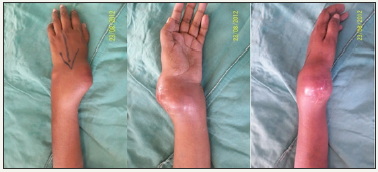
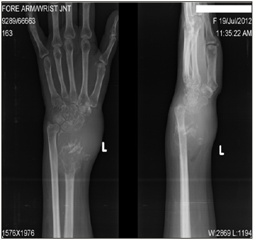
On examination, she had a swelling of nearly 8x10x15cms over dorsal and volar aspect of left wrist which was associated with warmth and tenderness. It was hard to firm in consistency. There was a scar on radial border of 5cms of left wrist which was consistent with the previous surgical scar mark of the open biopsy done before. Range of movements pre-operatively showed no dorsiflexion or palmar flexion possible and with no pronation or supination and no ulna deviation or radial deviation possible at the wrist joint (Figure 1). She was then investigated with an x-ray which showed osteolytic lesion in the distal radius metaphysis with thinned out edges with no evidence of destruction of articular cartilage (Figure 2). She was worked up for metastasis with bone scan which was negative and normal haemogram work up as per pre-operative protocol.
Figure 1: Range of movements pre-operatively showed no dorsiflexion or palmar flexion possible and with no pronation or supination and no ulna deviation or radial deviation possible at the wrist joint.

Figure 2: Range of movements pre-operatively showed no dorsiflexion or palmar flexion possible and with no pronation or supination and no ulna deviation or radial deviation possible at the wrist joint.

Procedure
Exposure
The left upper limb was painted and draped. Tourniquet was inflated up to 250mmHg. Incision was made from radial border of left forearm and was extended upto the 3rd metacarpal shafting in cooperating the previously made surgical scar for biopsy in it.
Resection
After careful dissection of the extensor retinaculum and the neurovascular bundle, the stretched extendons were separated and cut and the distal radius with the tumour was exposed circumferentially. The hand was dislocated surgically. An ostetotome was used and the radius was osteotomised 3cms away from the tumour margin which was calculated pre -operatively.
Reconstruction
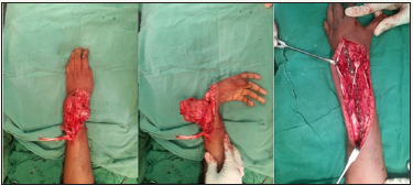
The ulna was osteotomised at the level of where the radius was cut and the ulna was transposed onto the radius. A favourable for wrist arthrodesis was obtained by nibbling on the lunate and the distal end of ulna so the cancellous bone ends are exposed. The ulna was then fixed to the 3rd metacarpal and the proximal third radius with an eighteen holed reconstruction plate and 3.5mm cortical screws. Alignment of the axis was checked under C-arm to see the axis from 3rd metacarpal to distal ulna to proximal third radial diaphysis. The extensor tendons were stitched back (Figure 3).
Figure 3: The extensor tendons were stitched back.

Post operative period
She was maintained on an above elbow slab immediately post operatively and after suture removal on the 14th post operative day was converted to an above elbow cast for a period of 1 month. Followed by a period of 1 month on a below elbow cast (Figure 4).
Figure 4: Followed by a period of 1 month on a below elbow cast.

According to histologic results, the surgical margins were free of disease. At 4 months follow-up, the patient was pain free and had started all her day to day activities. She had 30° of supination and 80° of pronation. The grip strength of the operated hand was 80% compared with the contralateral side. Radiographs of the wrist and forearm showed incorporation of the ulnar graft distally and proximally with maintenance of wrist position. At 1 yr follow up showed union of the radius and the ulna at site of osteotomy and union at the level of the crapus. Confirmed by radiographs of wrist and forearm showed incorporation of ulnar graft distally and proximally with maintenance of wrist position. Patient was able to do all her daily activities with the forearm with minimal restriction in daily activity of inability to lift heavy weights. At 2 years post operative now, she is symptom free with no recurrence and x-rays show good union of the ulnar graft proximally and distally.
Discussion
Treating a distal radius giant cell tumour is a big challenge in its way considering the extent of neuro vascular structures which pass through the wrist joint giving the functionality of the hand. This makes it essential to excise the tumour with utmost care but at the same time keeping in mind the high chance of local recurrence and distant metastasis. In this case it was a Campannaci Grade III tumour and with a size so large it was essential to assess the extent of tendons involved.
According to literature, it was noted that previously giant cell tumours of distal end of radius with a Campanacci Grade III tumour was associated with resection of the tumour and reconstruction of the distal end of radius with vascularised fibula graft. Later with advances in research it was found that non vascularised fibula grafts were used with bone iliac crest bone grafts and was found to produce the same functional result.
Wide resection with fibular grafting also caries the risk of complications related to the graft such as non union, graft fracture, residual subluxation of the carpus, degenerative oeteoathrosis, limited wrist movement and pain. There can also be donor site complications such as chronic leg pain, lateral ligament laxity, leg dysesthesia and foot drop. The rate of recurrence at the bone graft sites made orthopaedic surgeons look for alternatives for surgery.
With this new technique, the complications which were faced were that of non union at the junction of the proximal radius and distal radius intersection necessitating the need for a 2nd surgery for bone grafting. Arthrodesis with the help of external fixator in this case was replaced with a long eighteen holed reconstruction plate which incorporated the ulna metacarpal arthrodesis and the radius and ulna joint proximally. This helped avoid the pin tract and chances of pin tract infection. With the avoidance of bone grafting, the chance of inducing a recurrence at the graft site is also avoided.
Conclusion
With this new technique it is advantageous that the anatomic axis and functional outcome is achieved with minimal surgical technique and the chance of recurrence is reduced at a distal site with no bone graft being used. Therefore the need of a functional hand is obtained by the deed of making it a single bone forearm.
References
- Hussin P, Singh VA (2008) Giant cell tumor of distal radius: a case report and description of surgical technique. The Internet Journal of Orthopedic Surgery 8(2): 1-4.
- Panchwagh Y, Puri A, Agarwal M, Anchan C, Shah M (2007) Giant cell tumor-distal end radius: Do we know the answer? Indian J Orthop 41(2): 139-145.
- Chalidis BE, Dimitriou CG (2008) Modified ulnar translocation technique for the reconstruction of giant cell tumor of the distal radius. Orthopedics 31(6): 608.
- Sherbiny ME (2003) Replacement of the distal radius after resection of primary bone tumours using nonvascularised fibula proximal fibula graft. Journal of the Egyptian Nat Cancer Inst 15(2): 163-168.
- Bassiony AA (2009) Giant cell tumour of the distal radius: wide resection and reconstruction by non-vascularised proximal fibular autograft. Ann Acad Med Singapore 38(10): 900-904.
© 2018 Bharath Raj R. This is an open access article distributed under the terms of the Creative Commons Attribution License , which permits unrestricted use, distribution, and build upon your work non-commercially.
 a Creative Commons Attribution 4.0 International License. Based on a work at www.crimsonpublishers.com.
Best viewed in


.jpg)


 Editorial Board Registrations
 Submit your Article
 Refer a Friend
 Advertise With Us
.jpg)


.jpg)


.bmp)
.jpg)
.png)
.jpg)


.jpg)


.png)

.png)


.png)


