- Submissions

Full Text
Open Journal of Cardiology & Heart Diseases
Three and One Method (Yasser’s Method) to Overcome Streptokinase-Induced Hypotension in Acute Myocardial Infarction; 20-Case Report Series and Retrospective- Observational Study
Yasser Mohammed Hassanain Elsayed*
Critical Care Unit, Kafr El-Bateekh Central Hospital, Damietta Health Affairs, Egyptian Ministry of Health (MOH), Egypt
*Corresponding author: Yasser Mohammed Hassanain Elsayed, Critical Care Unit, Kafr El-Bateekh Central Hospital, Damietta Health Affairs, Egyptian Ministry of Health (MOH), Damietta, Egypt
Submission: January 17, 2023;Published: June 01, 2023

ISSN 2578-0204Volume4 Issue2
Abstract
Aim of the study: The study aimed to clarify how to overcome streptokinase-induced hypotension during
acute myocardial infarction intravenous infusion?
Background: Streptokinase is the cheapest approved thrombolytic agent. Streptokinase is commonly
associated with hypotension. The delay in giving a thrombolytic agent for acute myocardial infarction
may be hazardous.
Method of study and patients: My study was an observational-retrospective twenty-case report series.
The study was conducted in Fraskour Central Hospital and Kafr El-Bateekh Central Hospital. The author
reported twenty cases of confirmed acute myocardial infarction with indications for thrombolytic over
about 34 months, starting on October 5, 2018, ended on July 25, 2021. Testing for the probability of
hypotension during infusion of streptokinase was done for all cases. Three and One Method (Yasser’s
Method) was only applied in the cases of hypotension during streptokinase intravenous infusion.
Results: The mean age in the current study is 60.6 with male sex predominance (85%). Acute inferior
myocardial infarction is the most common (55%) infarction. Pre-testing for the probability of hypotension
during infusion of streptokinase was only applied in (50%) with equal positive probability and negative
probability test was (50%). Yasser’s Methods was applied in (75%) in response in (100%).
Conclusion: Three and One Method (Yasser’s Method) is an innovative clinical and therapeutic method
in cardiovascular science. The method is used in cases of acute myocardial infarction. It is indicated in
the cases of hypotension during the intravenous infusion of streptokinase. The Three and One Method
(Yasser’s Method) is effective, safe, and time saving for cases of acute myocardial infarction.
Keywords: Three and one method; Yasser’s Method; Streptokinase; Hypotension; Myocardial infarction; Ischemic heart disease; Thrombolytic
Abbreviations:ABG: Arterial Blood Gases; AMI: Acute Myocardial Infarction; BP: Blood Pressure; CAS: Coronary Artery Spasm; CBC: Complete Blood Count; CCBs: Ca2+ Channel Blockers; ECG: Electrocardiography; ED: Emergency Department; EF: Ejection Fraction; GCS: Glasgow Coma Scale; HR: Heart Rate; ICU: Intensive Care Unit; IHD: Ischemic Heart Disease; IVI: Intravenous Infusion; LBBB; Left Bundle Branch Block; MI: Myocardial Infarctions; NSR: Normal Sinus Rhythm; O2: Oxygen; PVCs: Premature Ventricular Contractions; RBBB: Right Bundle Branch Block; RBS: Random Blood Sugar; RR: Respiratory Rate; SCD: Sudden Cardiac Death; SK: Streptokinase; SK-hta: Streptokinase-Induced Hypotension; STEMI: ST-Elevation Myocardial Infarction; UA: Unstable Angina; VR: Ventricular Rate
Background
Introduction and historical bit
Streptokinase (SK) is traditional old thrombolytic therapy activating plasminogen by nonenzymatic mechanism [1]. FDA had approved the SK in the management of acute STSegment Elevation Myocardial Infarction (STEMI), arterial thrombosis or embolism, Deep Vein Thrombosis (DVT), Pulmonary Embolism (PE), and arteriovenous cannula occlusion [2]. The World Health Organization (WHO) mentioned SK in the “List of Essential Medicines” [3]. In the early 19th century, Acute Myocardial Infarction (AMI) was exceptionally considered a medical inquisitiveness [4]. The SK era dates back to 1933 when Dr. Tillett WS and Sherry S. revealed the development of SK and its application to acute coronary thrombosis [5]. An accidental discovery by Tillett WS in 1933, and later by his student Sherry S, considered the use of SK as a thrombolytic medication in the treatment of AMI. The drug was previously applied in the treatment of fibrinous pleural exudates, hemothorax, and Tuberculous (TB) meningitis [6]. In 1952, Johnson & Tillett [7] enabled the SK to lyses artificially induced vascular thrombus in the marginal ear veins of rabbits. In 1958, Sherry and colleagues [8] started to use SK in AMI and converted the directory of treatment from conservative to cure. Intravenous Infusion (IVI) streptokinase was used in primary trials but with contradictory results. In 1979, Rentrop and colleagues stated that the SK intracoronary infusion was a primary innovative approach. Subsequently, larger trials of intracoronary infusion achieved reperfusion rates ranging from 70% to 90% [6].
Significance and advantages
The cost of medical therapy for unpredictable principal events such as AMI is often a problem in countries with low income [6]. Randomized Clinical Trials (RCT) showed the role of SK in the reduction of mortality rate, particularly if the physician started the SK within 6 hours of symptom presentation [9,10]. In the US, statistically, over 50,000 deaths yearly are due to medication errors. The evidence-based medicine showed that non-bolus fibrinolytic therapy is accompanied by a 15% to 20% error rate with likely fatal events such as intracranial hemorrhage and Sudden Cardiac Death (SCD) [11]. Both published Oxford based ISIS 2, GISSI study, and the Netherlands Interuniversity study revealed that IVI of SK produced a significant reduction in early Mortality Rate (MR) post-MI [12]. However, AMI in Europe, the UK, and more than 2 billion in Asia will be lucky to the benefits of SK [13]. The GISSI study and ISIS- 2 showed that an IVI of 1.5mu of SK over about one hour is not exceptionally costly or worrisome to give customary [13]. Indeed, reperfusion could be established in between 55% and 80% of cases if SK is given in either intracoronary or IVI [14].
Administration
Streptokinase is administered intravenously at a dose of 1.5mu given over 1 hour [13,15]. Traditional administration of streptokinase is given via intravenous infusion, by diluting the vial 1,500,000 IU with add 5Ml NaCl- injection, or 5% dextrose dilution is given in 45ml of sodium chloride, then infuse it over 60 min [16]. Slowing of infusion due to hypotension may be required. So, close monitoring for Blood Pressure (BP) during the infusion is essential [17,18]. The rate of an IV infusion should be reduced regarding the degree of hypotension [18].
Advantages
Streptokinase is the cheapest approved thrombolytic agent [18,19]. Streptokinase is indicated for the management of AMI in adults aiming for the dissolution of intracoronary thrombi, the improvement of ventricular function, and the reduction of AMIassociated MR. This will occur if SK is given via either the IVI or the intracoronary route. Moreover, reduction of infarct size and Congestive Heart Failure (CHF) associated with AMI if SK will be administered by the IV route [16].
Patients and Methods
The author reported twenty cases of acute myocardial infarction. The study was conducted in both an observationalretrospective twenty-case report series. The study was conducted in Fraskour Central Hospital and Kafr El-Bateekh Central Hospital. The author reported twenty cases of acute myocardial infarction indicating thrombolytic over about 34 months, starting on October 5, 2018, and ending on July 25, 2021. The study is an observational retrospective case report series (Table 1). Males and females were included. The adult age is only involved. There was a different diagnosis for all cases. All selected cases were associated with acute myocardial Infarction. All cases are immediately exposed to traditional treatment of acute myocardial infarction according to the current international cardiovascular guidelines. All study cases were reported as sporadic case reports. All included cases were undergone to measure blood pressure on presentation or just before streptokinase intravenous infusion, during infusion, and after infusion. Testing for the probability of hypotension during infusion of streptokinase was done for all cases. “Three and One Method (Yasser’s Method)” was only applied in the cases of hypotension during streptokinase intravenous infusion. The serial ECG tracings were reported before and after streptokinase intravenous infusion. The initial ECG tracings were only provided. The troponin test and echocardiography were nearly requested for all cases. CBC, liver enzymes, RBS, renal function tests, and sometimes ABG, ionized calcium, were done as routine investigations in most cases. ABG and ionized calcium were done in selected cases. History, clinical data, testing for the probability of hypotension during infusion of streptokinase, and application of the “Three and One Method (Yasser’s Method)” to overcome streptokinase-induced hypotension were recorded. For more details, you can see (Table 2&3).
Table 1:shows remarks on the study method and data.
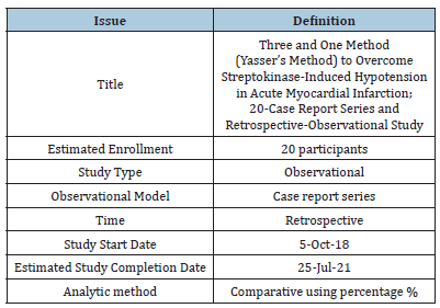
Table 2:Summary of the history, clinical, and management data for streptokinase-inducing hypotension; n:20.
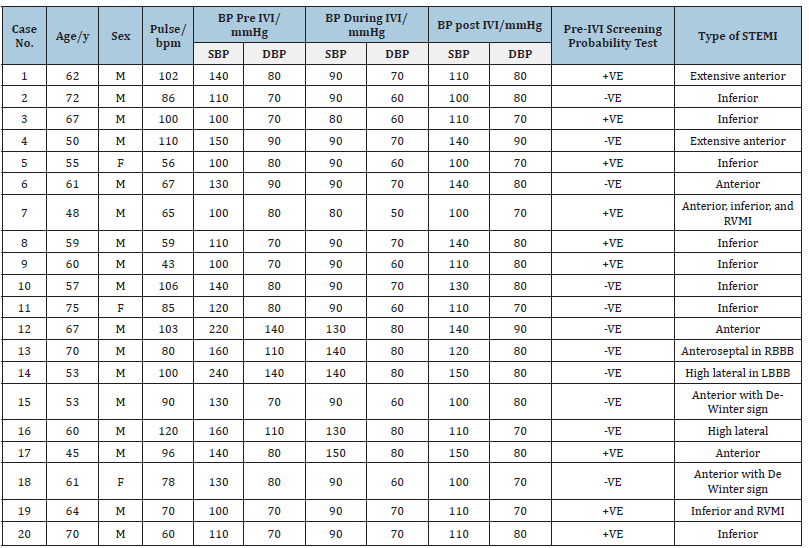
Abbreviations: BP: Blood Pressure; DBP: Diastolic Blood Pressure; F: Female; LBBB: Left Bundle Branch Block; M: Male; RBBB; Right Bundle Branch Block; RVMI: Right Ventricular Myocardial Infarction; SBP: Systolic Blood Pressure; STEMI: ST Segment Elevation Myocardial Infarction.
Table 3:Statistical summary of age, sex, types o MI, pre-test, Yasser’s Methods, and response; n:20.
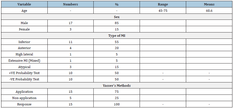
Abbreviations: MI: Myocardial Infarction.
Suggesting hypothesis and research objectives
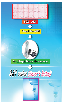
A. Suggesting hypothesis: How to overcome streptokinaseinduced hypotension during IVI in acute myocardial infarction? B. The research objectives are to clarify the efficacy of the “Three and One Method (Yasser’s Method)” to overcome streptokinase-induced hypotension during IVI acute myocardial infarction. The “Three and One Method (Yasser’s Method)” can be summarized in Figure 1.
Figure 1:The author’s diagrammatic presentation for Three and One Method (Yasser’s Method).
Eligibility criteria:
a) Inclusion criteria: All cases of acute myocardial infarction
indicate immediate thrombolytic.
b) Exclusion criteria: 1. Non-ST-elevation myocardial infarction
(non-STEMI)
c) Coronary artery spasms.
d) Left Bundle Branch Block (LBBB) with lower Sgarbossa
criteria.
e) Assessment of the study cases with pre-testing for the
probability of hypotension during infusion of streptokinase.
The response was done with the presence of either:
f) Positive Probability Test (+VE Probability); it indicates the
application of the “Three and One Method (Yasser’s Method)”
to overcome streptokinase-induced hypotension during IVI
acute myocardial infarction.
g) Negative Probability Test (-VE Probability); it does not indicate
application of the “Three and One Method (Yasser’s Method)”.
h) The study limitations were the absence of tilting test.
Cases Presentation
Acute extensive anterior MI with signs with Wavy double and Wavy triple signs of hypocalcemia (Yasser’s signs) post-sildenafil
A 62-year-old, Carpenter, cigarette smoker, Egyptian, married, male patient was admitted to the Intensive Care Unit (ICU) with acute severe anginal, compressible, intolerable chest pain within 2 hours of swallowing sildenafil tablet (25mg). Profuse sweating and facial flushing were the associated symptoms. Upon general physical examination, generally, the patient was anxious, severely sweaty, had hot extremities, with a regular Heart Rate (HR) of 102bpm, Blood Pressure (BP) of 140/80mmHg, Respiratory Rate (RR) of 16bpm, temperature of 37.1 °C, and pulse oximeter of O2 saturation of 96%. The measured random blood sugar (RBS) was 119mg/dl. The troponin test was positive (8ng/L). The echocardiographic report showed anterior hypokinetic segments with an ejection fraction (EF) of 51%. Three and One Method (Yasser’s Method) was applied (Figure 2).
Figure 2:Initial tracing showing sinus tachycardia (VR; 102) with extensive anterior MI (I, aVL, and V1-6) STsegment elevation myocardial infarction (red arrows) and reciprocal ST-depression changes in leads (II, III, and aVF; lime arrows). There is a Wavy double sign (orange and light blue arrows; V3 and V4) and a Wavy triple sign (orange, green, and light blue arrows; V6) of hypocalcemia (Yasser’s signs).

Acute inferior MI and old anterior MI in a diabetic with fronto-temporo-parietal ischemic cerebral infarction
A 72-year-old, retired Gov. officer, tobacco smoker, Egyptian, married, male patient was admitted to the ICU with acute severe anginal chest pain for 12 hours. Profuse sweating was the associated symptom. He is a diabetic on oral hypoglycemic tablets with a recent history of fronto-temporo-parietal ischemic infarction. Upon general physical examination, generally, the patient was anxious, severely sweaty, and had hot extremities, with a regular HR of 86 bpm, BP of 110/70mmHg, RR of 18 bpm, the temperature of 36.5 °C, and pulse oximeter of O2 saturation of 98%. The measured RBS was 256mg/dl. The troponin test was positive (77.54ng/L). The echocardiographic report showed inferior hypokinetic segments with an EF of 59%. The Three and One Method (Yasser’s Method) was applied (Figure 3).
Figure 3:Initial tracing showing normal sinus rhythm (VR; 82) with acute inferior MI (II, III, and aVF; red arrows) and reciprocal ST-depression changes in high lateral leads (I and aVL; lime arrows) leads. There is evidence of old anterior MI (pathological Q waves in V1-3, dark blue arrows).
Acute inferior myocardial infarction with RV involvement post-sildenafil
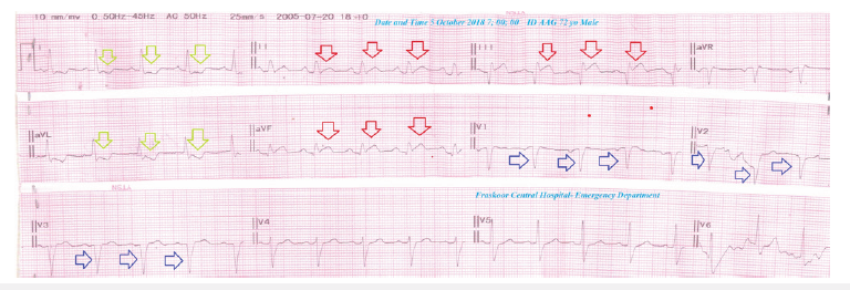
A 67-year-old, Driver, tobacco smoker, Egyptian, married, male patient was admitted to the ICU with acute severe anginal chest pain within 1 hour of swallowing of Sildenafil tablet (50mg). Profuse sweating and facial flushing were the associated symptoms. Upon general physical examination, generally, the patient was anxious, severely sweaty, and had hot extremities, with a regular HR of 100bpm, BP of 100/70mmHg, RR of 15bpm, a temperature of 37 °C, and pulse oximeter of O2 saturation of 96%. The measured RBS was 88mg/dl. The troponin test was positive (2.5ng/L). The echocardiographic report showed inferior hypokinetic segments with an EF of 59%. Three and One Method (Yasser’s Method) was applied (Figure 4).
Figure 4:Initial tracing showing sinus tachycardia (VR; 104) with acute inferior MI (II, III, and aVF; red and dark blue arrows) and reciprocal ST-depression changes in all anterior leads (I and aVL, and V1-6; lime arrows) leads. There is evidence of widespread tremor artifacts (small brown arrows).
Acute extensive anterior myocardial infarction with quadrigeminy PVCs
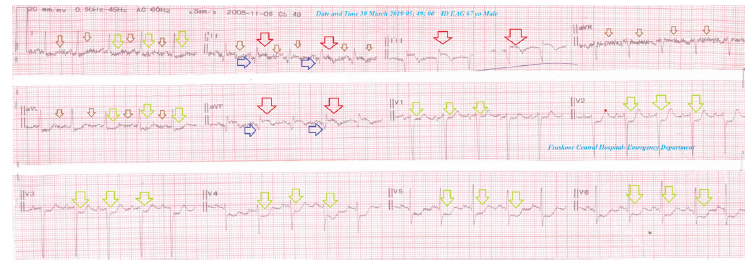
A 50-year-old, Worker, cigarette smoker, Egyptian, married, male patient was admitted to the ICU with acute severe anginal, chest pain for 6 hours. Profuse sweating and palpitations were the associated symptom. He gave a recent history of psyco-famolial stress. Upon general physical examination, generally, the patient was anxious, severely sweaty, and had hot extremities, with a regular HR of 110 bpm, BP of 150/90mmHg, RR of 14bpm, a temperature of 36 °C, and pulse oximeter of O2 saturation of 95%. The measured RBS was 70mg/dl. The troponin test was positive (13ng/L). The echocardiographic report showed anterior hypokinetic segments with an EF of 61%. Three and One Method (Yasser’s Method) was applied (Figure 5).
Figure 5:Initial tracing showing sinus tachycardia (VR; 110) with extensive anterior MI (I, aVL, and V1-6; red and blue arrows) and reciprocal ST-depression changes in leads (II, III, and aVF; lime arrows). There is evidence of quadrigeminy PVCs (orange arrows).
Acute inferior myocardial infarction with bradycardia
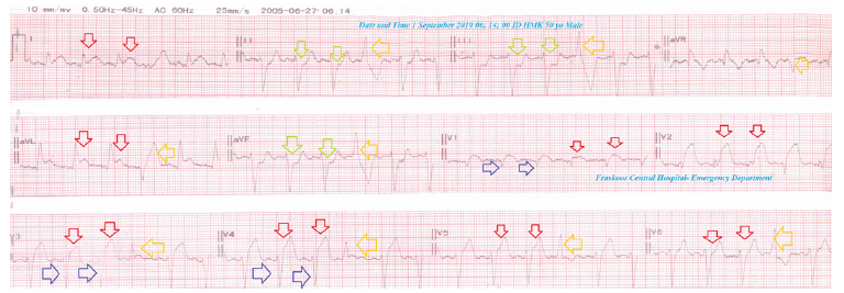
A 55-year-old, Housewife, Egyptian, married, a female patient was admitted to the ICU with acute severe anginal chest pain for 6 hours. Tachypnea and profuse sweating were the associated symptoms. She gave a recent history of psyco-famolial stress. Upon general physical examination, generally, the patient was anxious, severely sweaty, and had cold extremities, with a regular HR of 56bpm, BP of 100/80 mmHg, RR of 24bpm, a temperature of 36 °C, and pulse oximeter of O2 saturation of 93%. The measured RBS was 186mg/dl. The troponin test was positive (74.2ng/L). The echocardiographic report showed inferior hypokinetic segments with an EF of 42%. The Three and One Method (Yasser’s Method) was applied (Figure 6).
Figure 6:Initial tracing showing sinus bradycardia (VR; 58) with acute inferior MI (II, III, and aVF; red arrows) and reciprocal ST-depression changes in all anterior leads (I and andaVL; lime arrows) leads.

Acute anterior myocardial infarction with Wavy triple sign of hypocalcemia (Yasser’s sign) post-sildenafil
A 61-year-old, married, male Egyptian, Worker, the patient was admitted to the ICU with acute severe anginal chest pain within 1 hour of swallowing of sildenafil tablet (50mg). Dizziness and profuse sweating were the associated symptoms. Upon general physical examination, generally, the patient was anxious, severely sweaty, and had hot extremities, with a regular HR of 67 bpm, BP of 130/90mmHg, RR of 18 bpm, a temperature of 36.5 °C, and pulse oximeter of O2 saturation of 97%. The measured RBS was 90 mg/ dl. The troponin test was positive (3.4ng/L). The echocardiographic report showed anterior hypokinetic segments with an EF of 55%. Three and One Method (Yasser’s Method) was applied (Figure 7).
Figure 7:Initial tracing showing normal sinus rhythm (VR; 67) with anterior MI (V1-6; red arrows) and reciprocal ST-depression changes in leads (II and aVF; lime arrows). There is a Wavy triple sign (aVR, aVL, and aVF; red, green, and light blue arrows) of hypocalcemia (Yasser’s sign).

Extensive acute myocardial infarction (anterior, inferior, and RV) in a heavy smoker
A 48-year-old, married, male Egyptian, Carpenter, a heavy smoker, the patient was admitted to the ICU with acute severe anginal chest pain for 4 hours. Dizziness, tachypnea and profuse sweating were the associated symptoms. He gave a recent history of psyco-famolial stress. Upon general physical examination, generally, the patient was anxious, and severely sweaty, with a regular heart rate of 65 bpm, BP of 100/80mmHg, RR of 23bpm, a temperature of 37 °C and pulse oximeter of O2 saturation of 92%. The measured RBS was 113 mg/dl. The troponin test was positive (22ng/L). The echocardiographic report showed global hypokinesia with an EF of 40%. Three and One Method (Yasser’s Method) was applied (Figure 8).
Figure 8:Initial tracing showing normal sinus rhythm (VR; 65) with inferior MI (II, III, and aVF; red arrows) anterior MI (V2-6; red arrows) and reciprocal ST-depression changes in leads (I, aVR, and aVL; lime arrows).

Acute inferior myocardial infarction with bradycardia post-NSAIDs in a heavy smoker
A 59-year-old, married, male Egyptian, Farmer, a heavy smoker, the patient was admitted to the ICU with acute severe musculoskeletal chest pain for 3 hours. Dizziness, tachypnea, and profuse sweating were the associated symptoms. Upon general physical examination, generally, the patient was anxious, and severely sweaty, with a regular HR of 59bpm, BP of 110/70mmHg, RR of 25bpm, a temperature of 36 °C and pulse oximeter of O2 saturation of 94%. The measured RBS was 86 mg/dl. The troponin test was positive (7ng/L). The echocardiographic report showed inferior hypokinesia with an EF of 52%. Three and One Method (Yasser’s Method) was applied (Figure 9).
Figure 9:Initial tracing showing bradycardia (VR; 58) with inferior MI (II, III, and aVF; red arrows) and anterior reciprocal ST-depression changes in leads (I, aVLand V1-6; lime arrows).

Acute inferior myocardial infarction with severe bradycardia in a heavy smoker
A 60-year-old, married, male Egyptian, Accountant and heavy smoker, the patient was admitted to the ICU with acute severe chest pain for 3 hours. Dizziness, syncope, and profuse sweating were the associated symptoms. He gave a recent history of psyco-famolial stress. Upon general physical examination, generally, the patient was anxious, and severely sweaty, with a regular HR of 43bpm, blood pressure of 100/70mmHg, RR of 14bpm, a temperature of 36.7 °C, and pulse oximeter of O2 saturation of 93%. The measured RBS was 70mg/dl. The troponin test was positive (1.8ng/L). The echocardiographic report showed inferior hypokinesia with an EF of 47%. Three and One Method (Yasser’s Method) was applied (Figure 10).
Figure 10:Initial tracing showing severe bradycardia (VR; 43) with inferior MI (II, III, and aVF; red arrows) and reciprocal ST-depression changes in leads (I, aVR, and aVL; lime arrows).

Acute inferior myocardial infarction with Wavy triple sign of hypocalcemia (Yasser’s sign) post-sildenafil
A 57-year-old, married, male Egyptian, Tailor, and heavy smoker, the patient was admitted to the ICU with acute severe anginal chest pain within 1 hour of swallowing of sildenafil tablet (50mg). Dizziness, palpitations, and profuse sweating were the associated symptoms. Upon general physical examination, generally, the patient was anxious, and severely sweaty, and had hot extremities, with a regular HR of 106bpm, BP of 140/80mmHg, RR of 16bpm, a temperature of 37.2 °C, and pulse oximeter of O2 saturation of 96%. The measured RBS was 101mg/dl. The troponin test was positive (72.29ng/L). The echocardiographic report showed inferior hypokinesia with an EF of 65%. Three and One Method (Yasser’s Method) was applied (Figure 11).
Figure 11:Initial tracing showing sinus tachycardia (VR; 110) with inferior MI (II, III, and aVF; red arrows) and reciprocal ST-depression changes in leads (I and aVL; lime arrows). There is a Wavy triple sign (orange, green, and light blue arrows; V1) of hypocalcemia (Yasser’s sign).

Acute inferior myocardial infarction with uncontrolled diabetic
A 75-year-old, married, female, Housewife, a heavy smoker, and Egyptian, the patient was admitted to ICU with acute tachypnea within 12 hours. Dizziness and profuse sweating were the associated symptoms. Upon general physical examination, generally, the patient was anxious, and severely sweaty, and had hot extremities, with a regular HR of 85bpm, BP of 120/80mmHg, RR of 22bpm, a temperature of 36.5 °C, and pulse oximeter of O2 saturation of 94%. The measured RBS was 135 mg/dl. The troponin test was positive (16ng/L). The echocardiographic report showed inferior hypokinesia with an EF of 59%. Three and One Method (Yasser’s Method) was applied (Figure 12).
Figure 12:Initial tracing showing normal sinus rhythm (VR; 85) with inferior MI (II, III, and aVF; red arrows) and reciprocal ST-depression changes in leads (I, aVL, and V2-6; lime arrows).

Acute anterior myocardial infarction and hypertensive crises in a heavy smoker
A 67-year-old, married, male Egyptian, Worker and a heavy smoker, the patient was admitted to the ICU with acute severe angina chest pain and tachypnea within 3 hours. Headache, palpitation, dizziness and profuse sweating were the associated symptoms. Upon general physical examination, generally, the patient was irritable, anxious and severely sweaty, with a regular tachycardia of HR of 103bpm, BP of 220/140mmHg, RR of 28bpm, a temperature of 36 °C, and pulse oximeter of O2 saturation of 91%. The measured RBS was 197mg/dl. The troponin test was positive (4.2ng/L). The echocardiographic report showed Left Ventricular Hypertrophy (LVH) and anterior hypokinesia with an EF of 54%. Three and One Method (Yasser’s Method) wasn’t applied (Figure 13).
Figure 13:Initial tracing showed sinus tachypnea (VR; 103) with anterior MI (V1-6; red and green arrows) and reciprocal ST-depression changes in leads (II, III, and aVF; lime arrows). T-wave inversion is present in high lateral leads (I and aVL; dark blue arrows). There is evidence of anterior AC artifacts (small brown arrows).

Acute myocardial infarction in Right Bundle Branch Block (RBBB) with tri fascicular block
A 70-year-old married, retired, heavy smoker and Egyptian male patient presented to the ICU with acute severe angina chest pain. Profuse sweating and headache were associated symptoms. Upon general physical examination, generally, the patient was anxious, severely sweaty, and had cold extremities, with a regular HR of 80bpm, BP of 160/110mmHg, RR of 22bpm, a temperature of 36.7 °C, and pulse oximeter of O2 saturation of 95%. The measured RBS was 189mg/dL. The troponin test was positive (385ng/L). Later echocardiography was mild anteroseptal and lateral hypokinesia with an EF of 65%. The Three and One Method (Yasser’s Method) wasn’t applied (Figure 14).
Figure 14:Initial ECG tracing showing RBBB, ST-segment elevations in V1-3, I, and aVL (red arrows) with Q-wave (black arrows) in V1-3 leads, reciprocal ST-segment depressions in II, III, and aVF (green arrows) leads, LAD (lemon and purple arrows), prolonged PR-interval (green rectangles) lead II, sinus arrhythmia, and PVCs in V1-3(lemon arrows).

Developing high lateral STEMI in higher sgarbossa criteria with Left Bundle Branch Block (LBBB) and hypertensive emergency
hypertensive emergency A 53-year-old heavy smoker, Egyptian, married male, Worker patient presented to the ICU with acute angina chest pain, palpitations, rapid breathing, and dizziness. The patient had a recent history of psycho-familial problems. The patient used furosemide (40mg once daily) and captopril (25mg twice daily) for previous episodes of chest pain and hypertension, respectively. Upon examination, the patient appeared irritable, sweaty, anxious, and tachypneic. His vital signs were as follows: BP: 240/140mmHg, HR: 100/minute, temperature: 36.2 °C, RR: 36/min, initial pulse oximetry: 92%. The troponin test was positive, and RBS was 223mg/ dl on admission. Echocardiography then revealed anterolateral hypokinesia with an EF of 63%. Three and One Method (Yasser’s Method) wasn’t applied (Figure 15).
Figure 15:ECG tracing was done on serial tracings within 2 hours of admission to the ICU showing LBBB with new concordant ST elevation>5mm in high lateral leads (I and aVL; blue arrows), with reciprocal ST segment depression in inferior leads (II, III, and aVF; black arrows). Red arrows indicate discordant ST elevation>5mm (V2-4) (one of Sgarbossa criteria).

De winter sign as a precursor to acute myocardial infarction
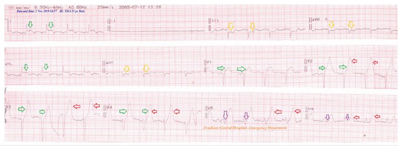
A 53-year-old male, heavy smoker, and Egyptian, Restaurant worker, a heavy smoker patient presented with acute severe angina chest pain and profuse sweating. The patient underwent coronary angiography due to angina during his stay in Kuwait three years ago. There was no detected abnormity. Upon general physical examination, the patient was anxious, and severely sweaty, with cold extremities, a regular HR of 90bpm, a BP of 130/70mmHg, a RR of 18bpm, a temperature of 36.4 °C, and a pulse oximeter of O2 saturation of 96%. The measured RBS was 134mg/dL. The troponin test was positive (271ng/L). Later echocardiography was mild anterior hypokinesia with an EF of 59%. Three and One Method (Yasser’s Method) was applied (Figure 16).
Figure 16:Initial ECG tracing was done in the ED showing tall, prominent, symmetrical T-waves in the V2-6 leads (red arrows), up-sloping ST-segment depressions in the V5-6 leads (purple arrows), ST-segment elevations in I, and aVL leads (green arrows), and reciprocal ST-segments depressions in III, aVF, and aVR leads (yellow arrows).
High lateral myocardial infarction with hypertension
A 60-year-old married, officer, heavy smoker, and Egyptian male the patient presented to the ICU with acute severe angina chest pain. Profuse sweating was the only associated symptom. Upon general physical examination, generally, the patient was anxious, severely sweaty, and had cold extremities, with an irregular HR of 120bpm, BP of 160/110mmHg, RR of 18 bpm, a temperature of 36.9 °C and pulse oximeter of O2 saturation of 97%. The measured RBS was 147mg/dl. The troponin test was positive (233ng/L). Later echocardiography was mild high lateral hypokinesia with an EF of 61%. The Three and One Method (Yasser’s Method) wasn’t applied (Figure 17).
Figure 17:Initial ECG tracing was done in the ICU showed high lateral (I, aVL, and V2) STEMI (green arrows) and reciprocal ST-depression changes in leads (III, aVF, aVR; purple arrows), multiple PVCs in V1-3 (blue arrows) and AF.

Sildenafil, tramadol, and hashish; the single or triple triggering cause of acute myocardial infarction with possible coronary spasm
A 45-year-old married male and Egyptian Worker patient presented to the ICU with acute severe agonizing ischemic chest pain. The patient gave a recent history of taking an oral sildenafil tablet (25mg), oral tramadol tablet (25mg), and two hashish cigarettes at a party since about 2 hours of the initial ECG tracing. Upon examination, the patient appeared distressed, sweaty, and anxious. His vital signs were as follows: BP of 140/80mmHg, HR of 96/minute; regular, temp. of 36.2 °C, RR of 16/min, and initial pulse oximetry of 98%. The initial emergency troponin I test was positive (76ng/l). RBS was 214mg/dl on admission. Later echocardiography showed inferior hypokinesia with an EF of 63 %. The Three and One Method (Yasser’s Method) wasn’t applied (Figure 18).
Figure 18:ECG tracing was done on the initial presentation of chest pain was done on ICU admission showing ST-segment elevations (red arrows) with pathological Q-waves (blue arrows) in inferior leads (II, III, and aVF), new ST-segment elevations in V1 and V3 (purple arrows), and reciprocal ST-segment depressions in I, aVL and V2 leads (lime arrows) of VR; 96 bpm.

Acute myocardial infarction with arrhythmogenic multiformed PVCs presentation developing inferior de-winter sign
A 61-year-old housewife, and Egyptian, married female the patient presented to the ICU with acute ischemic chest pain and palpitations for 2 days. Sweating and dizziness were associated symptoms. Currently, she gave a history of psycho-familial troubles. Upon general physical examination, generally, the patient was distressed and anxious, with a regular HR of VR; 78bpm, BP of 130/80mmHg, RR of 18bpm, a temperature of 36.7 °C and pulse oximeter of Oxygen (O2) saturation of 97%. The WBCs; 13 *103/ mm3. RBS; 143mg/dl. The troponin test was positive (16.8U/L). CK-MB was high (74U/L). The echocardiography was done on the presentation showing extensive anterior regional wall motion abnormalities and grade II LV diastolic dysfunction with an EF of 56%. The Three and One Method (Yasser’s Method) was applied (Figure 19).
Figure 19:ECG was done on serial tracings with double calibration within 80 minutes of the ICU admission and 30 minutes after starting streptokinase IVI showing acute anterior STEMI (red arrows) with a regular rhythm of VR of 92. Reciprocal ST-segment depression was seen in the inferior leads (II, III, and aVF leads; light blue arrows). There is a De-Winter sign in V4-6 leads (green arrows).

QRS-complex fragmentations with RV and inferior MI with wavy triple an ECG sign (yasser’s sign)
A 64-year-old, Farmer, heavy smoker and Egyptian, married, male the patient presented with acute severe angina chest pain. Profuse sweating, palpitations and dizziness were the only associated symptoms. Upon general physical examination, generally, the patient was anxious, severely sweaty, and had cold extremities, with a regular HR of 70bpm, BP of 100/70mmHg, RR of 22bpm, the temperature of 36.5 °C and pulse oximeter of O2 saturation of 95%. The measured RBS was 169mg/dl. The troponin test was positive (88ng/L). The initial echocardiography was done after hospital discharge within 4 days of the presentation showing LV dilation with both systolic (an EF of 49%;) and diastolic dysfunction, mild MR, mild TR, trivial AR, and PHT. The Three and One Method (Yasser’s Method) was applied (Figure 20).
Figure 20:ECG tracing was done on the ICU admission showing acute inferior STEMI (II, III, and aVF; blue arrows) and reciprocal ST-depression changes in leads (I, aVL, V2, and V3; orange arrows). There are QRS fragmentations in I, II, III, aVL, and aVF; small red arrows), a Wavy triple an ECG sign (Yasser’s sign; red, blue, and green arrows), and AC artifacts (small brown arrows).

COVID-19 inducing RV and inferior MI
A 70-year-old, heavy smoker, Farmer, Egyptian, married, male the patient was admitted to the ICU with acute severe angina chest pain. Profuse sweating, tachypnea, generalized body aches, and fatigue were the associated symptoms. He gave a history of fever, cough, and generalized body aches one week ago. There was a recent positive history of contact with a COVID-19 confirmed patient. Upon general physical examination, generally, the patient was anxious, severely sweaty and had cold extremities, with a regular HR of 60bpm, BP of 110/70mmHg, RR of 24bpm, a temperature of 37 °C, and pulse oximeter of O2 saturation of 93%. The measured RBS was 137mg/dl. The troponin test was positive (2.26ng/L). D-dimer was high (876ng/ml). CRP was high (28g/ dl). Ferritin was high (511ng/ml). LDH was high (593U/L). The echocardiographic report showed hypokinetic inferior and basal inferoseptal segments with mild to moderate MR, mild AR, mild TR, and grade I diastolic dysfunction with an EF of 60%. The Three and One Method (Yasser’s Method) was applied (Figure 21). For more details for all the study cases (Table 2).
Figure 21:Initial ECG tracing was done on the presentation in the ICU showing an acute inferior STEMI (II, III, and aVF; red arrows) and reciprocal ST-depression changes in leads (I, aVL, and V2-V5; blue arrows.

Age:Averages; Range; 45-75 years, Mean; 60.6, Minimal; 45 years, Maximal; 75 years (Table 2&3).
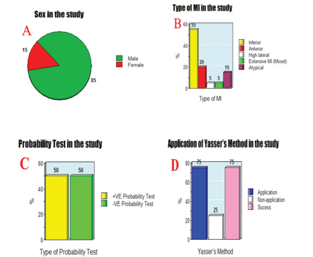
Sex:Male sex; 85% (17 cases), Female sex; 15% (3 cases) (Figure 22-A).
Type of MI:Inferior; 55% (11 cases), Anterior; 20% (4 cases), High lateral; 5% (1 case), Extensive MI (Mixed); 5% (1 case), Atypical; 15% (3 cases) (Table 3) (Figure 22-B).
Probability Test:Positive Probability Test (+VE Probability); 50% (10 cases), Negative Probability Test (-VE Probability); 50% (10 cases) (Table 3) (Figure 22-C).
Yasser’s Method:Application; 75% (15 cases), Nonapplication; 25% (5 cases) (Table 3) (Figure 22-D).
Response:Application; 100% (15 cases) (Table 3).
Figure 22:A-Pie chart presentation showing the percentage of Sex in the study. B- Bar chart presentation showing the types of MI in the study. C- Bar chart presentation showing the probability test in the study. D- Bar chart presentation showing the application of Yasser’s Method in the study.
Discussion
The mean age in the current study is 60.6 with male sex predominance (85%). Acute inferior myocardial infarction is the most common (55%) infarction. Pre-testing for the probability of hypotension during infusion of streptokinase was only applied in (50%) with equal positive probability and negative probability test was (50%). Yasser’s Methods was applied in (75%) in response in (100%).
Hypotension during infusion of streptokinase is the problem
Despite hypotension during infusion of streptokinase being problematic but there is no relevant studies. Only, just slow the rate of streptokinase IVI with monitoring of blood pressure. Also, there is no specific reported regimen, technique, or maneuver to overcome hypotension during infusion of streptokinase. Indeed, the most common side effects of streptokinase include nausea, headache, dizziness, hypotension, mild fever, bleeding from wounds or gums, rash, itching, flushing, muscle or bone pain, shivering, and allergic reactions [16]. Hypotension is the most common allergic reactions to streptokinase [20]. Allergic reactions and bleeding are common adverse events related to streptokinase. The GISSI study demonstrated a 3.4% incidence of minor bleeding. The ISSI- 2 study demonstrates a 0.4% incidence of significant bleeding. Patients may experience transient bradycardia or hypotension, with an incidence of 10%. Streptokinase is derived from bacterial proteins and thus can result in allergic reactions. Allergic reactions have been noted in up to 4.4% of patients and may present with fever, shivering, or rash. In rare cases, anaphylaxis may occur, which appears to be Ige mediated. Patients who develop anaphylactic signs and symptoms should promptly discontinue treatment and receive epinephrine [21,22]. Patients may experience transient bradycardia or hypotension, with an incidence of 10%2. Hypotension, sometimes severe, not secondary to bleeding or anaphylaxis has been observed during i.e., streptokinase infusion in 1 to 10% of patients [16]. Patients should be monitored closely and should symptomatic or alarming hypotension occur, appropriate treatment should be administered. This treatment may include a decrease in the IV streptokinase infusion rate. Smaller hypotensive effects are common [23]. Streptokinase-induced hypotension is a specific effect of this thrombolytic agent [24]. Despite a very high incidence (44.55%), the Streptokinase-Induced Hypotension (SKhTA) did not have a detrimental effect in treated patients with the acceleration of SK for STEMI [24]. Streptokinase can be rapidly administered without an increased risk [24]. Hypotension was more commonly noted in AMI patients given streptokinase. The Mean Arterial Blood Pressure (MAP) tends to decrease in the first 30 minutes after commencing the SK infusion. It is thus possible to conclude that the hypotension was at least partly due to SK and is probably a rate-related phenomenon [25]. When streptokinase is administered intravenously, a large dose is necessary to overcome antibody resistance [4,16]. The use of corticosteroids to avoid allergic reactions is no longer recommended [4,16].
Pre-testing for the probability of hypotension during infusion of streptokinase
A. Assessment of the study cases with pre-testing for the
probability of hypotension during streptokinase IVI is essential.
The target aimed to triage the suggested hypotension during
streptokinase IVI.
B. Probability of hypotension in the author’s opinion in pretesting
for the probability is meaning that borderline blood
pressure to more near hypotension such as 100/80, 100/70,
100/60, 110/70, 110/60, 110/80, etc.…
C. Hypertension or hypertensive crises is not needed to undergo
pre-testing for the probability of hypotension during infusion
of streptokinase such as 150/90, 180/100, 190/130, etc.…
D. Tolerable normal blood pressure is also not needed to undergo
pre-testing for the probability of hypotension during infusion
of streptokinase such as 140/80, 130/80, 120/80, etc.…
E. The response was done with the presence of either:
F. Positive Probability Test (+VE Probability); it indicates the
application of the “Three and One Method (Yasser’s Method)”
to overcome streptokinase-induced hypotension during IVI
acute myocardial infarction.
G. Negative Probability Test (-VE Probability); it does not indicate
application of the “Three and One Method (Yasser’s Method)”.
H. However, streptokinase is widely and still used in some
countries for acute myocardial infarction. So, if streptokinase
causes hypotension, it so difficult to use the alternative
thrombolytic because of their higher cost. Thus, the problem
should be solved.
Three and one method (Yasser’s method)
a) It is an innovative clinical and therapeutic method in
cardiovascular science.
b) The method is used in cases of acute myocardial infarction.
c) It is indicated in the cases of hypotension during the IV infusion
of streptokinase.
d) The method is effective in overcoming and passing the
hypotension during the IV infusion of streptokinase.
e) The Three and One Method (Yasser’s Method) is effective, safe,
and time-saving for cases of acute myocardial infarction.
f) Pre-testing application for the probability of hypotension
during infusion of streptokinase is essential.
g) Principal and the Method: This method is based on two points.
h) Stoppage of streptokinase IV infusion for 3 minutes (Three) if
there is hypotension happened.
i) Start again of streptokinase IV infusion and let it to the rapid
infusion (or run flow) for only one minute (One).
j) After the end of the last infusion minute the blood pressure
starts to be low.
k) So, stoppage of streptokinase IV infusion for 3 minutes (Three)
as above
l) Repeat the above steps till the end of infusion.
m) Continuing blood pressure monitoring during streptokinase IV
infusion is essential.
n) Time accuracy for both (Three) and (One) is needed.
Interpretation
A. Hypotension during the IV infusion of streptokinase. So,
three minutes (Three) of stoppage of the infusion will be
enough to spontaneous recovery blood pressure or circulatory
compensation to the normal level (Resting phase)
B. Rapid infusion (or run flow) for only one minute (One) is
enough to start to produce hypotension (Acceleration phase)
C. Streptokinase can be rapidly administered without an
increased risk [24].
D. The streptokinase-induced hypotension (SK-hTA) did not have
a detrimental effect [24].
E. Disadvantages:
F. There are no known reported drawbacks to the method.
Conclusion and Recommendation
a) Three and One Method (Yasser’s Method) is an innovative
clinical and therapeutic method in cardiovascular science.
b) The method is used in cases of acute myocardial infarction. It
is indicated in the cases of hypotension during the intravenous
infusion of streptokinase.
c) Three and One Method (Yasser’s Method) is effective, safe, and
time saving for cases of acute myocardial infarction.
d) Widening the research for use of the “Three and One Method
(Yasser’s Method)” in the cases of hypotension during the
intravenous infusion of streptokinase will be recommended.
Acknowledgment
I wish to thank Dr. Ameer Mekkawy (M.sc.) and Eng. Ahmed Alghobary (B.sc.) for their technical support. I hope to thank the team nurses of both emergency and critical care departments in Fraskour Central Hospital and Kafr El-Bateekh Central Hospital who make extra ECG copy for helping me. Also, I want to thank my wife for saving time and improving the conditions for supporting me.
References
- Jan M, Martin T, David B, Jiri D (2019) Structural biology and protein engineering of thrombolytics. Computational and Structural Biotechnology Journal 17: 917-938.
- Edwards Z, Nagalli S (2022) Streptokinase. Treasure Island, Robert LS.
- (2019) World Health Organization model list of essential medicines: 21st list 2019. World Health Organization, Geneva.
- Braunwald E (1998) Evolution of the management of acute myocardial infarction: A 20th century saga. Lancet 352(9142): 1771-1774.
- Sherry S (1981) Personal reflections on the development of thrombolytic therapy and its application to acute coronary thrombosis. Am Heart J 102(6 Pt 2): 1134-1138.
- Sikri N, Bardia A (2007) A history of streptokinase use in acute myocardial infarction. Tex Heart Inst J 34(3): 318-327.
- Johnson AJ, Tillett WS (1952) The lysis in rabbits of intravascular blood clots by the streptococcal fibrinolytic system (streptokinase). J Exp Med 95(5): 449-464.
- Sherry S, Titchener A, Gottesman L, Wasserman P, Troll W (1954) The enzymatic dissolution of experimental arterial thrombi in the dog by trypsin, chymotrypsin and plasminogen activators. J Clin Invest 33(10): 1303-1313.
- Becker RC, Spencer FA (2006) Fibrinolytic and antithrombotic therapy: Theory, practice, and management 2nd (edn), Fibrinolytic Agents, 59-75.
- (1986) Effectiveness of intravenous thrombolytic treatment in acute myocardial infarction. Gruppo Italiano per lo Studio della Streptochinasi nell'Infarto Miocardico (GISSI). Lancet 1(8478): 397-402.
- Gurwitz JH, Gore JM, Goldberg RJ, Barron HV, Breen T, et al. (1998) Risk for intracranial hemorrhage after tissue plasminogen activator treatment for acute myocardial infarction, participants in the national registry of myocardial infarction 2. Ann Intern Med 129(8): 597-604.
- Oldershaw P (1988) Thrombolytic therapy in acute myocardial infarction. Postgraduate Medical Journal 64(758): 915-918.
- Khan MG (2007) Cardiac Drug Therapy Thrombolytic Therapy. 7th (edn), Humana Press, pp. 189-190.
- Yusuf S, Collins R, Peto R, Furberg C, Stampfer MJ, et al. (1985) Intravenous and intracoronary fibrinolytic therapy in acute myocardial infarction: Overview of results on mortality, reinfarction and side-effects from 33 randomized controlled trials. Eur Heart J 6(7): 556-585.
- Maskell NA, Christopher WHD, Andrew JN, Emma LH, Fergus VG, et al. (2005) UK Controlled trial of intrapleural streptokinase for pleural infection. N Engl J Med. 352(9): 865-874.
- (2021) Streptase. Rxlist.
- Menyar ELAA, Omar MA, Mohamed MG, Wafer D, Ali AA, et al. (2006) Clinical and biochemical predictors affect the choice and the short-term outcomes of different thrombolytic agents in acute myocardial infarction. Coron Artery Dis 17(5): 431-437.
- Antman EM, Daniel TA, Paul WA, Eric RB, Lee AG, et al. (2004) ACC/AHA guidelines for the management of patients with ST-elevation myocardial infarction-executive summary: A report of the American college of cardiology/American heart association task force on practice guidelines (writing committee to revise the 1999 guidelines for the management of patients with acute myocardial infarction). J Am Coll Cardiol 44(3): 671-719.
- Elliott M (2007) ST-elevation myocardial infarction; Management antman, Braunwald's Heart Disease: A Textbook of Cardiovascular Medicine, 8th (edn) 51:1249.
- Ryan TJ, Antman EM, Brooks NH, Califf RM, Hillis LD, et al. (1999) ACC/AHA guidelines for the management of patients with acute myocardial infarction: A report of the American college of cardiology/American heart association task force on practice guidelines (committee on management of acute myocardial infarction). J Am Coll Cardiol 34(3): 890-911.
- Goa KL, Henwood JM, Stolz JF, Langley MS, Clissold SP (1990) Intravenous streptokinase, A reappraisal of its therapeutic use in acute myocardial infarction. Drugs 39(5): 693-719.
- Tisdale JE, Stringer KA, Antalek M, Matthews GE (1989) Streptokinase-induced anaphylaxis. DICP 23(12): 984-987.
- Streptase® (Streptokinase Injection). Product Monograph, CSL Behring Canada, Inc.
- Tatu CG, Cristina T, Monica D, Manuela G, Căpraru P, et al. (2004) Streptokinase-induced hypotension has no detrimental effect on patients with thrombolytic treatment for acute myocardial infarction. A substudy of the Romanian study for accelerated streptokinase in acute myocardial infarction (ask-Romania). Rom J Intern Med 42(3): 557-573.
- Lateef F, Anantharaman V (2000) Hypotension in acute myocardial infarction patients given streptokinase. Singapore Med J 41(4): 172-176.
© 2023 Yasser Mohammed Hassanain Elsayed. This is an open access article distributed under the terms of the Creative Commons Attribution License , which permits unrestricted use, distribution, and build upon your work non-commercially.
 a Creative Commons Attribution 4.0 International License. Based on a work at www.crimsonpublishers.com.
Best viewed in


.jpg)


 Editorial Board Registrations
 Submit your Article
 Refer a Friend
 Advertise With Us
.jpg)


.jpg)


.bmp)
.jpg)
.png)
.jpg)


.jpg)


.png)

.png)


.png)


