- Submissions

Full Text
Open Journal of Cardiology & Heart Diseases
The Rare Case of Family Hereditary Disease Recurrent Myxoma of the Heart
Abdumadzhidov Kh A1*, Uraqov Sh T1, Buranov Kh Zh1 and Isomitdinov B Sh1
1Republican Specialized Scientific and Practical Medical Center for Surgery, Bukhara State Medical Institute, Uzbekistan
*Corresponding author: Abdumadzhidov KH A, Republican Specialized Scientific and Practical Medical Center for Surgery, Bukhara State Medical Institute, Uzbekistan
Submission: July 01, 2022;Published: November 30, 2022

ISSN 2578-0204Volume4 Issue1
Introduction
Myxoma of the heart is a primary benign tumor that mainly affects the chambers of the heart, most often occurs in the Left Atrium (LA). The frequency of mixomas, papillary fibroelastoma, rhabdomyomas is 0.0017-0.02% in the structure of cardiac pathology. According to the literature, myxomas account for up to 50% of all benign tumors of the heart, among which, in addition to myxoma, there are lipomas, fibromas, hemangiomas, papillary fibroelastomas, and rhabdomyomas. F. Gerbode summarized the literature in 1978 and noted that myxomas are found in more than 1/3 of cases among 600 different heart tumors. According to the histological structure, myxomas are classified as benign tumors, however, according to the clinical course, due to frequent complications (embolism, circulatory failure, sudden death due to obstruction of the blood inflow and outflow tracts), they are malignant. Myxomas are more often (75%) localized in the LA than in the right atrium (RA) - 15-20%, much less often in the ventricles of the heart and simultaneously on both sides of the interatrial septum (biatrial myxoma). Most mixomas are isolated, but in 7% of cases they are an integral part of hereditary syndromes with autosomal dominant inheritance. In our practice, there was a case of familial myxomatosis of the heart with repeated recurrence of the tumor. In the specialized literature.
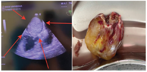
Figure 1&2:Giant LA myxoma obturates the mitral orifice.

Isolated cases of repeated operations for tumor recurrence, but the family variant of the disease with repeated recurrence of the disease, we think, is of interest to cardiosurgeons involved in this pathology [1-3]. In our practice, there was a case of a family disease with myxoma of the heart: the father of the family, a 48-year-old man, was the first to apply, he was admitted to the cardio surgical department in a serious condition, with obvious signs of hemodynamic disturbances, circulatory decompensation, complicated by anasarca, ascites, bilateral exudative pleuritites, severe edema in the lower extremities. The state at admission was assessed as extremely serious, he was hospitalized in the intensive care unit, where a large tumor was diagnosed, occupying almost the entire LA cavity, with obstruction in the left atrioventricular hole (Figure 1 & 2), creating a congestion of the pulmonary circulation, pre- edema of the lungs, high pulmonary hypertension with moderate tricuspid insufficiency.
Creating a congestion of the pulmonary circulation, pre-edema of the lungs, high pulmonary hypertension with moderate tricuspid insufficiency. It is decided to operate the patient according to vital urgent indications. The next morning the patient was urgently operated on. A huge LA myxoma obstructing the mitral orifice was removed [4]. After sternotomy, opening of the pericardium and cannulation of the aorta, vena cava, artificial sirculation was started, cardioplegia to the aortic root - antegrade, asystole. Opened RA. Significant stress is determined in the cavity of the LA, RA and raight ventriculum. The oval hole is also significantly tense, expanded, the latter is opened longitudinally. By revision, a large myxoma was established, a jelly-like structure, similar in appearance to bunches of grapes, partly obturates the mitral orifice significantly, during removal, a palm technique was used, which facilitated the removal of a huge tumor without practical fragmentation. The size of the tumor is specified on the table. 12x8cm, mixed consistency, jellylike, dark crimson color, really looks like bunches of grapes.
The base of the tumor is the oval fossa of the atrial septum, wide, about 2 cm in diameter, carefully excised, treated with betadine, the cavities were washed with saline, the revision revealed the absence of other pathologies, the fibrous ring of the mitral valve was somewhat enlarged, the hydraulic test showed the competence of the mitral valve. The atrial septum was sutured with a two-row blanket suture. The revision of the tricuspid valve revealed the absence of organic changes, the absence of morphological insufficiency, the valve was assessed as competent. Warming, suturing the wall of the RA with a double-row suture. Prevention of aeroembolism, the clamp was removed from the aorta. Cardiac activity recovered on its own. Myocardial electrode on the right ventricle. After stabilization of hemodynamics, a phased exit with artificial circulation. About 2 liters of exudate was evacuated from the pleural cavities. Recovery of the wound of the sternum - approximation of the sternum, layerby- layer suturing of the wound. Aseptic sticker. After stabilization of hemodynamics with medication, the patient was transferred to the intensive care department.
Within 12 hours, intensive cardiac therapy was carried out, the restoration of consciousness, the functions of other organs, the patient was extubated in a stable condition. The next day, the patient was transferred to the department, planned therapy for 9 days, the patient is discharged in a stable condition, further treatment at the place of residence in the Andijan Cardiovascular Surgery Clinic. After 5 years, the patient again comes to us, to the cardiosurgical department, again in a serious condition, the clinical picture is almost similar to the first admission. In the Andijan clinic, a recurrent Myxoma was diagnosed, but with a completely different localization. A tumor was found in the RA, in the area of the dome of the RA, in the right atrioventricular hole with obstruction of the blood flow, further visualization is difficult due to the severity of the patient’s condition. Clinically, a severe degree of heart failure, circulatory decompensation, the patient is stretcher, passive, in anasarcoma, edema of heart failure, ascites, bilateral pleuritites, etc.
Urgent preparation for reoperation in the intensive care unit, intensive care unit. The next day, the patient was again reoperated according to vital indications [4]. Under conditions of artificial circulation and cardioplegia, a second operation was performed - removal of the RA mixoma (dome: tricuspid valve). Antegrade cardioplegia. Asystole. Opened RA. Revision: a tumor with several fragments was found - in the region of the oval window on the right, a large tumor descending into the tricuspid valve, obturating it, continuing into the right ventricle, 13x9 cm in size, mixed consistency, like a bunch of grapes, dark red, the second tumor is located on RA dome, apparently applique, 6x5 cm in size, identical consistency, tightly fixed on the PP dome. The tumors were removed step by step: the tumor of the foramen ovale on the right, TC, and RVOT were removed first. The base of the tumor is about 2.5 cm, with excision of the foramen ovale of the MPP. Removal of the second tumor - the RA dome, with excision of a part of the RA dome.
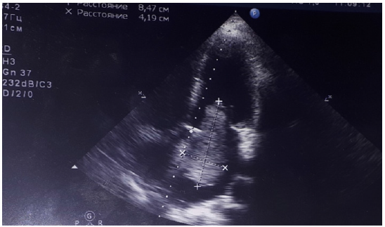
Figure 3:Myxoma LA, 5x4 cm in size, partially obturates the mitral orifice.

The audit established the absence of other pathologies, the TC is competent, the MPP was restored by applying an autopericardial patch 3x3 cm, the dome of the PP was sutured with a double-row twist suture, the tightness was restored. Treatment of tumor bases with betadine, alcohol. Washing. Sewing up the walls of the PP with a two-row seam. The tightness has been checked. Warming up to 37gr. Triple air embolism prophylaxis, aortic clamp removed. Cardiac activity recovered on its own. Postoperative course with symptoms of heart failure, the latter was successfully stopped by medical means within 10 days. The patient is discharged in a relatively stable condition, with a recommendation to continue conservative therapy at the place of residence in the Andijan clinic. After 10 years, a 26-year-old patient is admitted to the clinic of the Andijan Medical Institute, with a diagnosis of LA myxoma in a typical place (oval window), of medium size - 6x4 cm, grapeshaped, partially obturating the mitral orifice (Figure 3), there is no high pulmonary hypertension, the patient in a stable condition was operated on there in a planned manner. Removal of myxoma LA (oval window) 5x4 cm in size, soft jelly-like consistency was performed, the tumor was removed without technical difficulties, without fragmentation
The region of the base of the tumor was discharged in a satisfactory condition after a week. It was treated with betadine, alcohol, and washed. No other pathologies were identified. The operation was completed without technical problems, the patient was discharged home in a satisfactory condition after 7 days. The problem of this case is that after 2 years the same patient went to the clinic of the Andijan Medical Institute, where she had previously been operated on - the removal of the LA myxoma was performed under the conditions of EC and CP. The LA myxoma from the foramen ovale was again diagnosed with partial obstruction of the mitral orifice. The size of the myxoma is 4x5 cm. The patient had no signs of circulatory decompensation, however, the progression of the tumor size over several months, partial obstruction of the mitral orifice, the risk of tumor fragmentation, were indications for surgery. The patient was referred to our clinic 3 months later. observations. Examination reveals the growth of the tumor, the dimensions are somewhat larger: the tumor is 5x6 cm, partially obturated the left atrioventricular orifice, the base of the tumor was in the region of the oval fossa in the LA.
Taking into account the negative dynamics of tumor growth, obstruction of the mitral orifice with moderate (II) degree of pulmonary hypertension, the possibility of tumor fragmentation, the patient was offered a second operation - repeated removal of the tumor recurrence under the conditions of artificial circulation, cardiac plegia. The patient and relatives agreed to reoperation. After appropriate preparation in a planned manner, the patient was reoperated. Repeated removal of the tumor from the oval window of the LA was performed under the conditions of artificial circulation, cardiac plegia [4]. During reoperation, the size of the tumor corresponded to the ECHO cardiograph data, 5x6 cm, it partially obturated the mitral hole, it was storm-shaped, jelly-like, dark brown in color, it was removed without technical difficulties with excision of the tumor base (1.5 cm on the ventricular septum), treatment with betadine, alcohol 95 %, washing of cavities. During the revision, no other pathologies were found. The operation was completed without technical problems. The patient was transferred to the intensive care unit in stable condition. 6 hours after the operation, the patient was extubated, the postoperative course was uneventful. The patient was discharged home on the 8th day.
The interest in this case is mixed in that these 2 patients (a 48-year-old man and a 26-year-old girl) are close relatives - father and daughter, from the same family. There is not only a similarity of diagnoses, but also the course of the postoperative period, in both cases a recurrence of the tumor was detected, despite the classical performance of the first operation, excision of the base of the tumor, treatment of the bases of the atrial septum with antiseptics. The duration of tumor recurrence is different, the course of the disease is also different. In both cases, the father has a rapid development of severe hemodynamic disorders with an aggressive course of tumor recurrence, and the daughter has a more favorable course of relapse. However, it should be noted that the daughter, 3 years after the second operation with us, was again re-operated at another clinic. According to the operator, the technical implementation of the third re-resternotomy and cardiolysis was fraught with technical difficulties. This time, the tumor grew again from other areas of the heart chambers (similar to the recurrence in the father), it seems that the tumor grew from the application areas of the heart [5-8].
According to the last operator, the tumor consisted of several fragments (like that of the father), was located in the area of the excised foramen ovale both on the right and on the left, a fixed tumor on the dome of the atrium, partial obturation of both the right and left atrioventricular orifices, the size of the tumor of medium size was about 4x5 cm. The third operation for the girl was completed successfully, without complications, although the technical difficulties of reresternotomy and cardiolysis with the removal of a widespread tumor were noted. At discharge, the patient was firmly explained the possibility of other recurrences, that this was by no means due to the performance of operations, but to the peculiarity of the tumor itself. Here, I involuntarily recalled a similar case of multiple recurrences of this tumor - myxoma in a patient who was reoperated by a cardiac surgeon 6 times (Stiven Westaby, from Great Britain), after the most difficult multiple reoperations, the author warned the patient that he would not re-operate on her again if a tumor recurrence was detected again. Fortunately, for the past 5 years, the author has not noted a recurrence of the tumor in this patient.
The purpose of our report is to share the experience of multiple surgical correction of recurrent myxoma of the heart, as well as to emphasize the possibility of a hereditary variant of the disease. Here, not only the diagnosis of the disease is identical, but also the features of tumor growth in two members of the same family - the application of a repeatedly recurrent variant of the pathology, which is indeed rare, but occurs in the practice of cardiac surgery clinics. The achievements of modern cardiac surgery still give grounds to perform repeated complex interventions on the heart under conditions of EC and CP with a favorable result.
References
- Bockeria LA, Malashenkov AI, Kavsadze VE, Serov RA (2003) Cardiooncology. p. 254s.
- Maytesyan Sh A, Mironenko VA, Mutema Ch A (2015) Threefold recurrence of multiple mixomas of the right and left atria. Mat.XX1 of the All-Russian Congress of Cardiovascular Surgeons. Russia, p. 32.
- Sato T, Watanabe H, Okawa M, Iino T, Iino K, et all. (2012) Right atrial giant myxoma occupying the right ventricular cavity. Ann Thorac Surgery 94(2): 643-648.
- Abdumajidov Kh A, Nazirova LA, Turgunov AI (2016) Features of diagnosis, clinical examination and surgical treatment of cardiac myxomas. Toolkit Tashkent, p. 40.
- Ikramov AI, Aliev Sh M, Juraeva NM, Pulatov LA (2015) MSCT angiography in the diagnosis of primary heart tumors. Mat.XX1 of the All-Russian Congress of Cardiovascular Surgeons. Russia, p. 32
- Lugovsky M K (2017) Myxomas of the heart: The results of surgical treatment and clinical and morphological characteristics: author. Dis Dr Med Sciences, Russia, p. 148.
- Merello L, Elton V, González D, Elgueta F, Rodrigo S, et al. (2020) Cardiac myxomas. Analysis of 78 cases. Rev Med Chil 148(1): 78-82.
- Vijan V, Vupputuri A, Nair RC (2006) Case report an unusual case of biatrial myxoma in a young female. Case Rep Cardiol p. 3.
© 2022 Abdumadzhidov Kh A. This is an open access article distributed under the terms of the Creative Commons Attribution License , which permits unrestricted use, distribution, and build upon your work non-commercially.
 a Creative Commons Attribution 4.0 International License. Based on a work at www.crimsonpublishers.com.
Best viewed in


.jpg)


 Editorial Board Registrations
 Submit your Article
 Refer a Friend
 Advertise With Us
.jpg)


.jpg)


.bmp)
.jpg)
.png)
.jpg)


.jpg)


.png)

.png)


.png)


