- Submissions

Full Text
Open Journal of Cardiology & Heart Diseases
Digital Gangrene with Subclavian Steal Syndrome Secondary to Takayasu Arteritis
Abhijeet Shelke*, Ramesh Kawade and Shivaraj SA
Department of Cardiology, Krishna Institute of Medical Sciences, India
*Corresponding author: Abhijeet Shelke, MD DNB Cardiology, Cardiac electrophysiologist, Department of Cardiology, Krishna Institute of Medical Sciences, Malkapur, Karad Dist Satara, Maharashtra, India
Submission: February 09, 2018;Published: April 18, 2018

ISSN 2578-0204Volume1 Issue4
Abstract
“Subclavian steal” refers to a phenomenon of flow reversal in a branch of the subclavian artery that is the result of an ipsilateral hemodynamically significant lesion of the proximal subclavian artery. Patients with subclavian stenosis do not require specific therapy as most oftenly they are asysmptomatic. “Subclavian steal syndrome” can become manifest in some patients with symptoms of arterial insufficiency affecting the brain, the upper extremity, or even the heart if part of the coronary circulation is supplied via an IMA graft. Digital infarction occur secondary to various systemic diseases like diabetes, thrombophilic states, vascular embolism, and medium or small vessel vasculitis or infections. Digital gangrene as an initial presentation in Takayasu’s areterities is very rare. According to our knowledge, this is the unique and the first case of subclavian steal syndrome secondary to takayasu arteritis leading to digital infarcts and was successfully treated with angioplasty to the subclavian artery.
Case Presentation
A 60 year old gentleman presented to the cardiology department with the history of repeated episodes of syncope since 4 months, severe pain in the left upper limb, which increased mainly during physical activity and radiated to index and middle fingers since 3 months, blackish discoloration of index finger since 7 days. He was not a known diabetic or hypertensive. There was no history of fever, rashes, joint pain, oral ulcer or Raynaud’s phenomenon. Patient is a chronic alcoholic and tobacco chewer since past 20 years.
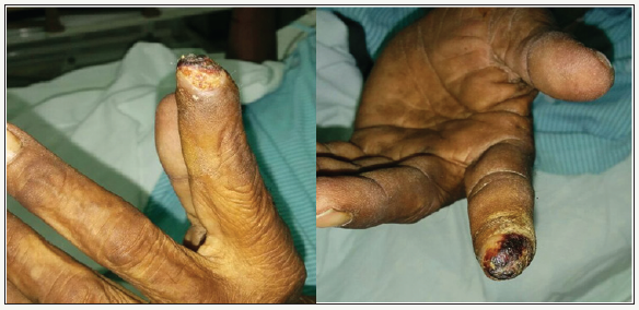
Figure 1:Showing digital infarction of index finger of left hand.

Family history was noncontributory
Clinical examination: pulse rate was 70beats/min, regular with brachial and radial pulse on left side found to be feeble compared to right side. There was left radial artery pulsation delay compared to right radial artery. Lower limb peripheral pulses were normal and equally palpable both side. Blood pressure was 110/70mmHg in the left arm, 136/90mmHg on right arm, and 148/80 and 144/80mmHg in right and left leg, respectively. There was a bruit audible over left infraclavicular region. No renal or abdominal bruit heard. Local examination of left hand showed, infarction of left index finger in the distal part with a line of demarcation between normal and infarcted tissue (Figure 1). Other systemic examination was normal.
Investigations: Haemoglobin was 11.0g/dL, total leukocyte count of 7,600/mm3 with normal differential count and platelet count. Erythrocyte sedimentation rate (ESR) was 60mm/1st hr, fasting and postprandial blood glucose of 90 and 136mg/dL, respectively. Liver, renal function, and coagulation profile were normal with lipid profile study showed Total cholesterol of 148mg/ dL, Triglyceride of 123 mg/dL, and Low density lipoprotein (LDL) of 55mg/dL and HDL of 30mg/dl. . Serum ANA, p-ANCA, and c-ANCA were negative. Anticardiolipin antibody was negative.
Cardiac evaluation: Electrocardiogram and two-dimensional (2D) transthoracic echocardiography were normal except left subclavian stenosis in supra sternal view.
Color Doppler study of arterial system of left upper limb showed- approximately 10x4 mm Hypoechoic plaque at left subclavian artery with 80 to 90% narrowing, causing significant hemodynamic compromise of distal part of left upper extremity. Flow velocity at narrowing was 288cm/s and is significantly raised. Invasive peripheral conventional angiography revealed 90 percent stenosis of the left subclavian artery proximal to the origin of the vertebral artery. Right subclavian artery, aorta and its other branches were normal.
Coronary angiography showed normal coronary arteries
Patient underwent percutaneous transluminal angioplasty of left subclavian artery. Patient tolerated the procedure well. Post procedure he was symptomatically better with blood pressure in the left upper limb of 120/70mmHg and right upper limb- of 114/74mmHg. Patient was discharged on dual antiplatelets and statins.
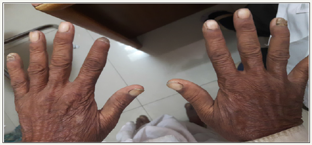
Follow up after 6 months- patient was asymptomatic. Pulse -80 beats/min regular, good volume, no radio radial or radio femoral delay, blood pressure- 110/70mmHg, equal in both upper limbs. Left index finger was completely healed with intact skin and no evidence of ischemia or necrosis (Figure 2). Follow up CT Aortogram showed normal flow in the bilateral subclavian arteries with patent stent in situ in the left subclavian artery (Figure 3).
Figure 2:Post angioplasty to left subclavian artery -showing healing of digital infarction.

Figure 3:CT angiography (6 month Post Angioplasty to left subclavian artery) showing patent stent.

Discussion
The term “subclavian steal” describes retrograde blood flow in the vertebral artery associated with proximal ipsilateral subclavian artery stenosis or occlusion, usually in the setting of subclavian artery occlusion or stenosis proximal to the origin of the vertebral artery. Alternatively, innominate artery disease has also been associated with retrograde flow in the ipsilateral vertebral artery, particularly where the subclavian artery origin is involved.
Subclavian steal phenomenon is frequently asymptomatic and may be discovered incidentally on ultrasound or angiographic examination for other indications, or it may be prompted by a clinical examination finding of reduced unilateral upper limb pulse or blood pressure. In some cases, patients may develop upper limb ischemic symptoms due to reduced arterial flow in the setting of subclavian artery occlusion, or they may develop neurologic symptoms due to posterior circulation ischemia associated with exercise of the ipsilateral arm [1-3].
Pathophysiology
The upper limb is supplied primarily via the axillary artery, the continuation of the subclavian artery that exits the thoracic outlet. On the right, the common carotid artery and the subclavian artery share a common trunk, known perplexingly as the in nominate artery. On the left, the subclavian artery typically arises directly from the aorta as the last supra-aortic trunk.
Proximal subclavian artery stenosis or occlusion, mainly due to atherosclerotic disease, causes insufficient flow distally. In this case, the branches of the subclavian artery may be recruited to provide collateral retrograde flow to the upper limb. Of greatest relevance for present purposes is the confluence of the vertebral arteries at the basilar artery and its subsequent communication with the circle of Willis, which allows the ipsilateral vertebral artery to provide flow in a retrograde manner from the contralateral vertebral artery or from the anterior cerebral circulation.
With exercise, innate and metabolite-induced vasodilatation leads to a drop in peripheral resistance in upper-limb vessels, and the mismatch between arterial inflow and metabolic demand may lead to claudication of the arm. Furthermore increased retrograde flow through the ipsilateral vertebral artery may “steal” blood away from the cerebral circulation. This may be more likely if there is concomitant stenotic disease of the other extracranial or intracranial vessels. In these patients, neurologic symptoms consistent with cerebral or brainstem ischemia may develop. In a 1991 study of 43 patients undergoing carotid duplex study who were incidentally found to have retrograde vertebral artery flow, 16% had posterior circulation symptoms (dizziness, vertigo, blurred vision, diplopia, and near-syncope) upon exercise of the ipsilateral arm; 30% had similar symptoms but present even at rest; 21% had anterior circulation hemispheric symptoms referable to a carotid territory; and 33% were asymptomatic at all times [2].
Takayasu arteritis (TA)
For diagnosis of TA, a criterion was formed by American College of Rheumatology. The diagnosis is made when three out of six points are present. The sensitivity and specificity for diagnosis TA are 91 and 98%, respectively.
Our patient met 3 out of 6 criteria.
American College of Rheumatology 1990 criteria for the classification of Takayasu’s arteritis
1. Age at onset of disease less than or equal to 40 years.
2. Claudication of extremities: development and worsening of fatigue and discomfort in muscles of 1 or more extremity while in use.
3. Decreased brachial artery pulse.
4. Systolic BP difference greater than 10mmHg between arms.
5. Bruit over subclavian arteries or abdominal aorta.
6. Arteriographic abnormality: narrowing or occlusion of the entire aorta, its primary branches or large arteries in the proximal upper or lower extremities, not due to arteriosclerosis, fibromuscular dysplasia, or similar causes; changes usually focal or segmental.
The patient had gangrene involving index finger of the left hand. Digital gangrene in a patient of Takayasu arteritis is very rare manifestation. Till now, few cases of digital gangrene involving lower limbs and only one case of digital gangrene involving hand were reported worldwide in TA [4-13]. Patient is nonsmoker and nondiabetic. There was no history of Raynaud’s phenomenon, rash, arthritis, oral ulceration, and hemoptysis. There was no evidence of sepsis. Serum ANA, p-ANCA, and c-ANCA were negative. Lipid profile and CIMT were normal. Prothrombin time and partial thromboplastin time were normal. Antiphospholipids antibody syndrome was excluded as anticardiolipin antibody was negative. Echocardiography did not show any vegetation or left atrial clot.
Takayasu arteritis can be divided into the following six types based on angiographic involvement:
1. Type I - Branches of the aortic arch
2. Type IIa - Ascending aorta, aortic arch, and its branches
3. Type IIb - Type IIa region plus thoracic descending aorta
4. Type III - Thoracic descending aorta, abdominal aorta, renal arteries, or a combination
5. Type IV - Abdominal aorta, renal arteries, or both
6. Type V - Entire aorta and its branches.
According to this classification our case belongs to type 1 Takayasu artertitis. Digital gangrene in upper limb is very uncommon as there is abundance of collaterals and rarity of atherosclerosis. So, this patient who was suffering from TA with digital gangrene is a very rare manifestation. One should always consider TA as a differential diagnosis of digital gangrene.
References
- Reivich M, Holling HE, Roberts B, Toole JF (1961) Reversal of blood flow through the vertebral artery and its effect on cerebral circulation. N Engl J Med 265: 878-885.
- Ochoa VM, Yeghiazarians Y (2011) Subclavian artery stenosis: a review for the vascular medicine practitioner. Vasc Med 16(1): 29-34.
- Hennerici M, Klemm C, Rautenberg W (1988) The subclavian steal phenomenon: a common vascular disorder with rare neurologic deficits. Neurology 38(5): 669-673.
- Verma S, Saraf S, Himanshu D, Singh S (2013) Takayasu’s arteritis presenting as digital gangrene of right hand. BMJ Case Rep 2013.
- Schatzl S, Karnik R, Gattermeier M (2013) Coronary subclavian steal syndrome: an extra coronary cause of acute coronary syndrome. Wien Klin Wochenschr 125(15-16): 437-438.
- Aboyans V, Aruna K, Allison MA, McDermott, Crouse JR, et al. (2010) The epidemiology of subclavian stenosis and its association with markers of subclinical atherosclerosis: the Multi-Ethnic Study of Atherosclerosis (MESA). Atherosclerosis 211(1): 266-270.
- Labropoulos N, Nandivada P, Bekelis K (2010) Prevalence and impact of the subclavian steal syndrome. Ann Surg 252(1): 166-170.
- Harper C, Cardullo PA, Weyman AK, Patterson RB (2008) Transcranial Doppler ultrasonography of the basilar artery in patients with retrograde vertebral artery flow. J Vasc Surg 48(4): 859-864.
- Rogers JH, Calhoun RF (2007) Diagnosis and management of subclavian artery stenosis prior to coronary artery bypass grafting in the current era. J Card Surg 22(1): 20-25.
- Clark CE, Taylor RS, Shore AC, Ukoumunne OC, Campbell JL (2012) Association of a difference in systolic blood pressure between arms with vascular disease and mortality: a systematic review and meta-analysis. Lancet 379(9819): 905-914.
- Betensky BP, Jaeger JR, Woo EY (2011) Unequal blood pressures: a manifestation of subclavian steal. Am J Med 124(8): e1-e2.
- (2011) Takayasu arteritis. Eur J Cardiothorac Surg 40: 1268.
- Vecera J, Vojtísek P, Varvarovský I, Lojík M, Másová K, et al. (2010) Noninvasive diagnosis of coronary-subclavian steal: role of the Doppler ultrasound. Eur J Echocardiogr 11(9): E34.
© 2018 Abhijeet Shelke. This is an open access article distributed under the terms of the Creative Commons Attribution License , which permits unrestricted use, distribution, and build upon your work non-commercially.
 a Creative Commons Attribution 4.0 International License. Based on a work at www.crimsonpublishers.com.
Best viewed in


.jpg)


 Editorial Board Registrations
 Submit your Article
 Refer a Friend
 Advertise With Us
.jpg)


.jpg)


.bmp)
.jpg)
.png)
.jpg)


.jpg)


.png)

.png)


.png)


