- Submissions

Full Text
Novel Research in Sciences
Microscopic Appearance of Synthetic DNA (E. coli) Crown Cells in Secondary Cultures
Shoshi Inooka*
Japan Association of Science Specialists, Japan
*Corresponding author: Shoshi Inooka, Japan Association of Science Specialists, Japan
Submission: October 26, 2022;Published: November 07, 2022
.jpg)
Volume12 Issue4November , 2022
Abstract
DNA crown cells (artificial cells), in which the outer membrane is covered with DNA, can be readily synthesized in vitro using Sphingosine (Sph)-DNA-adenosine mixtures. Previous experiments demonstrated that assemblies of synthetic DNA (E. coli) crown cells formed naturally or in response to the presence of inorganic substances, and that modified synthetic DNA crown cells were produced within such assemblies. Moreover, the cells in these assemblies formed crystal-like substances and the cells were regenerated with further monolaurin treatment. Recent reports examined whether DNA crown cells, including regenerated synthetic DNA (E. coli) crown cells, could be cultivated in vitro. The cultivation of synthetic DNA (E. coli) crown cells was performed using egg white as a culture medium and it was found that such cells exit in medium after culturing for 7 days.
The aim of this study was to obtain evidence on whether such DNA crown cells could be cultivated further. Cells including related objects that had been cultivated in primary cultures were transferred to new medium (egg white) and incubated for a further 7 days at 37 C. Then, their microscopic appearance of such cells and related objects were observed in medium. Here, it was demonstrated that cells including related objects exist in the medium after secondary culture and that synthetic DNA (E. coli) crown cells could be cultivated. Here, the microscopic appearance of these cells and related objects are shown.
Keywords:Synthetic DNA crown cells; Sphingosine-DNA; Cell proliferation; In vitro; Cultures
Introduction
Cells covered with DNA and referred to as DNA crown cells, have been described previously [1-3]. Synthetic DNA crown cells can be prepared using Sphingosine (Sph)-DNA and Adenosine-Monolaurin (A-M) compounds [4,5]. DNA crown cells can be generated by incubating synthetic DNA crown cells in egg white. Unlimited numbers of synthetic DNA crown cells and DNA crown cells can be prepared [6-8]. In a previous experiment [9], assemblies of synthetic DNA (E. coli) crown cells were produced using commercial salt, or spontaneously. Such assemblies formed crystal-like substances after the addition of monolaurin [10]. Cells can be regenerated from the assemblies [11], which formed crystal-like substances. In a previous report [12], to clarify whether such regenerated cells could be produced, these cells and synthetic DNA crown cells were cultivated using egg white for 7 days at 37 C. The findings showed that such cells or related objects existed in the medium after cell culture.
In the present study, to obtain more evidence, cells and cell-related objects that had been in primary culture medium for 7 days were cultivated. These cells were transferred into new medium (egg white) and incubated for a further 7 days at 37 C and cells and related objects in culture medium were observed. The findings showed that cells and related subjects could be observed in medium after secondary cultures were performed and that these cells and related subjects could be cultivated. Here, the microscopic appearance of these cells and related objects are shown.
Materials and Methods
Materials
The following materials were used: Sph (Tokyo Kasei, Japan); DNA (E. coli B1 strain, Sigma-Aldrich, USA); adenosine (Sigma- Aldrich and Wako, Japan); and monolaurin (Tokyo Kasei), A-M compound synthesized from a mixture of adenosine and monolaurin [5]. Monolaurin solutions were prepared to a concentration of 0.1M in distilled water. Egg white was obtained from eggs bought at local markets.
Methods
Preparation of DNA crown cells: The generation of artificial cells using Sph-DNA-A-M was performed as described previously [5]. Briefly, 180μL of Sph (10mM) and 90L of DNA (1.7g/L) were combined, and the mixture was heated and cooled twice. A-M solution (100L) was added, and the mixture was then incubated at 37 0C for 15 minutes. Next, 30μL of monolaurin solution was added and the mixture was incubated at 37 0C for another 5 minutes. The resulting suspension was diluted with distilled water two times and used as the synthetic DNA (E. coli) crown cells.
Sample preparation and procedures
a. A total of 25μL of synthetic DNA crown cells was incubated
for 18 hours at 37 0C
b. A total of 25μL of monolaurin was added to synthetic DNA
crown cells
c. The mixture was incubated for 18, 36 and 54 hours at
37 C. Sample No. 1 was incubated for 18 hours, sample No. 2 was
incubated for 36 hours, sample No. 3 was incubated for 54 hours.
d. Then, 25μL of monolaurin was added to sample No. 1, No.
2 and No. 3 and incubated for 40-60 minutes at 37 C.
Culture
A total of 20l of each sample was added to 200l of egg white and incubated for 7 days at 37 C. Then, 20l of the cultured fluids was added to 200l of new egg white and incubated for 7 days at 37 C (secondary cultures).
Microscopic observations
A total of 20l of sample was placed on a slide glass in to cover an area of 1cm2, which was then covered with a cover glass. The slides were observed under a light microscope.
Results and Discussion
General microscopic appearance of synthetic DNA (E. coli) crown cells in secondary cultures
As described in a previous report [9], when synthetic DNA (E. coli) crown cells were incubated for 18 hours, assemblies were spontaneously formed which changed into crystal-like substances [10] in the presence of monolaurin. Moreover, cells proliferated in the presence of monolaurin. Thus, samples with specific characteristics were cultured in primary cultures. However, it is unclear whether there were clear differences in primary cultures among the samples. On the other hand, it is very important that cells that develop from the samples that were treated with further monolaurin stimulation be studied. Therefore, in secondary cultures, all of the samples that were treated twice with monolaurin were selected and cultured.
The procedure used for the secondary cultures was as follows:
a. A total of 20l of each of these samples was added to
200l of egg white and incubated for 7 days at 37 C.
b. Then, 20l of the fluid was added to 200l of new egg
white, which was then incubated for 7 days at 37 C before being
observed under a microscope.
Cells and cell-like objects were observed in all samples tested. The objects which consist of several rode objects like stick and were rarely observed in primary cultures, were often observed, suggesting that these might be produced by cells. On the other hand, the sample used was diluted more than 100-fold compared to the original sample. The findings showed that DNA crown cells were cultivated. On the other hand, in secondary cultures, it is unclear whether there were clear differences in cultures among samples (No.1, No.2 and No.3) used.
Microscopic appearance of twice monolaurin-treated synthetic DNA crown cells and related objects in secondary cultures
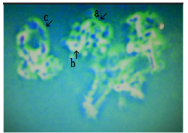
Cells or related objects were observed in all samples. The color in the Figures did not show specific substances, but different colors were used to show different substances (Figures 1-4). Cells or related objects in cultures were yellow-green and orange in color. The yellow-green objects are called outside structures (o-structures) and the orange spaces are called middle structures (m-structures). Most objects, including cells, consisted of o-structures which enclosed m-structures. The objects in (Figure 5) were typical DNA crown cells. Because the outside of the synthetic DNA crown cells consists of sphingosine-DNA, it was considered that the o-structure components were comprised of sphingosine-DNA. On the other hand, synthetic DNA crown cells consist of sphingosine, DNA, adenosine, and monolaurin (Figures 6-10). Therefore, the substances in m-structure components may be composed of the materials that were used in the preparation of synthetic DNA crown cells; adenosine monolaurin and compounds of adenosine-monolaurin. Moreover, the components of egg white used in medium may be used in the formation of such substances. Also, based on the observed microscopic structures, it may be possible propose new mechanisms for the multiplication of DNA crown cells.
Figure 1:Microscopic appearance of secondary cultures of synthetic DNA crown cells twice treated with monolaurin and incubated for 36 hours (Sample No. 2). Many round cells were observed (Figure 1 arrows a, b, c, d, and e). The size of the structure shown by arrow e is approximately 8-10m.
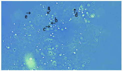
Figure 2:Enlargement of Figure 1 (arrow a). The yellowgreen objects are called outside structures (o-structures) and the orange spacses are called middle structures (m-structures). The outer surfaces of the objects consist of o-structures (arrow a) and the inner surfaces of objects consist of m-structures (arrow b).
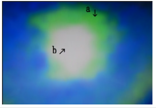
Figure 3:Enlargement of Figure 1 (arrows b). Three differently shaped objects were observed: o-structures (arrow a), m-structures (arrow b). O-structures (arrow d) were derived from m-structures (arrow b). Also, o-structures (arrow c) were derived from o-structures (arrow a).
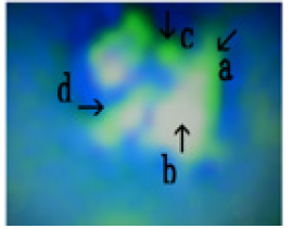
Figure 4:Enlargement of Figure 1 arrow c. Two cells that may have fused with o-structures (arrow c). The outside of cell was covered with fluffy o-structures (arrow a). M-structures were observed within o-structures (arrow b).
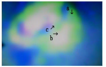
Figure 5:Enlargement of Figure 1 arrow d. Two cells were observed with outsides comprised of o-structures that enclosed m-structures (arrows a and b).
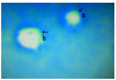
Figure 6:Enlargement of Figure 1 (arrow e). The outside consists of o-structures (arrow a). It is not clear whether the substances were involved (arrow b).
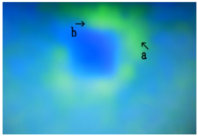
Figure 7:Another picture in Sample No. 2. A cluster of several cells (arrow a) was observed. Also, an object was observed (arrow b).
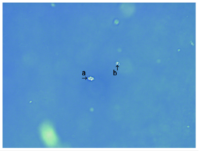
Figure 8:Enlargement of Figure 7 (arrow a). Several cells consisting of o-structures (arrow a) and m-structures (arrow b). were showed. Cells were connected by o-structures (arrow c).
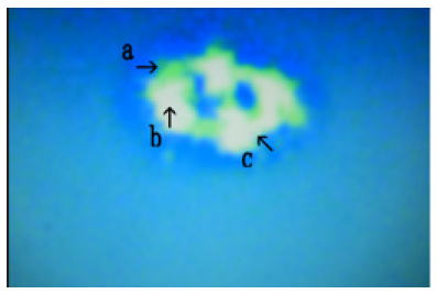
Figure 9:Enlargement of Figure 7 (arrow b). Three objects of various shapes were observed: o-structures (arrow a), m-structures (arrow b), and objects that may form ramifications (arrow c). The object shown by arrow d may be derived from the object shown by arrow b.
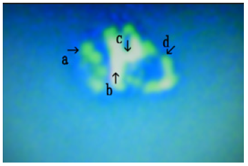
Figure 10:Microscopic appearance of secondary cultures of synthetic DNA crown cells twice treated with monolaurin and incubated for 18 hours (Sample No. 1). Numerous objects with several protuberances (arrows a, b, and c) measuring approximately 20-30m) were observed (arrow b).
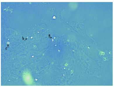
In previous and present experiments, it was demonstrated that several kinds of objects including cells were observed after culturing and most of these objects consists of o-structures and m-structures. Moreover, o-structures may be sphingosine-DNA, and m-structures may be adenosine-monolaurin and include components of egg white which was contained in the culture medium. Microscopic appearances which o-structures were grown were observed (Figure 11 arrows a and b). Moreover, growing o-structures enclosed m-structures and formed cell-like objects (Figures 12 & 13 arrows d and e). The findings suggest that the development of new cells begins with the growth of o-structures, which seem to be sphingosine-DNA fibers (Figures 14-17). These findings on the mechanisms of multiplication in cultivated synthetic DNA crown cells will clarify whether synthetic DNA crown cells can be cultivated, and if so, whether strains can be developed.
Figure 11:Enlargement of Figure 10 (arrow a). Two objects with several protuberances were observed (arrow a). The objects consist of o-structures (arrow b) and m-structures (arrow c).
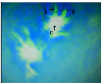
Figure 12:Enlargement of Figure 10 (arrow b). A cell with an exterior composed of o-structures (Figure 12 arrow a) was shown and m-structures in the middle (arrow c). Objects comprised of o-structures enclosing m-structures were observed (arrow b).
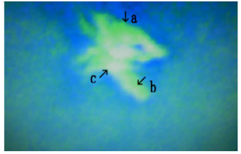
Figure 13:Enlargement of Figure 10 (arrow c). A large object composed of o-structures (arrow a) and m-structures (arrow b). Numerous objects that branched from the large object were observed (arrow c). The objects comprised of o-structures enclosed m-structures (arrows d and e).
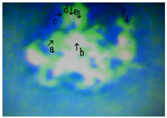
Figure 14:Microscopic appearance of secondary cultures of synthetic DNA crown cells twice treated with monolaurin and incubated for 72 hours and treated with monolaurin (Sample No. 3). A large assembly was observed (arrows a and c). Objects with several outgrowths were observed (arrows b and d). The size is approximately 20-30μm (arrow b).
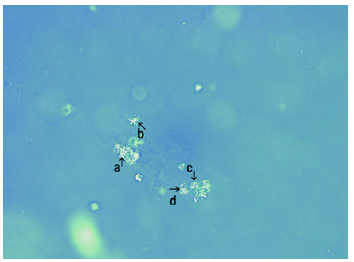
Figure 15:Enlargement of Figure 14 (arrow a). Many objects with various shapes consisting of o-structures (arrow a) enclosing m-structures (arrow b) were observed. Objects that may have been produced by cells were observed (arrows c and d).
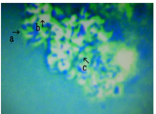
Figure 16:Enlargement of Figure 14 (arrow c). An object (arrow b) with many ramifications (arrows a, c, and d) was observed. The ramifications were composed of o-structures which enclosed m-structures (arrow b).
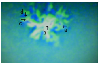
Figure 17:Enlargement of Figure 14 (arrows c and d). Several objects consisting of o-structures (arrow a) and m-structures (arrow b) were observed. Object like stick or rode that were composed of o-structures and m-structures were observed (arrow c)
References
- Inooka S (2012) Preparation and cultivation of artificial cells. App Cell Biol 25: 13-18.
- Inooka S (2016) Preparation of artificial cells using eggs with sphingosine-DNA. J Chem Eng Process Technol 7(1): 1-5.
- Inooka S (2016) Aggregation of sphingosine-DNA and cell construction using components from egg white. Integr Mol Med 3(6): 1-5.
- Inooka S (2017) Biotechnical and systematic preparation of artificial cells (DNA Crown Cells). The Global Journal of Researches in Engineering 17(c1): 1-11.
- Inooka S (2017) Systematic preparation of artificial cells (DNA Crown Cells). J Chem Eng Process Technol 8(1): 1-4.
- Inooka S (2017) Systematic preparation of bovine meat DNA crown cells. App Cell Biol 30: 13-16.
- Inooka S (2018) Systematic preparation of DNA (Akoya pearl oyster) crown cells. App Cell Biol 31: 21-34.
- Inooka S (2019) Preparation of DNA (Nannochloropsis species) crown cells using eggs, sphingoshin and DNA, and subsequent cell recovery. App Cell Biol 32: 55-64.
- Inooka S (2022) Assemblies formation of synthetic DNA ( coli) crown cells with salt. Reconstruction and regeneration of synthetic DNA crown cells. American Journal of Biomedical Science & Research 16(1): 1-10.
- Inooka S (2022) The assembly in synthetic DNA ( coli) crown cells with inorganic salts and the transformation of DNA crown cells to crystal-like substances in the presence of monolaurin. Novel Research Sciences 11(2): 1-8.
- Inooka S (2022) Cell proliferation from the assembly of synthetic DNA ( coli) crown cells with stimulation by monolaurin twice. Curr Tr Biotech & Microbio 2(5): 493-498.
- Inooka S (2022) Microscopic appearance of synthetic DNA ( coli) crown cells in primary cultures. App Cell Bio, Japan.
© 2022 Shoshi Inooka. This is an open access article distributed under the terms of the Creative Commons Attribution License , which permits unrestricted use, distribution, and build upon your work non-commercially.
 a Creative Commons Attribution 4.0 International License. Based on a work at www.crimsonpublishers.com.
Best viewed in


.jpg)


 Editorial Board Registrations
 Submit your Article
 Refer a Friend
 Advertise With Us
.jpg)


.jpg)


.bmp)
.jpg)
.png)
.jpg)


.jpg)


.png)

.png)


.png)


